- Protocols

- Articles and Issues
- For Authors
- About
- Become a Reviewer

Past Issue in 2018
Volume: 8, Issue: 3
Biochemistry
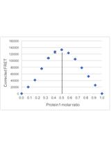
FRET-based Stoichiometry Measurements of Protein Complexes in vitro


Cell Biology
Tracking Endocytosis and Intracellular Trafficking of Epitope-tagged Syntaxin 3 by Antibody Feeding in Live, Polarized MDCK Cells
Ex vivo Analysis of Lipolysis in Human Subcutaneous Adipose Tissue Explants
Measurement of Lysosomal Size and Lysosomal Marker Intensities in Adult Caenorhabditis elegans
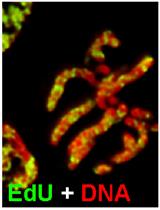
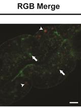
Analysis of Chromosome Condensation/Decondensation During Mitosis by EdU Incorporation in Nigella damascena L. Seedling Roots

Developmental Biology
How to Catch a Smurf? – Ageing and Beyond…
In vivo Assessment of Intestinal Permeability in Multiple Model Organisms

Preparation of Precisely Oriented Cryosections of Undistorted Drosophila Wing Imaginal Discs for High Resolution Confocal Imaging

Microbiology
Extraction of DNA from Murine Fecal Pellets for Downstream Phylogenetic Microbiota Analysis by Next-generation Sequencing
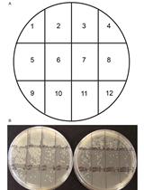
Quantification of Neisseria meningitidis Adherence to Human Epithelial Cells by Colony Counting
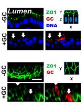
Immunofluorescence Analysis of Human Endocervical Tissue Explants Infected with Neisseria gonorrhoeae
Molecular Biology
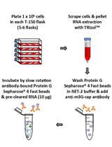
Immunoprecipitation of Tri-methylated Capped RNA

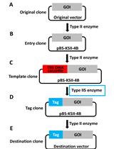
Precision Tagging: A Novel Seamless Protein Tagging by Combinational Use of Type II and Type IIS Restriction Endonucleases

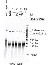
Measuring Nucleosome Assembly Activity in vitro with the Nucleosome Assembly and Quantification (NAQ) Assay
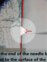
Medaka-microinjection with an Upright Microscope

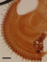
Determination of DNA Damage in the Retina Photoreceptors of Drosophila
.JPG)
Neuroscience
Culture and Nucleofection of Postnatal Day 7 Cortical and Cerebellar Mouse Astroglial Cells
Plant Science
Investigating Localization of Chimeric Transporter Proteins within Chloroplasts of Arabidopsis thaliana
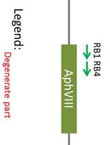
Identification of Insertion Site by RESDA-PCR in Chlamydomonas Mutants Generated by AphVIII Random Insertional Mutagenesis

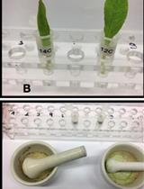
Radioactive Tracer Feeding Experiments and Product Analysis to Determine the Biosynthetic Capability of Comfrey (Symphytum officinale) Leaves for Pyrrolizidine Alkaloids
Systems Biology
Extraction and Analysis of Pan-metabolome Polar Metabolites by Ultra Performance Liquid Chromatography–Tandem Mass Spectrometry (UPLC-MS/MS)