- Protocols

- Articles and Issues
- For Authors
- About
- Become a Reviewer

Past Issue in 2017
Volume: 7, Issue: 12
Biochemistry
Thermostability Measurement of an α-Glucosidase Using a Classical Activity-based Assay and a Novel Thermofluor Method


Fluorophore Labeling, Nanodisc Reconstitution and Single-molecule Observation of a G Protein-coupled Receptor
Biophysics
Modification and Application of a Commercial Whole-body Plethysmograph to Monitor Respiratory Abnormalities in Neonatal Mice

Cancer Biology

Soft Agar Colony Formation Assay as a Hallmark of Carcinogenesis
Cell Biology
Functional ex-vivo Imaging of Arterial Cellular Recruitment and Lipid Extravasation


Developmental Biology
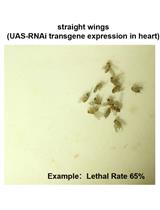
Validating Candidate Congenital Heart Disease Genes in Drosophila


Microbiology
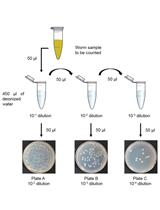
Culturing Bacteria from Caenorhabditis elegans Gut to Assess Colonization Proficiency


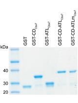
Producing GST-Cbx7 Fusion Proteins from Escherichia coli

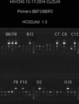
Single Genome Sequencing of Expressed and Proviral HIV-1 Envelope Glycoprotein 120 (gp120) and nef Genes

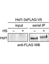
Serial Immunoprecipitation of 3xFLAG/V5-tagged Yeast Proteins to Identify Specific Interactions with Chaperone Proteins

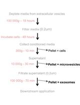
Loading of Extracellular Vesicles with Chemically Stabilized Hydrophobic siRNAs for the Treatment of Disease in the Central Nervous System
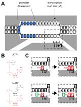
RNA Capping by Transcription Initiation with Non-canonical Initiating Nucleotides (NCINs): Determination of Relative Efficiencies of Transcription Initiation with NCINs and NTPs

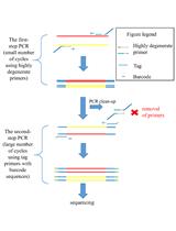
Tagged Highly Degenerate Primer (THDP)-PCR for Community Analysis of Methane- and Ammonia-oxidizing Bacteria Based on Copper-containing Membrane-bound Monooxygenases (CuMMO)

Molecular Biology
Single-molecule RNA Fluorescence in situ Hybridization (smFISH) in Caenorhabditis elegans
Dense sgRNA Library Construction Using a Molecular Chipper Approach
Modification of 3’ Terminal Ends of DNA and RNA Using DNA Polymerase θ Terminal Transferase Activity
In vitro Assay to Measure DNA Polymerase β Nucleotide Insertion Coupled with the DNA Ligation Reaction during Base Excision Repair
Neuroscience

Stereotaxic Surgery for Suprachiasmatic Nucleus Lesions in Mice

Optogenetic Stimulation and Recording of Primary Cultured Neurons with Spatiotemporal Control
Contusion Spinal Cord Injury Rat Model

Transplantation of Embryonic Cortical Tissue into Lesioned Adult Brain in Mice
Representation-mediated Aversion as a Model to Study Psychotic-like States in Mice

Plant Science
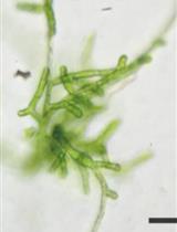
Phototaxis Assay for Chlamydomonas reinhardtii


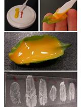
Estimation of Stomatal Aperture in Arabidopsis thaliana Using Silicone Rubber Imprints

Targeted Mutagenesis Using RNA-guided Endonucleases in Mosses
Analysis of 3D Cellular Organization of Fixed Plant Tissues Using a User-guided Platform for Image Segmentation
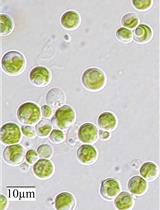
Extraction and Activity of O-acetylserine(thiol)lyase (OASTL) from Microalga Chlorella sorokiniana


Root Aliphatic Suberin Analysis Using Non-extraction or Solvent-extraction Methods

