- Protocols

- Articles and Issues
- For Authors
- About
- Become a Reviewer

Past Issue in 2015
Volume: 5, Issue: 6
Cancer Biology

Detection of Phospho-KRAS by Electrophoretic Mobility Change in Human Cell Lines and in Tumor Samples from Nude Mice Grafts
Microbiology
RNA Isolation from Synechocystis
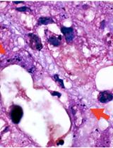
Gram Stain for Intestinal Bacteria

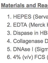
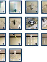
Neutrophil Isolation from the Intestines
Rapid Preparation of Unsheathed Bacterial Flagella

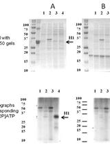
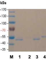
Substrate Specificity of Recombinant Ser/Thr Protein Kinase
Plant Science
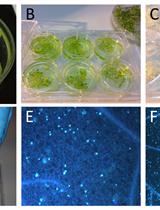
Staining of Callose Depositions in Root and Leaf Tissues

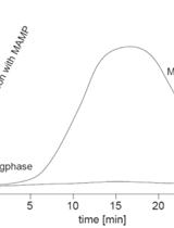
Chemiluminescence Detection of the Oxidative Burst in Plant Leaf Pieces
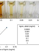
Lignin Extraction and Quantification, a Tool to Monitor Defense Reaction at the Plant Cell Wall Level

Scanner-based Time-lapse Root Phenotyping


A Chemiluminescence Based Receptor-ligand Binding Assay Using Peptide Ligands with an Acridinium Ester Label
Airbrush Infiltration Method for Pseudomonas syringae Infection Assays in Soybean


