- Protocols

- Articles and Issues
- For Authors
- About
- Become a Reviewer

Past Issue in 2015
Volume: 5, Issue: 5
Immunology
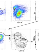
Isolation of Splenic Dendritic Cells Using Fluorescence-activated Cell Sorting

Microbiology
Vesicle Isolation from Bacillus subtilis Biofilm
Differentiation of Naturally Produced Extracellular Membrane Vesicles from Lipid Aggregation by Glucuronoxylomannan Immunogold Transmission Electron Microscopy in Bacillus subtilis
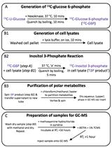
A Gas Chromatography-Mass Spectrometry-Based Two Stage Assay for Measurement of in vitro myo-Inositol 3-phosphate Synthase (INO1) Activity

Neuroscience
Thirty-Second Net Stressor Task in Adult Zebrafish


Plant Science
An Efficient Procedure for Protoplast Isolation from Mesophyll Cells and Nuclear Fractionation in Rice

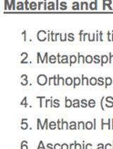
Citrus Fruit Ascorbic Acid Extraction and Quantification by HPLC


Isolation of Polysome-bound mRNA from Rice Solid Tissues Amenable for RT-PCR and Profiling Experiments
Extraction of Small RNA and qPCR Validation of miRNAs in Vigna mungo


Measurement of Cellular Redox in Pollen with Redox-Sensitive GFP (roGFP) Using Live Cell Imaging


RNA Editing Detection by Direct Sequencing