Past Issue in 2015

Volume: 5, Issue: 4

left

Dec

20

Dec

5

Nov

20

Nov

5

Oct

20

Oct

5

Sep

20

Sep

5

Aug

20

Aug

5

Jul

20

Jul

5

Jun

20

Jun

5

May

20

May

5

Apr

20

Apr

5

Mar

20

Mar

5

Feb

20

Feb

5

Jan

20

Jan

5

right

Biochemistry

Determination of Protein-DNA (ZMYND11-DNA) Interaction by a Label-Free Biolayer Interferometry Assay

Determination of Protein-DNA (ZMYND11-DNA) Interaction by a Label-Free Biolayer Interferometry Assay

YL Yuan-Yuan Li
HW Hong Wen
XS Xiao-Bing Shi
Hai-Tao Li Hai-Tao Li
12972 Views
Feb 20, 2015
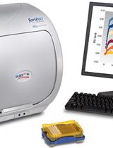
This protocol describes a robust technique for the measurement of ZMYND11-DNA interaction by a label-free Biolayer Interferometry (BLI). ZMYND11 is a novel histone reader protein that specifically recognizes H3.3K36me3 via its tandem Bromodomain, zinc-finger and PWWP domain (BP). ZMYND11 links the histone-variant-mediated transcription elongation control to tumour suppression and may therefore represent a novel class of drug targets. Like other PWWP domains, ZMYND11 PWWP domain shows highly positively charged surface and interacts with DNA. Previously reported methods include NMR, FP or EMSA. Biolayer interferometry (BLI) is an emerging technology for analyzing all kinds of biomolecular interactions, such as protein-protein and protein-DNA binding. BLI allows for the real time monitoring of the interactions between biomolecules without the need for reagents with enzymatic, fluorescent, or radioactive labels. The technology is based upon the changes in interference pattern of light reflected from the surface of an optical fiber when materials bind to the tip of the fiber. The technique represents an alternative to technologies such as surface plasmon resonance, providing a simple platform that enables label-free monitoring of biomolecular interactions without the use of flow cells. Label-free biosensor methods provide information on binding, kinetics, concentration, and the affinity of an interaction.
In vitro Chitin Binding Assay

In vitro Chitin Binding Assay

Geneviève Ball Geneviève Ball
Frédéric Cadoret Frédéric Cadoret
Romé Voulhoux Romé Voulhoux
12380 Views
Feb 20, 2015
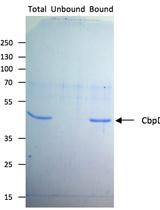
Chitin is polymer of N-acetylglucosamine (GlcNAc) found in the exoskeleton of arthropods and the fungal cell wall. GlcNAc is also implicated in bacterial development, adherence, and signal transduction but can also be used as a carbon source. In vitro chitin binding assay is performed to determine the affinity of a purified protein to the chitin molecule. The principle is based on the co-sedimentation of chitin-binding proteins together with chitin-coated beads.

Immunology

Isolation of Lung Infiltrating Cell in Mice

Isolation of Lung Infiltrating Cell in Mice

EA Eduardo P. Amaral
EL Elena B. Lassunskaia
MD Maria Regina D’Império-Lima
19897 Views
Feb 20, 2015
Inflammatory lung diseases induce strong leukocyte recruitment into the organ, culminating in pneumonia area formation. Here, we describe the protocol for isolation of lung infiltrating cells. Using this assay, we analyzed the lung cell phenotyping by flow cytometry and spontaneous cytokine production by cultivating lung cells ex vivo (Amaral et al., 2014).
Macrophage Phagocytosis Assay of Staphylococcus aureus by Flow Cytometry

Macrophage Phagocytosis Assay of Staphylococcus aureus by Flow Cytometry

QY Qin Yan
SA Sun Hee Ahn
VF Vance G. Fowler Jr
15256 Views
Feb 20, 2015
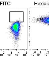
This protocol describes a straightforward technique to evaluate the phagocytotic capacity of murine macrophages for Staphylococcus aureus (S. aureus). By staining S. aureus with Hexidium Iodide and staining murine bone marrow-derived macrophages (BMDMs) with FITC, the macrophage bacterial up-taking ability can be rapidly analyzed by flow cytometry. S. aureus is a Gram-positive bacteria causing severe human and animal infections. Host immune cells such as macrophages serve to eliminate S. aureus by phagocytosing the pathogen and save the host from life-threatening diseases. Study of host macrophage ability to phagocytose S. aureus is important for understanding the host-pathogen interaction and can help to elucidate the pathogenesis of S. aureus infection. This protocol may also be applied for macrophage phagocytotic assay of other gram-positive bacteria.

Neuroscience

Preparation of Synaptosomes from the Motor Cortex of Motor Skill Trained Mice

Preparation of Synaptosomes from the Motor Cortex of Motor Skill Trained Mice

Anand Suresh Anand Suresh
Anna Dunaevsky Anna Dunaevsky
14163 Views
Feb 20, 2015
Learning and memory are thought to occur due to changes in synaptic strength. Strengthening of synapses due to Long Term Potentiation mechanisms are mediated by increases in synaptic α-amino-3-hydroxy-5-methyl-4-isoxazolepropionic acid receptor (AMPAR) levels. Here we describe a protocol to isolate and quantify synaptic AMPAR subunit GluA1 levels from the motor cortex of mice which have undergone motor skill training.

Plant Science

Protein Degradation Assays in Arabidopsis Protoplasts

Protein Degradation Assays in Arabidopsis Protoplasts

Filip Mituła Filip Mituła
Anna Kasprowicz-Maluśki Anna Kasprowicz-Maluśki
MM Michał Michalak
MM Małgorzata Marczak
KK Konrad Kuczyński
Agnieszka Ludwików Agnieszka Ludwików
15662 Views
Feb 20, 2015
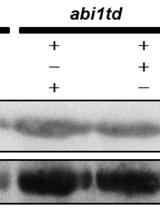
Plant transformation and exogenous protein expression is essential for molecular biology and biotechnology. Current approaches of stable plant transformation might be problematic and very time-consuming. Because of this, transient expression in protoplasts has become valuable alternative, being less cost and time-effective at the same time. Excellent for eukaryotic proteins, representing a natural cell habitat, protoplast isolation is widely used in protein interaction visualization techniques, like BiFC (Bimolecular fluorescence complementation) and FRET (Förster resonance energy transfer). In this protocol we present a another use of Arabidopsis protoplast in protein degradation assay, proving its high versatility as a tool in proteomics.
Isolation of Chloroplast Inner and Outer Envelope Membranes

Isolation of Chloroplast Inner and Outer Envelope Membranes

SW Shiwen Wang
Lina Yin Lina Yin
JM Jun’ichi Mano
KT Kiyoshi Tanaka
14187 Views
Feb 20, 2015
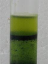
The chloroplast is an important organelle found in plant cells that conduct photosynthesis. It is enclosed by a pair of closely spaced membranes, the double-membrane envelope, consisting of the inner membrane bounding the matrix or stroma and the outer membrane in contact with the cytoplasm. Like many bio-membranes, the chloroplast envelope plays an important role in mediating the complex interactions between the chloroplast and the cytoplasm. The envelope is also the site of various biosynthetic reactions, including the formation of the galactolipids, which are the major components of both envelope and the thylakoid membranes. The inner and outer envelope membranes have differences in both structure and function. For example, the outer membrane exhibits lower density of intramembranous particles than the inner membrane dose, suggesting that the protein content of the outer membrane is low. Also, the outer membrane is nonspecifically permeable to low molecular weight compounds, whereas the inner is impermeable to such compounds and contains several translocator systems for the transport of metabolites. To prepare the envelope membranes, it is necessary to isolate intact chloroplasts first. Then the inner and outer envelope membranes are separated by: 1) the protease-treatment method and 2) the centrifuge method which based on the fact that the outer envelope is lighter and the inner membrane heavier. Both methods need to isolate the intact chloroplasts firstly. However, the centrifugal separation can get the pure inner and outer envelope preparations, which therefore are suitable to the subsequent analyses. Also, the centrifuge method can avoid the destruction of inner envelope polypeptides during the protease treatment, because some of the protease may gain access to the inner membrane. Moreover, the centrifuge method is easy to operate and to get the complete enveloped that contain less of the adhesion regions of the outer and inner envelope membranes. Here we describe a reliable method for isolation of the inner and outer envelope membranes of the chloroplasts from tobacco, which is the plant that relatively not easy to use for envelope isolation.
Phenol-based Total Protein Extraction from Lily Plant Tissues

Phenol-based Total Protein Extraction from Lily Plant Tissues

YC Yun-Chu Chen
YH Yu-Chun Hsiao
CW Co-Shine Wang
11285 Views
Feb 20, 2015
The phenol-based total protein extraction method is unique in that water-soluble components such as polyphenolic compounds and nucleic acids can be easily removed. Thus, total protein is free from contaminants and allows for high quality two–dimensional gel electrophoresis. The phenol-based extraction of total protein was used in various lily organs and may likely apply to other plants whose content of polyphenolics is high (Note 1). An additional advantage of this extraction method is that nucleic acids can be easily removed and thus, avoid adverse effects of nucleic acids on protein resolution in the gel. This method is modified from that of Hurkman and Tanaka (1986).
Transmission Electron Microscopy for Tobacco Chloroplast Ultrastructure

Transmission Electron Microscopy for Tobacco Chloroplast Ultrastructure

Lina Yin Lina Yin
SW Shiwen Wang
NS Norihiro Shimomura
KT Kiyoshi Tanaka
10843 Views
Feb 20, 2015
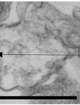
The chloroplast is the site of photosynthesis that enabled and sustains aerobic life on Earth. Chloroplasts are relatively large organelles with a diameter of ~5 μm and width of ~2.5 μm, and so can be readily analysed by electron microscopy. Each chloroplast is enclosed by two envelope membranes, which encompass an aqueous matrix, the stroma and the thylakoids. Components of stroma include starch granules and plastoglobuli, which can be observed by electron microscopy. And the thylakoids consist of stromal thylakoid, granal thylakoid and as well as granum (a stack of thylakoids). These structure components are quite sensitive to developmental changes and environmental variations, such as drought, salinity, cold, high temperature and others. Transmission electron microscopy (TEM) is a powerful technique for monitoring the effects of various changing parameters or treatments on the development and differentiation of these important organelles. Here we describe a reliable method for the analysis of plastid ultrastructure in tobacco plant by TEM.
Separation of Microspores from Anthers of Lilium longiflorum (Lily) and Subsequent RNA Extraction

Separation of Microspores from Anthers of Lilium longiflorum (Lily) and Subsequent RNA Extraction

ML Ming-Che Liu
YC Yun-Chu Chen
CW Co-Shine Wang
10008 Views
Feb 20, 2015
This protocol has been designed in order to facilitate the isolation and extraction of total RNA from microspores collected from lily anther sacs. This protocol allows the extraction of high amounts of high quality RNA, as observed in agarose gels.

Systems Biology

Whole Genome Bisulfite Sequencing and DNA Methylation Analysis from Plant Tissue

Whole Genome Bisulfite Sequencing and DNA Methylation Analysis from Plant Tissue

Daniela Pignatta Daniela Pignatta
GB George W. Bell
MG Mary Gehring
20177 Views
Feb 20, 2015
This protocol describes whole genome bisulfite-sequencing library preparation from plant tissue and subsequent data analysis. Allele-specific methylation analysis and genome-wide identification of differentially methylated regions are additional features of the analysis procedure.