- Protocols

- Articles and Issues
- For Authors
- About
- Become a Reviewer

Past Issue in 2015
Volume: 5, Issue: 2
Immunology
Phenotyping of Live Human PBMC using CyTOFTM Mass Cytometry

Isolation of CNS-infiltrating and Resident Microglial Cells
Microbiology
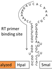
Determination of the Secondary Structure of an RNA fragment in Solution: Selective 2`-Hydroxyl Acylation Analyzed by Primer Extension Assay (SHAPE)
In vitro Dynamic Model of a Catheterized Bladder and Biofilm Assay


Loading of Cells with Fluorescent Probe to Study Intracellular Acid-base Homeostasis in Lactic Acid Bacteria
Mitochondrial Biogenesis Assay after 5-day Treatment in PC-3 Cells


Molecular Biology
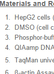
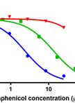
Determination of Mitochondrial DNA Upon Drug Treatment
Plant Science
Camalexin Quantification in Arabidopsis thaliana Leaves Infected with Botrytis cinerea


Stem Cell
PBMC-MSC Co-cultures for Induction of Treg Generation

Monocyte-MSC Co-cultures