


A Simplified Workflow for 3D Segmentation of Mouse Embryo Nuclei Using Biom3D

(§Technical contact: herve.alegot@uca.fr) 发布: 2026年11月20日第16卷第22期 DOI: 10.21769/BioProtoc.5878 浏览次数: 31
评审: Raniki KumariShensheng ZhaoAnonymous reviewer(s)
 PDF
 Q&A





Abstract
Three-dimensional segmentation of cell nuclei is a fundamental step in quantitative studies of morphogenesis and early embryonic development, enabling the analysis of cellular organization and behavior in complex biological tissues. However, accurate segmentation of densely packed nuclei in large volumetric microscopy datasets remains technically challenging, as touching or closely apposed nuclei are frequently merged by instance-based segmentation approaches. Several deep learning–based approaches have been developed to address this problem, including convolutional neural network architectures and shape-based segmentation methods. Although these techniques have improved segmentation performance, their adoption is often limited by complex software installation procedures, demanding dataset annotation requirements and computational expertise. Here, we present a user-oriented workflow for 3D segmentation of nuclei in mouse embryos designed to simplify and speed up image annotation, requiring little manual intervention. The protocol combines a dual-channel imaging strategy, in which a high-resolution channel optimized for nuclear boundary detection is used to generate ground-truth annotations, while a lower-resolution DAPI channel serves as the training input. Rather than Cellpose, which is difficult to train and slow in 3D, or StarDist, which is difficult to use without programming skills, we suggest training Biom3d in its default nnCore version. The workflow includes semi-automated generation of training datasets, model training and evaluation through a preconfigured Biom3d environment, and application of the trained model to new volumetric datasets. Automated preprocessing, prediction, and post-processing steps are integrated into the pipeline to facilitate high-throughput analysis. The trained model was evaluated with a Dice score of 0.81. This protocol provides an accessible and reproducible solution for non-specialist users by minimizing manual annotation efforts and eliminating most software configuration requirements. The combination of dual-channel training data generation and a preconfigured deep learning framework enables scalable and robust 3D nuclear segmentation with limited computational expertise.
Key features
• Semi-automated generation of 3D nuclear ground-truth annotations using dual-channel fluorescence imaging and automatic segmentation of nuclear envelopes.
• End-to-end workflow covering dataset preparation, model training, evaluation, prediction, and downstream quantitative analysis of embryo imaging data using a Fiji macro.
• Preconfigured Biom3d nnCore environment requiring no programming skills and minimal deep-learning expertise for model training and deployment.
• Optimized for densely packed nuclei in fixed mouse preimplantation and peri-implantation embryos acquired by confocal microscopy.
Keywords: 3D nuclear morphology Deep learning
Image analysis
Nucleus
Biom3d
nnCore
Dual-channel imaging
Graphical overview
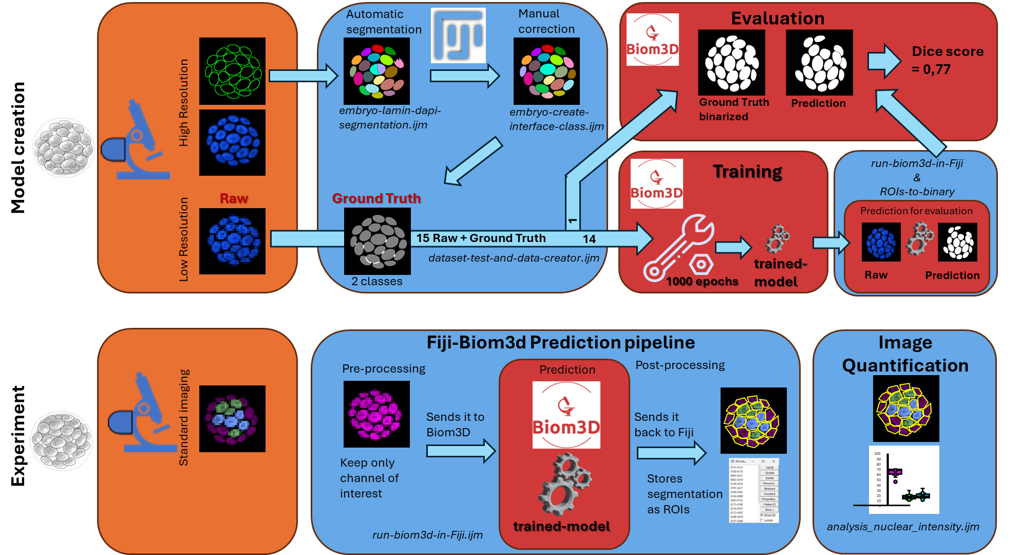
Workflow from the embryo to nuclei masks. The protocol consists of two parts: the creation of a dataset to train and evaluate a model (Model creation), and the segmentation of nuclei in an experiment of biological interest (Experiment). The left side represents the sample preparation steps, which include the immunostaining of the lamina for the training dataset and the selection of antibodies for the experiments. In orange boxes, the acquisitions for the training dataset include high-resolution images. In blue boxes are two workflows using Fiji, with various macros listed in italics and the .ijm file extension. Red boxes are the sections pertaining to the Biom3d deep learning software. The protocol includes quantification of the segmented images to evaluate the procedure.
Background
Three-dimensional imaging has become an essential tool for studying morphogenesis, embryonic development, and tissue organization at cellular resolution. Quantitative analysis of these datasets requires accurate segmentation of individual nuclei, which serves as the basis for cell counting, lineage reconstruction, spatial organization studies, and measurements of gene expression or signaling activities. However, segmentation of densely packed nuclei in volumetric microscopy datasets remains challenging because of heterogeneous fluorescence intensity, variable nuclear morphology, and close cell–cell contacts, especially in some samples like early embryos or embryoid bodies.
Recent advances in deep learning have substantially improved 3D nuclear segmentation. Convolutional neural network–based approaches and shape-based segmentation algorithms, such as Cellpose [1] and StarDist [2], can achieve high segmentation accuracy in complex biological samples. In the specific context of mouse embryo imaging, QCA-Net [3] has become a reference tool, combining a nuclear segmentation network with a nuclear detection network followed by marker-based watershed post-processing to separate touching nuclei. Nevertheless, the adoption of these methods by experimental biology laboratories remains limited. Existing workflows often require extensive manual annotation, complicated software installation procedures, management of multiple software dependencies, and manual tuning of preprocessing and training parameters, which are exacerbated by the reproducibility crisis in published software: Out of 104 published deep learning–based nuclei segmentation methods, only 6 are accompanied by source code, documentation, necessary computing environment, and training datasets (including Cellpose, StarDist, and QCA-Net) [4]. Finally, creating a training dataset, which involves manually labeling images to establish ground truth, often proves too time-consuming for the user. These requirements can represent a significant barrier for researchers without computational expertise.
The protocol presented here addresses these limitations through two complementary innovations. First, it uses a dual-channel imaging strategy [5] in which a high-resolution nuclear-envelope marker enables semi-automated generation of ground-truth annotations, substantially reducing the manual effort required to create training datasets. Second, it relies on the Biom3d framework [6] in its preconfigured nnCore mode, which provides automated preprocessing, model configuration, training, and prediction within an environment designed for non-programmer users. In a previous benchmark, nnCore demonstrated accuracy on par with the aforementioned software to segment plant nuclei [6], along with ease of use for non-experts, which is why we chose to include it in this workflow.
Compared with previously published workflows, this protocol reduces the technical expertise required to develop and deploy custom deep learning models while maintaining the flexibility to train models on user-generated datasets. Annotation, dataset creation, prediction, and image analysis steps include Fiji macros to help automate as many steps as possible. It is particularly advantageous for studies involving large collections of confocal images of mouse embryos, where manual segmentation would be impractical. The protocol also integrates downstream analysis tools that facilitate quantitative measurements after segmentation.
The protocol does, however, have several limitations. It was developed and validated using fixed mouse embryos imaged by confocal microscopy and therefore performs best on datasets acquired under similar imaging conditions. Model performance may decrease when applied to images generated with different microscopy modalities, staining strategies, or voxel sizes. In such cases, additional training data may be required to retrain or fine-tune the model. Although demonstrated here for segmentation of nuclei in mouse embryos, the workflow can be adapted to other biological systems provided that suitable training data are available. The same strategy could be applied to organoids, developing tissues, or other three-dimensional microscopy datasets. In addition, the dual-channel annotation approach can potentially be extended to the segmentation of other biological structures for which one imaging channel facilitates automated annotation while another represents the experimental signal of interest.
Materials and reagents
Biological materials
1. Wild-type CD1 mice (Janvier Labs, Le Genest-Saint-Isle, France; strain CD1, catalog number 7SN-SWISS-F); animals should be maintained under standard specific pathogen-free conditions according to institutional and national regulations
2. Mouse embryos collected at embryonic day (E) 3.5 and E4.5 from naturally mated CD1 mice (generated in-house)
Reagents
1. Paraformaldehyde (PFA), 16% p/v (Thermo Scientific Chemicals, catalog number: 043368.9M); store according to manufacturer’s instructions
2. Phosphate-buffered saline (PBS) 10×, liquid, OmniPur® grade (Sigma-Aldrich, catalog number: 6505-OP)
3. Triton X-100 (Sigma-Aldrich, catalog number: 10789704001)
4. Fetal bovine serum (FBS) (Eurobio, catalog number: CVFSVF00-01); store according to the manufacturer’s instructions
5. Lamin B1 antibody (B-10) is an IgG1 κ mouse monoclonal Lamin B1 antibody (Santa Cruz Biotechnology, catalog number: sc-374015); store according to the manufacturer’s instructions
6. SOX2 primary antibody in rabbit (Invitrogen/eBioscience, catalog number: 14-9811-82); store according to the manufacturer’s instructions.
7. GATA6 primary antibody in rat (Cell Signaling Technology, catalog number: 5851P); store according to the manufacturer’s instructions
8. Donkey anti-rat Alexa 488 secondary antibody (Life Tech, catalog number: A21208); store at 4 °C protected from light
9. Donkey anti-rabbit Cy5 secondary antibody (Jackson ImmunoResearch, catalog number: 711-175-152); store at 4 °C protected from light
10. Donkey anti-mouse Alexa Fluor 488 secondary antibody (Life Tech, catalog number: A21202); store at 4 °C protected from light
11. 4′,6-Diamidino-2-phenylindole (DAPI) (Sigma-Aldrich, catalog number: s9542); store according to the manufacturer’s instructions and protect from light
12. RapiClear® clearing reagent (SUNJin Lab, catalog number: RC147001); store according to the manufacturer’s instructions
Solutions
1. PBS 1× (see Recipes)
2. PFA 4% (see Recipes)
3. PBST 0.1% (see Recipes)
4. PBST 0.5% (see Recipes)
5. Blocking solution (see Recipes)
6. Primary antibody dilution (see Recipes)
7. Primary antibody incubation solution (see Recipes)
8. Secondary antibody staining solution (see Recipes)
9. DAPI 1/1000 (see Recipes)
Recipes
1. PBS 1×
| Reagent | Final concentration | Quantity or volume |
|---|---|---|
| PBS 10× | 1× | 100 mL |
| H2O | 900 mL | |
| Total | 1 L |
2. PFA 4%
| Reagent | Final concentration | Quantity or volume |
|---|---|---|
| PBS 1× | / | Up to 5 mL |
| PFA 16% | 4% | 1.25 mL |
| Total | 5 mL |
Store at 4 °C and use within 1 week after preparation.
3. PBST 0.1%
| Reagent | Final concentration | Quantity or volume |
|---|---|---|
| PBS 1× | / | Up to 50 mL |
| Triton X-100 | 0.1% | 50 μL |
| Total | 50 mL |
4. PBST 0.5%
| Reagent | Final concentration | Quantity or volume |
|---|---|---|
| PBS 1× | / | Up to 50 mL |
| Triton X-100 | 0.5% | 250 μL |
| Total | 50 mL |
5. Blocking solution
| Reagent | Final concentration | Quantity or volume |
|---|---|---|
| FBS | 10% | 500 μL |
| PBST 0.1% | / | Up to 5 mL |
| Total | 5 mL |
6. Primary antibody dilution
| Reagent | Final concentration | Quantity or volume |
|---|---|---|
| Primary antibody: α GATA6 or αSOX2 | 1/100 | 1 μL |
| Blocking Solution | / | Up to 100 μL |
| Total | 100 μL |
Store for up to 1 week at 4 °C.
7. Primary antibody incubation solution
| Reagent | Final concentration | Quantity or volume |
|---|---|---|
α GATA6 dilution 1/100 α SOX2 dilution 1/100 | 1:1,000 (αGATA6) 1: 3,000 (αSOX2) | 1 μL GATA6 (1/10th) 0.3 μL SOX2 (1/30th) |
| Blocking solution | / | Up to 10 μL |
| Total per well | 10 μL |
8. Secondary antibody staining solution (for 300 μL)
| Reagent | Final concentration | Quantity or volume |
|---|---|---|
| Secondary antibodies | 1:300 | 1 μL each |
| Blocking solution | / | Up to 300 μL |
| Total | 300 μL |
9. DAPI 1/1000
| Reagent | Final concentration | Quantity or volume |
|---|---|---|
| DAPI | 1:1,000 | 1 μL |
| Blocking solution | / | Up to 1 mL |
| Total | 1 mL |
Laboratory supplies
1. iSpacer, 50 μm thickness (SUNJin Lab, catalog number: IS315)
2. Terasaki plate (Thermo Scientific, catalog number: 163118)
Equipment
1. Laser scanning confocal microscopy (CSLM) (Zeiss, LSM980 with Airyscan 2, with 40× N.A.1.3 and 63× N.A.1.4 objectives).
Note: Several instruments can be used, such as laser scanning confocal microscopy (CSLM) or confocal spinning disk microscopy. The important thing is to use a lens with a high numerical aperture (NA) of 1.3–1.4. The microscope should also be equipped with high-end sensitive photodetectors or a digital camera (e.g., sCMO) with high sensitivity (high quantum efficiency, low e-read noise) suited for low-level signal acquisition in the spectral range of the fluorescent probes.
2. Computer (Dell, model: Precision 7670)
a. OS: Windows 11 Pro (x86_64/AMD64) or macOS 14 (Sonoma). If you are on Windows, you can consider that you are on “x86_64” architecture. However, if you want to confirm, open a terminal by entering “cmd” in the search bar and press Enter. Then, paste “echo %PROCESSOR_ARCHITECTURE%” and press Enter. If it prints AMD64, you are on x86_64. If not, your computer is not compatible with the installation method given here; refer to the Biom3d documentation (https://biom3d.readthedocs.io/en/latest/installation.html). On macOS, you should be on the “arm64” architecture (since M1). However, if you want to check, click the Launchpad icon in the Dock, type “Terminal” in the search field, then click Terminal. Then, paste “uname -m” and press Enter. If it prints arm64, you are on the correct architecture. If not, your computer is not compatible with the installation method given here; refer to the Biom3d documentation.
b. CPU: Intel Core i7-12800HX (or i5-12600HX) and an allocated computer memory of at least 16GB RAM (64-bit).
c. RAM: 32 GB DDR5 (64-bit, minimum 16 GB).
d. GPU: NVIDIA RTX A5000 (16 GB VRAM, CUDA 12.x) or Apple MacBook Pro (M2 Pro/Max, arm64). If you do not know the CUDA version, Biom3d will automatically detect it if it is compatible. Biom3d will automatically check your GPU for compatible CUDA versions; however, if you want to check before trying to install it, follow this process:
Open a command prompt, paste “nvidia-smi,” and press Enter. If the command is not recognized, it means that you do not have an Nvidia GPU; otherwise, it displays something similar to Figure 1.
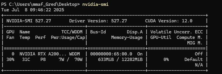
Figure 1. Result of the command “nvidia-smi” in a command prompt. The CUDA version is written in the top-right corner.
The CUDA version is written in the top-right corner of the header. The VRAM is written in the second column on the second line (without counting the header); here, 12282 MiB, so 12 GB.
Disk space: Biom3d will require 3 GB without a GPU or 7 GB with a GPU; but take into account the sizes of image datasets and future trained models.
Below the minimum system requirements, the software Biom3d may still function depending on the size of the images (dimensions and number), with longer processing times. Biom3d runs without a GPU, but the performance is significantly improved with a compatible GPU.
Software and datasets
| Type | Software/dataset/resource | Version | Date | License | Access (free or paid) |
|---|---|---|---|---|---|
| Image datasets | dataset | 2025 | CC-BY | Free | |
| Fiji | Software | 1.54p | G.P.L. | Open source | |
| Biom3d | Software | 0.0.40 | G.P.L. | Open source | |
| MorpholibJ plugin | Software | 1.6.5 | G.P.L. | Open source | |
| Omero batch plugin | Software | 2.1.1 | G.P.L. | Open source |
1. Fiji/ImageJ [7]. Fiji is just ImageJ with some useful modifications. It is an open-source image analysis software. https://Fiji.net/software/fiji/downloads
2. Default Biom3d framework nnCore: Biom3d is an open-source deep learning framework to segment 2D and 3D images [6]. Its default version, nnCore, can be installed with all dependencies using an installer function. https://github.com/GuillaumeMougeot/biom3d. Permanent link: https://archive.softwareheritage.org/browse/origin/directory/?origin_url=https://github.com/GuillaumeMougeot/biom3d.
3. MorpholibJ plugin [8]: The MorpholibJ plugin is a very useful plugin. It is a collection of mathematical morphology methods and plugins for ImageJ.
4. Omero_batch-plugin [9]: The Omero Batch Process plugin allows treating a dataset with a Fiji macro in a batch process. The image manager OMERO [10] is not required here. Version used here: 2.0.2. https://github.com/GReD-Clermont/simple-omero-client. Permanent link: GReD-Clermont/omero_batch-plugin: 2.1.1 | Zenodo
5. Download the following Fiji Macros from the folder “macros”: https://github.com/GReD-Clermont/biom3d-mouse-embryo-training
6. Seven datasets are available on a public repository of Université Clermont Auvergne: https://doi.org/10.18145/t2dq-1j51
Procedure
登录 / 注册后阅读全文
文章信息
稿件历史记录
提交日期: Jun 25, 2026
接收日期: Oct 14, 2026
在线发布日期: Nov 2, 2026
出版日期: Nov 20, 2026
版权信息
© 2026 The Author(s); This is an open access article under the CC BY license (https://creativecommons.org/licenses/by/4.0/).
如何引用
Alégot, H., Safarbati, S., Mougeot, G., Pouchin, P., Allègre, N., Boyer, T., Colmerauer, C., Chazaud, C., Osteil, P. and Desset, S. (2026). A Simplified Workflow for 3D Segmentation of Mouse Embryo Nuclei Using Biom3D. Bio-protocol 16(22): e5878. DOI: 10.21769/BioProtoc.5878.

 Download Citation in RIS Format
分类
生物信息学与计算生物学
生物科学 > 生物技术
您对这篇实验方案有问题吗?
在此处发布您的问题,我们将邀请本文作者来回答。同时,我们会将您的问题发布到Bio-protocol Exchange,以便寻求社区成员的帮助。
 发布问题


