


A Multiplexed Assay for High-Throughput Evaluation of In Vitro A-to-I Editing

发布: 2026年12月05日第16卷第23期 DOI: 10.21769/BioProtoc.5876 浏览次数: 38
 PDF
 Q&A





Abstract
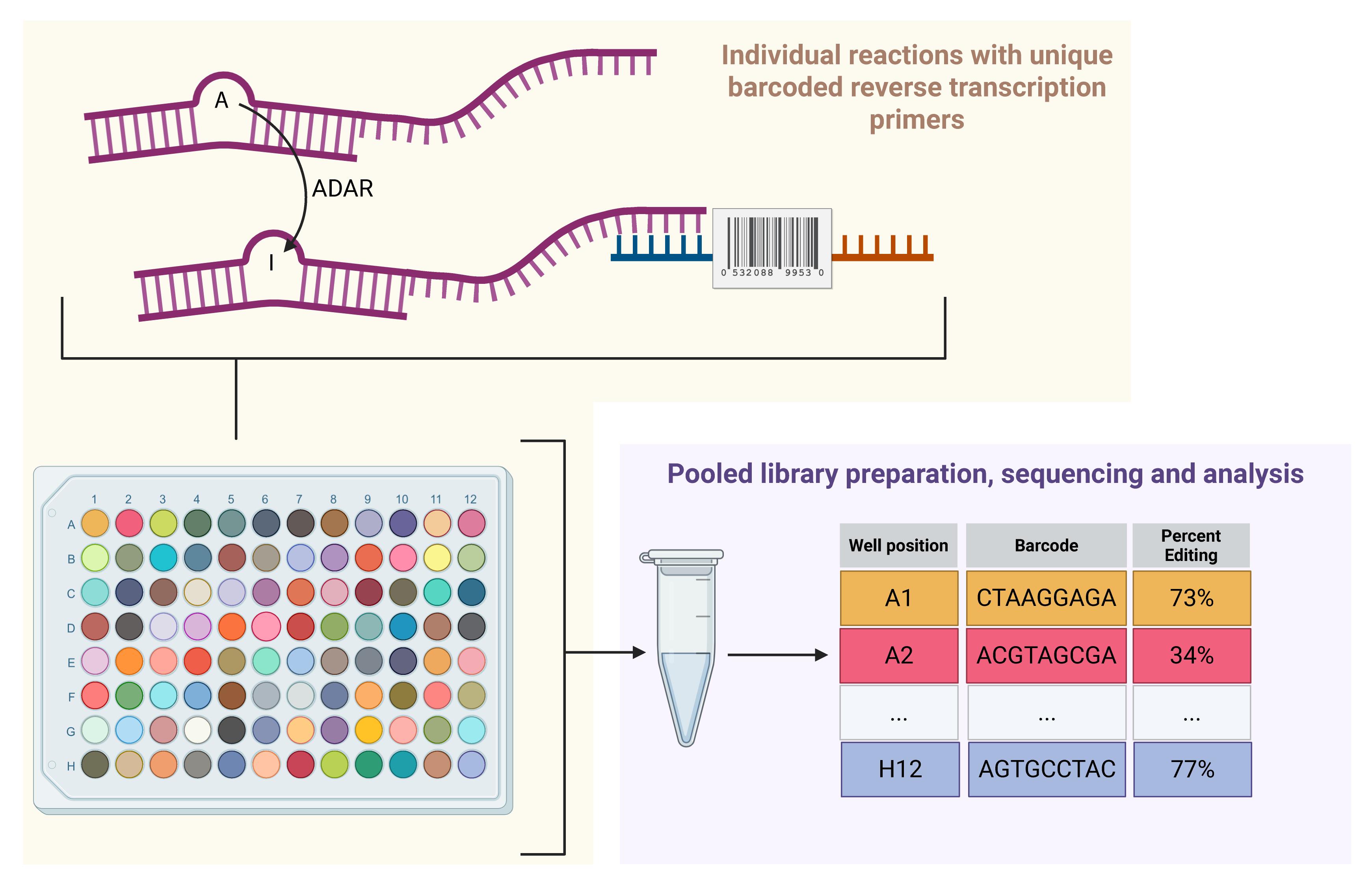
Adenosine-to-inosine (A-to-I) RNA editing, catalyzed by ADAR1, occurs on double-stranded RNA (dsRNA) and plays a key role in innate immunity. Editing of endogenous dsRNAs prevents aberrant activation of dsRNA sensors and resulting inflammation. This function of ADAR1 is essential in many cancers, making ADAR1 a promising therapeutic target. Current approaches for measuring A-to-I editing in biochemical assays, which often rely on Sanger sequencing, are limited in throughput. Thus, there is a need to develop high-throughput A-to-I editing assays to enable efficient screening of potential ADAR1 inhibitors. We developed a new high-throughput A-to-I editing assay that uses barcoded reverse-transcription primers to enable pooling and sequencing of multiple assays performed in parallel. Starting from an existing Sanger-based method, we optimized aspects of the A-to-I editing reaction, cDNA synthesis, and downstream processing to facilitate high-throughput assays and substantially lower per-reaction costs. In brief, immediately following the A-to-I editing reaction, cDNA is synthesized using well-specific barcoded reverse-transcription primers. Subsequently, cDNA from all reactions is pooled and processed as one sample for next-generation sequencing, with the potential for further multiplexing by indexed sequencing adapters. Our approach enables parallel measurement of RNA editing under multiple conditions in a plate-based format while significantly reducing the time, labor, and costs required compared to traditional methods. This high-throughput assay provides a scalable platform for screening ADAR1 inhibitors and may accelerate the development of targeted therapies for cancer.
Key features
• Percent A-to-I editing determined by count-based next-generation sequencing.
• Utilizes a widely used A-to-I editing substrate.
Keywords: RNA modification A-to-I editing
RNA-binding proteins
In vitro transcription
High-throughput screening
Graphical overview
Background
The adenosine deaminase acting on RNA (ADAR) family of proteins, including human ADAR1 and ADAR2, use their deaminase activity to edit adenosine to inosine in double-stranded RNAs (dsRNA), in a process known as A-to-I editing. In humans, ADAR1 edits millions of sites, primarily targeting inverted Alu repeats [1,2]. Editing by ADARs affects RNA structure, processing, localization, translation, and stability, and can even recode mRNAs [3]. These functions make both ADAR1 and ADAR2 essential: ADAR2 for its role in recoding the mRNA encoding GRIA2 [4,5], and ADAR1 for its role in suppressing dsRNA sensing [6–8]. Editing by ADAR1 is thought to destabilize the secondary structure of RNAs, making them less double-stranded and thus preventing detection by the dsRNA sensor MDA5 [6–8].
Inhibition of ADAR1 has therapeutic potential both in cancer and viral infection. Due to ADAR1’s role as a suppressor of dsRNA sensing and the elevated expression of dsRNA sensors in some cancer cells, ADAR1 has become a promising therapeutic target for multiple cancers [9–11]. Similarly, ADAR1 has pro-viral functions in infection by measles and several other viruses [12,13]. As such, there is considerable interest in identifying potent inhibitors of ADAR1. While multiple ADAR1 inhibitors have been reported in the literature, most have not proven to be selective inhibitors of ADAR1 in follow-up studies [14–18].
Conversion of adenosine to inosine by ADAR can be easily detected by multiple methods [19–26]. Common approaches rely on either sequencing of a cDNA copy of an edited RNA or enzymatic cleavage of edited RNA using the inosine-specific endonuclease V (EndoV). Sequencing benefits from reverse transcriptase incorporating a “C” opposite of “I” in the edited RNA. Thus, A-to-I editing events are easily detected as A-to-G substitutions in sequencing data. Next-generation (Illumina or other modalities) and Sanger sequencing are commonly used to measure A-to-I editing, with Sanger typically being used to evaluate editing at a small number of sites in RNA isolated from cells or tissues, or to evaluate editing in in vitro biochemical assays. Enzymatic cleavage of edited RNA by EndoV can be used in combination with next-generation sequencing to evaluate editing across the transcriptome or to evaluate editing in biochemical assays when combined with electrophoresis to quantify cleavage. Neither Sanger sequencing nor EndoV-based approaches are well-suited for high-throughput screening for ADAR1 inhibitors. Sanger sequencing is relatively labor-intensive and cost-prohibitive at larger scales. Similarly, the combination of EndoV and electrophoresis to quantify cleavage would be highly labor-intensive. A luciferase-based cellular assay was previously developed to enable high-throughput screening to identify ADAR inhibitors [27]. This system uses an mRNA encoding two luciferases separated by a stop codon that, when edited by ADAR1, allows translation of the downstream luciferase. Inhibition of A-to-I editing would thus result in reduced signal from the downstream luciferase. While this approach has been used successfully to identify putative ADAR1 inhibitors, it has two main drawbacks: 1) it is a cellular assay and thus will miss compounds that have poor membrane permeability, and 2) it has a high false-positive rate, as determined by rescreening of initial hits using the same assay or an orthogonal approach [18,27].
Here, we describe a high-throughput biochemical assay for A-to-I editing by ADAR1 (Figure 1). This assay builds upon prior approaches that measure editing by Sanger sequencing of PCR-amplified cDNA generated from reverse transcription of an ADAR1 substrate RNA [28,29]. To enable readout by next-generation sequencing, our assay uses barcoded reverse-transcription primers. Reverse transcription takes place immediately following and in the same well as A-to-I editing. Subsequently, cDNA from all reactions is pooled and used to generate an Illumina sequencing library. Analysis of A-to-I editing is performed using a custom analysis pipeline. Overall, this assay is considerably less expensive than the Sanger-based alternative. The cost savings arise from less labor, reagents, and sequencing costs. This assay is well-suited for the evaluation of putative ADAR1 inhibitors or to study ADAR1 mutants.
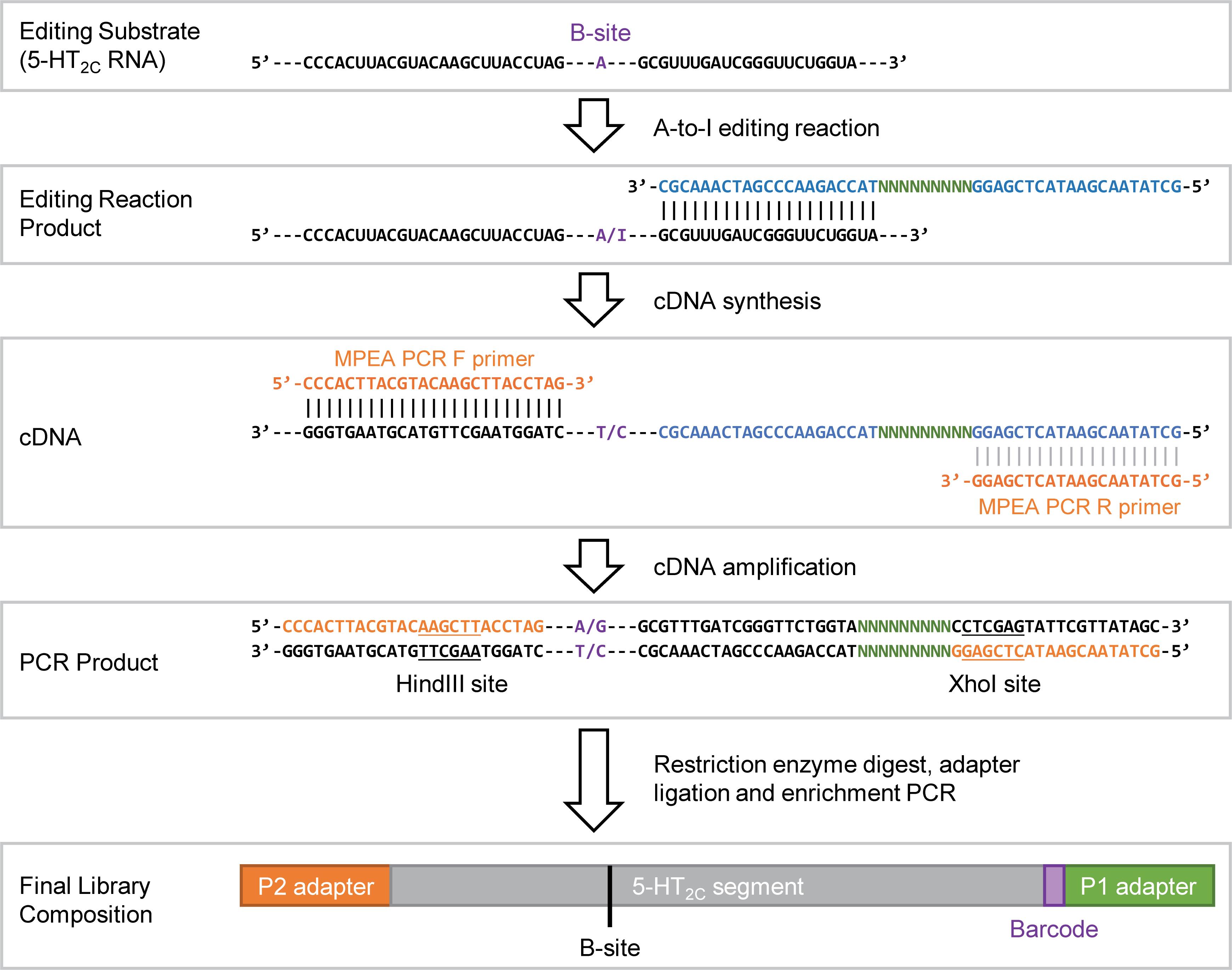
Figure 1. Diagram describing the assay. The diagram depicts how the barcoded reverse transcription primers are used to track RNA editing occurring in each well of the assay. Some portions of the editing substrate, its cDNA copy, and the sequencing adapters are not shown due to the length of those molecules; the sequence that is missing is represented by “---”. For the B-site, “A/I” in the RNA, or “T/C” or “A/G” in the cDNA or PCR product represents the possible identities of the nucleotide at that position; for the RNA, “I” for inosine if the B-site is edited, or “A” if no editing occurs.
Materials and reagents
Reagents
1. RNase inhibitor murine (New England BioLabs, catalog number: M0314L)
2. Yeast tRNA (Thermo Fisher Scientific, catalog number: AM7119)
3. HiScribe T7 Quick High Yield RNA Synthesis Kit (New England Biolabs, catalog number: E2050S); includes 100 mM DTT, T7 RNA polymerase mix, NTP buffer mix, and DNase I (RNase-free)
4. Deoxynucleotide triphosphate (dNTP) solution mix (New England BioLabs, catalog number: N0447L)
5. PrimeSTAR Max Premix (2×) (TaKaRa, catalog number: R045)
6. pTwist-5HT (TwistBioscience, special order, clonal gene inserted into the vector “pTwist Amp High Copy,” see File S1 for annotated plasmid sequence)
7. pTwist-5HT_edit (TwistBioscience, special order, clonal gene inserted into the vector “pTwist Amp High Copy,” see File S2 for annotated plasmid sequence)
8. ADAR1 (p150), FLAG-tag recombinant (BPS Bioscience, catalog number: 100472-1)
9. KpnI-HF (New England Biolabs, catalog number: R3142S)
10. XhoI (New England Biolabs, catalog number: R0146S)
11. HindIII-HF (New England Biolabs, catalog number: R3104S)
12. T4 DNA ligase (New England Biolabs, catalog number: M0202L)
13. Protoscript II reverse transcriptase (New England Biolabs, catalog number: M0368S), includes 5× ProtoScript II reaction buffer, 100 mM DTT, and ProtoScript II reverse transcriptase, 200,000 units/mL
14. rCutSmart (New England Biolabs, catalog number: B6004S)
15. 10× T4 DNA ligase buffer (New England Biolabs, catalog number: B0202S)
16. T4 polynucleotide kinase (New England Biolabs, catalog number: M0201S)
17. Qubit 1× dsDNA Broad Range Assay kit (Invitrogen, catalog number: Q33265)
18. Qubit RNA Broad Range Assay kit (Invitrogen, catalog number: Q32852)
19. Nuclease-free water, HyPure molecular biology–grade water (Cytiva, catalog number: SH30538.03)
20. Agarose (Fisher, catalog number: BP160500)
21. 100 bp DNA ladder (New England Biolabs, catalog number: N3231L)
22. 1 kb DNA marker (New England Biolabs, catalog number: N3232S)
23. Gel loading dye, purple (6×) (New England BioLabs, catalog number: B7024S)
24. Buffer kit, RNase-free (Invitrogen, catalog number: AM9010); includes 1 M Tris-HCl pH 7, 1 M Tris-HCl pH 8, 2 M potassium chloride (KCl), 0.5 M ethylenediaminetetraacetic acid (EDTA), and 5 M NaCl
25. NP-40 (Nonidet P-40 substitute) (Research Products International, catalog number: N59000-500.0)
26. Glycerol (Sigma-Aldrich, catalog number: G5516-100ML)
27. Potassium glutamate, L-Glutamic acid potassium salt monohydrate (Sigma-Aldrich, catalog number: G1149-100G)
28. Tris base (Fisher, catalog number: BP154-1)
29. Boric acid (Fisher, catalog number: A73-3)
30. 0.5 M EDTA pH 8.0 (Thermo Scientific, catalog number: AAJ15694AP)
31. SeeGreen Nucleic Acid Stain 20,000× (miniPCR bio, catalog number: RG-1550-10)
32. Monarch Spin RNA Cleanup kit (New England Biolabs, catalog number: T2040S)
33. Monarch PCR & DNA Clean Up kit (New England Biolabs, catalog number: T1030L)
34. Monarch Spin DNA Gel Extraction kit (New England Biolabs, catalog number: T1120L)
35. Ultrapure water from a Milli-Q or similar water purification system
36. Oligonucleotides (Table 1)
Table 1. Oligonucleotides (* phosphorothioate bond, italicized nucleotides are barcode sequences)
| Name | Sequence (5′ – 3′) |
|---|---|
| MPEA Test RT primer | GCTATAACGAATACTCGAGGGCTCTGATGTACCAGAACCCGATCAAACGC |
| MPEA Library RT primers | See Table S1 |
| MPEA PCR F primer | CCCACTTACGTACAAGCTTACCTA*G |
| MPEA PCR R primer | GCTATAACGAATACTCGAG*G |
| P1 XhoI F (Illumina adapter) | AATGATACGGCGACCACCGAGATCTACACTCTTTCCCTACACGACGCTCTTCCGATCTGCTCGAT |
| P1 XhoI R (Illumina adapter) | T*CGAATCGAGCAGATCGGAAGAGCGTCGTGTAGGGAAAGAGTGTAGATCTCGGTGGTCGCCGTATCATT |
| P2 HindIII F (Illumina adapter) | A*GCTAGATCGGAAGAGCACACGTCTGAACTCCAGTCACAGGTCACGCATCTCGTATGCCGTCTTCTGCTTG |
| P2 HindIII R (Illumina adapter) | CAAGCAGAAGACGGCATACGAGATGCGTGACCTGTGACTGGAGTTCAGACGTGTGCTCTTCCGATCT |
| Il enrich F primer | AATGATACGGCGACCACCGAG |
| Il enrich R primer | CAAGCAGAAGACGGCATACGA |
Solutions
1. 2× template annealing buffer (see Recipes)
2. 2× reaction buffer solution (see Recipes)
3. 1.5% agarose in TBE solution (see Recipes)
4. 50% glycerol solution (see Recipes)
5. 1 M potassium glutamate (see Recipes)
6. 10% NP-40 (see Recipes)
7. 5× and 1× TBE (see Recipes)
8. 10 μg/mL yeast tRNA working solution (see Recipes)
9. TE buffer (see Recipes)
10. DNA marker working solution (see Recipes)
Recipes
1. 2× template annealing buffer (10 mL)
| Reagent | Final concentration | Quantity or volume |
| 1 M Tris-HCl (pH 7.0) | 20 mM, pH 7.5 | 140 μL |
| 1 M Tris-HCl (pH 8.0) | 60 μL | |
| 0.5 M EDTA pH 8.0 | 2 mM | 40 μL |
| 5 M NaCl | 200 mM | 400 μL |
| Nuclease-free water | N/A | 9.4 mL |
Add water, followed by the remaining ingredients (in any order) to a 50 mL Falcon tube and vortex to mix. Store at room temperature. This solution does not need to be freshly prepared.
2. 2× reaction buffer solution (20 mL)
| Reagent | Final concentration | Quantity or volume |
| 1 M Tris-HCl (pH 7.0) | 15 mM, pH 7.5 | 420 μL |
| 1 M Tris-HCl (pH 8.0) | 180 μL | |
| 2 M KCl | 52 mM | 520 μL |
| 1 M potassium glutamate | 80 mM | 1.6 mL |
| 0.5 M EDTA pH 8.0 | 3 mM | 120 μL |
| 10% NP-40 | 0.006% (v/v) | 12 μL |
| 50% glycerol | 8% (v/v) | 3.2 mL |
| Nuclease-free water | N/A | 13.95 mL |
Add water, followed by the remaining ingredients (in any order) to a 50 mL Falcon tube and vortex to mix. Store at room temperature. This solution does not need to be freshly prepared.
3. 1.5% agarose in TBE solution
| Reagent | Final concentration | Quantity or volume |
|---|---|---|
| Agarose | 1.5% (w/v) | 7.5 g |
| 1× TBE | 1× | 500 mL |
| Total | N/A | 500 mL |
Add agarose and TBE into a glass media bottle. With the cap on loosely, microwave and swirl until the agarose has fully dissolved into solution. Store at room temperature or use to pour a gel while melted; allow the solution to cool enough to hold the bottle before pouring a gel. If stored at room temperature, microwave again to melt before pouring a gel. Add a nucleic acid stain (SeeGreen, final concentration of 1×) to the gel during casting, immediately after pouring the gel. The gel solution without nucleic acid stain does not need to be freshly prepared.
4. 50% glycerol solution
| Reagent | Final concentration | Quantity or volume |
|---|---|---|
| Glycerol | 50% (v/v) | 20 mL |
| Nuclease-free water | N/A | 20 mL |
| Total | N/A | 40 mL |
Prepare in a 50 mL Falcon tube. Add water first, followed by glycerol. Glycerol is thick and challenging to pipette. Use a 25 mL serological pipette and slowly draw up the desired volume. Add directly to the water and pipette up and down to mix and remove glycerol from the pipette. Vortex thoroughly and store at room temperature. This solution does not need to be freshly prepared.
5. 1 M potassium glutamate solution
| Reagent | Final concentration | Quantity or volume |
|---|---|---|
| L-Glutamic acid potassium salt monohydrate | 1 M | 30.48 g |
| Nuclease-free water | N/A | Up to 150 mL |
| Total | N/A | 150 mL |
Prepare in a glass beaker with a magnetic stir bar. Add water followed by glutamic acid. Mix on a stir plate until fully dissolved. Filter and store at room temperature. This solution does not need to be freshly prepared.
6. 10% NP-40
| Reagent | Final concentration | Quantity or volume |
|---|---|---|
| NP-40 | 10% | 1 mL |
| Nuclease-free water | N/A | 9 mL |
| Total | N/A | 10 mL |
Prepare in a 15- or 50-mL conical tube. Add water first, then NP-40. NP-40 is a thick solution. Use a serological pipette and pipette slowly. Add the NP-40 directly to the water and pipette up and down several times to remove the NP-40 from the pipette. Vortex to mix. Store at room temperature. This solution does not need to be freshly prepared.
7. 5× and 1× TBE
| Reagent | Final concentration | Quantity or volume |
|---|---|---|
| Tris base | 446 mM | 108 g |
| Boric acid | 446 mM | 55 g |
| 0.5 M EDTA, pH 8.0 | 10 mM | 40 mL |
| Ultrapure water | N/A | Up to 2 L |
| Total | N/A | 2 L |
Prepare in a beaker sufficient to hold 2 L. Add 1 L of water and a stir bar to the beaker on a stir plate. Slowly add the Tris base and boric acid. Once dissolved, add the EDTA. Transfer the solution to a 2-L graduated cylinder and bring to a total of 2 L with water. A final pH adjustment is not needed. Mix well and filter. Can be stored at room temperature. To make 1× TBE, dilute 5× TBE with ultrapure water to 1×. This solution does not need to be freshly prepared.
8. 10 μg/mL yeast tRNA working solution
| Reagent | Final concentration | Quantity or volume |
|---|---|---|
| Yeast tRNA (10 mg/mL) | 10 μg/mL | 1 μL |
| Nuclease-free water | N/A | 999 μL |
| Total | N/A | 1 mL |
Prepare in a 1.7 mL microfuge tube. Add water first. Vortex to ensure the solution is fully mixed. Store at -20 °C. This solution does not need to be freshly prepared.
9. TE buffer
| Reagent | Final concentration | Quantity or volume |
|---|---|---|
| 1 M Tris pH 8.0 | 10 mM | 0.5 mL |
| 0.5 M EDTA | 0.1 mM | 10 μL |
| Water | N/A | 49.49 mL |
| Total | N/A | 50 mL |
Prepare in a 50 mL conical tube. Add water first. Vortex to ensure the solution is fully mixed. Store at room temperature. This solution does not need to be freshly prepared.
10. DNA marker working solution
| Reagent | Final concentration | Quantity or volume |
|---|---|---|
| 100 bp or 1 kb DNA marker (500 μg/mL) | 10 μg/mL | 50 μL |
| 6× Purple Loading Dye | 1× | 170 |
| TE buffer | N/A | 780 μL |
| Total | N/A | 1 mL |
Prepare in a 1.7 mL microfuge tube. Vortex to ensure the solution is fully mixed. Store at -20 °C. Use 20 μL per lane. This solution does not need to be freshly prepared.
Laboratory supplies
1. 1.7 mL microfuge tubes (Genesee, catalog number: 24-282)
2. 96-well PCR plate (MidSci, catalog number: PR-PCR2196F)
3. Repeating pipette tips (Eppendorf, catalog numbers: 0030089634, 0030089618)
4. Reagent reservoir (Fisher, catalog number: 01-670-454)
5. 50 mL conical tubes (Genesee, catalog number: 21-108)
6. 15 mL conical tubes (Genesee, catalog number: 21-103)
7. 150 mL filter bottle unit (Fisher, catalog number: FB12566500)
8. 1 L filter bottle unit (Fisher, catalog number: FB12566506)
9. Pipette tips, filtered, nuclease-free (p10, p20, p200, p1000)
10. PCR plate seals (Genesee, catalog number: 12-170)
11. Foil plate seals (Corning, catalog number: 6570)
12. PCR strip tubes (Genesee, catalog number: 27-125)
13. Spatula
14. Weighing boats
15. Magnetic stir bar
16. Glass media bottles (250 mL, 500 mL, 1 L)
17. Glass beakers (200 mL, 500 mL, 2 L)
18. Graduated cylinders (500 mL, 1 L, 2 L)
Equipment
1. Freezer (-20 °C)
2. Ultra-low freezer (-80 °C)
3. Refrigerator (4 °C)
4. Tube racks (microcentrifuge, 15 and 50 mL conicals)
5. Microcentrifuge (Eppendorf, model: 5420)
6. Mini microcentrifuge (Corning, catalog number: 6770)
7. Thermocycler (Bio-Rad, model: T100)
8. Vortex mixer, vortexer (Corning, catalog number: 6775)
9. ChemiDoc, gel imaging system (Bio-Rad, catalog number: 12003153)
10. Power supply (Bio-Rad, catalog number: 1645050)
11. Salad spinner (Oxo, catalog number: 1071497)
12. Repeating pipette (Eppendorf, model: E3)
13. Horizontal gel electrophoresis rig (Fisher, catalog number: FB-SB-710)
14. Nanodrop (Thermo Fisher Scientific, model: Onec)
15. Qubit fluorometer (Invitrogen, model: Qubit 4)
16. Microwave
17. Stir plate
18. Balance
19. Pipettes (0.1–2.5, 0.5–10, 2–20, 20–200, 100–1000 μL)
Software and datasets
1. Microsoft Excel or Google Sheets
2. Custom analysis scripts (analyze_fastq.sh and analyze_fastq_w-adjustment.sh) freely available here: https://github.com/cottrellka/Barcoded_AtoI_assay
Procedure
登录 / 注册后阅读全文
文章信息
稿件历史记录
提交日期: Jul 22, 2026
接收日期: Oct 14, 2026
在线发布日期: Nov 3, 2026
出版日期: Dec 5, 2026
版权信息
© 2026 The Author(s); This is an open access article under the CC BY-NC license (https://creativecommons.org/licenses/by-nc/4.0/).
如何引用
Gardner, E. N. and Cottrell, K. A. (2026). A Multiplexed Assay for High-Throughput Evaluation of In Vitro A-to-I Editing. Bio-protocol 16(23): e5876. DOI: 10.21769/BioProtoc.5876.

 Download Citation in RIS Format
您对这篇实验方案有问题吗?
在此处发布您的问题,我们将邀请本文作者来回答。同时,我们会将您的问题发布到Bio-protocol Exchange,以便寻求社区成员的帮助。
 发布问题


