


Mouse Intratongue Injection With Cancer Cells—An Orthotopic Model for Head and Neck Squamous Cell Carcinoma

发布: 2026年11月05日第16卷第21期 DOI: 10.21769/BioProtoc.5861 浏览次数: 36
评审: Sergii RomanenkoAnonymous reviewer(s)
 PDF
PDF  Q&A
Q&A
参见作者原研究论文
The authors used this protocol in:
May 2026





Abstract
Syngeneic and orthotopic injection of cancer cells in mice is a gold standard model in pre-clinical cancer research. Herein, we detail a protocol of orthotopic intratongue injection as a model for upper respiratory tract cancer studies. This model recapitulates tumor growth and invasion in an orthotopic site, the oral cavity mucosa. It can be experimentally designed in a syngeneic context or used to study patient-derived cells in immunocompromised animals. Procedurally, mice are anesthetized and injected with a small volume of cancer cell suspension near the lateral border of the tongue, right under the mucous membrane. Post-injection, animal welfare is carefully attended to by appropriate analgesia, while providing soft food for the duration of the experiment to ensure the ability of the mice to eat ad libitum. Tongues and potential metastatic organs are dissected and can be sampled and analyzed using relevant state-of-the-art methods, including -omics and imaging approaches. We describe a broad range of distinct tumor profiling and analysis methods applicable to this model. These include physical measurements of elasticity with atomic force microscopy on fresh tissue, hematoxylin-eosin staining and standard immunohistochemistry, and confocal microscopy applied to paraffin-embedded tongue sections. This model is orthotopic, accessible without surgery, easy to monitor, and rapid to perform.
Key features
• Orthotopic and clinically relevant head and neck squamous cell carcinoma (HNSCC) model.
• Rapid, reproducible, and minimally invasive procedure.
• Compatible with multimodal downstream analyses.
• Versatile platform for preclinical studies.
Keywords: Preclinical cancer model Orthotopic mouse model
Head and neck squamous cell carcinoma (HNSCC)
Intratongue injection
Tumor microenvironment
Tumor growth
Tumor invasion
Orthotopic mouse model
Head and neck squamous cell carcinoma (HNSCC)
Intratongue injection
Tumor microenvironment
Tumor growth
Tumor invasion
Background
This protocol was inspired by previous work [1–3] demonstrating the preclinical relevance of tongue orthotopic injection in the study of head and neck cancers. Myers et al. [1] implanted human oral cancer cell lines directly into the tongue, where tumors formed more efficiently and more closely reproduced the histology and metastatic behavior of human disease than more classic subcutaneous models. Of note, this observation was confirmed and further studied recently from an immune landscape perspective [4]. Myers’ seminal work accelerated the development of metastatic progression studies, either focusing on the local tumor microenvironment characterization [5–7] or the factors influencing the formation of lung metastases. As such, Benaich et al. [2] focused on the role of miR-203, and Pascual et al. [3] studied how lipid metabolism affects survival and metastatic initiation.
Our protocol was developed and optimized for the completion of two studies: Punovuori et al. [8] focused on the contribution of extracellular matrix composition and cancer-associated fibroblasts in tumor progression, and Kaivola et al. [9] focused on the role of tissue stiffening in vocal fold cancer progression. We employed mouse intratongue injection as an in vivo model system to compare the growth of advanced-stage T3 head and neck squamous cell carcinoma (HNSCC) cells and their therapeutic response to treatment with a clinically relevant YAP-TEAD inhibitor. We also recently used this protocol in Punovuori et al. [8], where we reprogrammed cancer cells through cancer-stroma (HNSCC cell–cancer-associated fibroblast) crosstalk in vitro using spheroid co-cultures and compared the growth of two distinct HNSCC models injected in mice tongues with or without cancer-associated fibroblasts.
Most previously reported applications of intratongue (also called intralingual) injection involve injecting cell-free solutions of chemicals (like sodium selenite [10] or choleratoxin B-saporin [11]), prions [12], or virus particles (AAV), often in the context of optogenetic studies [13–18]. Further, it has also been used for acute pain studies using large needles and volumes under isoflurane anesthesia [19,20].
In contrast, there are only a few carcinogenesis [21] and tumor xenograft studies published for this orthotopic injection site, first proposed by the Fidler lab [1] (2002). Technically, downstream applications involved histology (tissue staining, immunohistochemistry, and immunostaining) and whole-animal non-invasive imaging (bioluminescence- or fluorescence-based). Intravital imaging [22] was also applied to the tongue using a non-invasive holder and 2-photon illumination post-intratongue injection to study tumor invasion and intravasation [23,24]. Of note, the experimental endpoint post-injection varies from two weeks to several months, depending on cell lines and animal models, as well as the variable cell numbers used. Lastly, similar to the orthotopic injection protocol described here, intramucosal injection in the mouse cheek has been described [25]. However, to the best of our knowledge, a detailed protocol for intratongue injection is still lacking. We believe that our refined protocol is compatible with high reproducibility and aligns extremely well with current ethics on animal welfare worldwide.
As a brief description of the protocol (Figure 1A), mice are first treated with analgesic and anti-inflammatory drugs. The injection mix for the intratongue injection is prepared. The mice are anesthetized with a ketamine/xylazine mix that will keep them unconscious for approximately 40 min. When ready, the injection needle is prepared, and the mice are suitably positioned to enable smooth intratongue injections (illustrated in Figure 1C). The injection procedure itself takes less than 1 min per animal. Mice are monitored until the anesthesia subsides. Analgesic and anti-inflammatory treatment is administered 6 h post-intratongue injections and continued for the following three days. Fresh, softened food is provided daily throughout the experiment. Cancer progression is closely monitored until the experimental objective is achieved and/or the animal reaches the humane endpoint. As this protocol includes injection inside the mouth, we typically observe a modest, transient decrease in body weight shortly after injection. Mice usually recover the lost weight in the first week post-injection. Then, when approaching the endpoint, animals lose weight rapidly and irreversibly (Figure 1B). After sacrifice, the tongues and potential metastatic organs are harvested and processed. As a control, in addition to the experimental controls, two non-injected animals are included to provide a reference to normal tissue.
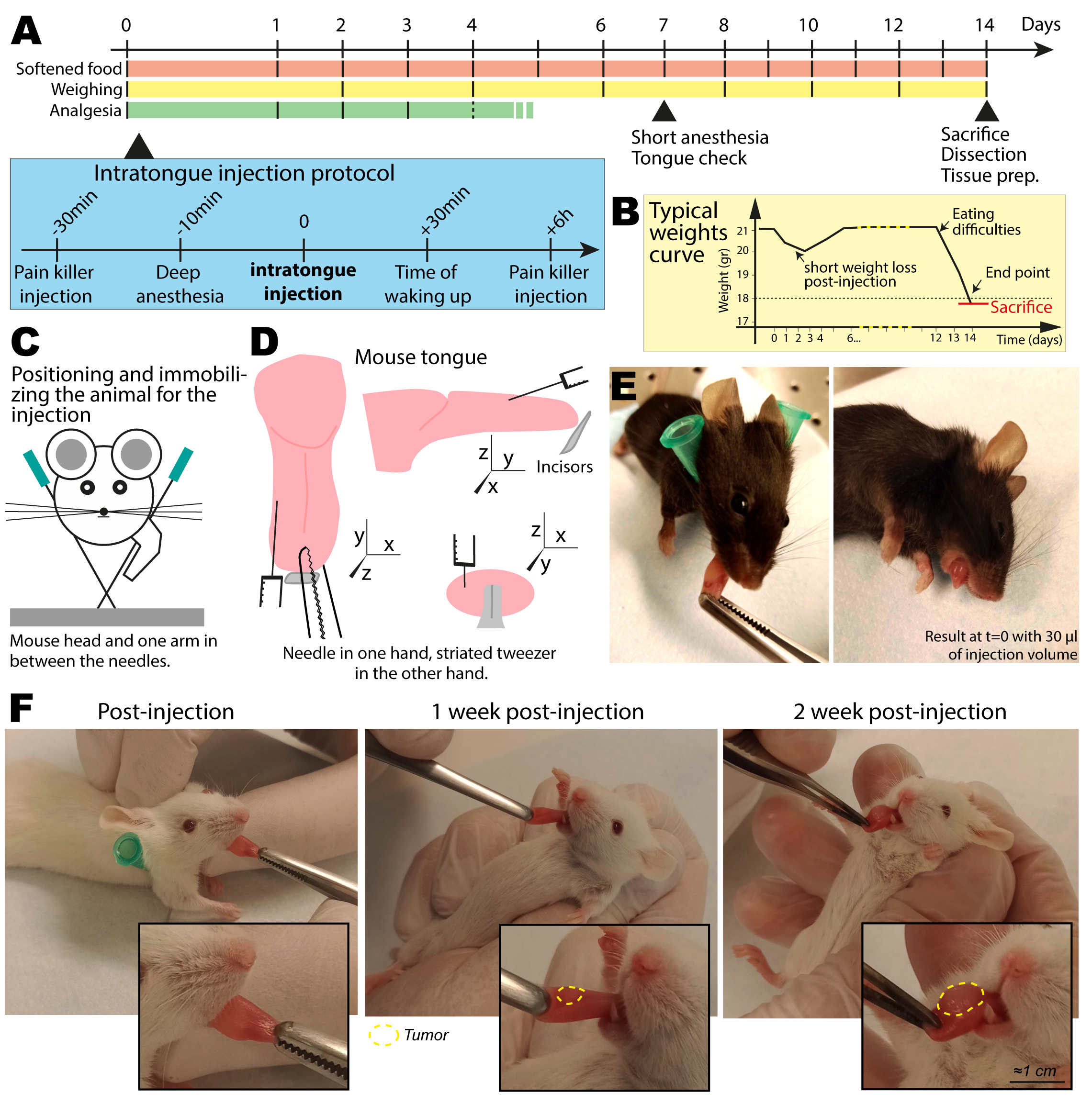
Figure 1. Experimental design for intratongue injection and subsequent monitoring. (A) Experimental timeline and summary of key steps. (B) Typical weight curve of the animals during the protocol. (C) Schematic of the position of the animal on the holder for intratongue injection. (D) Schematic of the tongue injection spot from three angles. The injection needle comes nearly parallel to the tongue, on the side, and the needle does not enter more than 2 mm into the tissue to stay in the submucosal compartment. (E) Representative pictures of the mouse tongue, directly post-injection (C57BL/6 mouse). (F) Representative pictures of the mouse tongue during the monitoring of the tongue (NSG mouse): directly post-injection, after 1 week, and after 2 weeks (endpoint).
Based on our expertise and existing literature, intratongue injection in mice is a relevant experimental system in research contexts ranging from fundamental to pre-clinical research. The tools, methods, and technical tips presented here are applicable to any tongue injection, with or without cells. In cancer research, it should be used as an orthotopic site of injection for upper respiratory tract cancer models. As previously reported [26], growing HNSCC lines in classical subcutaneous assays in the flank of mice is challenging. In our hands, all tested HNSCC cell lines grew a tumor in the tongue [8,9]. However, it has, a priori, no added value for other cancers, in which other orthotopic sites or more classical subcutaneous injection are more relevant and/or safer for the animals.
Our protocol is compatible with any kind of cellular mixes (cancer, immune, or stromal cells, separately or together). We believe that the addition of Matrigel is instrumental for the early engraftment of the tumor cells post-injection, but one can work without it if the cellular model allows. If well-positioned, liquid solutions do not leak out of the tongue when you pull the needle out. This model is suitable for distinct systemic pre-treatment or treatment during the experiment, and therapeutic agents can be administered in different ways (drinking water, gavage, injections, etc.). Although we did not perform live imaging, this approach is possible after the injection, as mentioned earlier (using bioluminescence, intravital imaging, etc.). Furthermore, tumor growth can be observed visually and with caliper measurements of animals under low isoflurane anesthesia. Visual inspection enables excellent quality control and evaluation of the progression of the experiment. After injection, the administered Matrigel–cell mixture appears like a small translucent lentil. After a week, a small whiter spot appears on the tongue, which grows over the weeks. Post-sacrifice, tongue dissection is relatively simple if well executed, and sample processing complexity is dependent on the experimental goal. Lastly, although depending on the aggressiveness of the cancer cell line and the cell number injected, the experiment duration will vary [9]; animals in our study reached the clinical endpoint in two weeks (precisely reproduced in independent experiments). We believe this is the shortest time to be expected.
This protocol requires training in animal experimentation. It uses intraperitoneal, subcutaneous, and intratongue injections. Experimenters must be familiar with animal anesthesia procedures and safety. It also requires a good appreciation of animal signs of discomfort, and significant dedicated time for animal monitoring (regular weighing and daily softened food change). Toward the endpoint of the experiment, mouse health will deteriorate very quickly; thus, careful and thorough monitoring is necessary. We recommend following the OBSERVE consensus guidelines to monitor the animals [27]. Based on our experience, this protocol is non-trivial and should be performed by advanced animal experimenters with potential support from animal facility personnel. Of note, the injection protocol presented here does not strictly require the presence of a second experimenter, although it is generally advisable to work in pairs during animal experimentation, as it can help during certain steps.
This protocol is not compatible with resection of the primary tumor for long-term relapse studies. Tumor growth impairs the animal’s capacity to eat and, ultimately, to breathe. Animal pain signs are visible soon after the injection (prompting treatment with analgesics) and are later replaced by low-level animal stress (hyperactive animals, sometimes more aggressive than non-injected counterparts). These are ethical, as well as experimental, concerns that must be considered before starting the experiment, with a clear definition of endpoints.
Last, since this procedure includes animal experimentation, all experiments should be performed in accordance with relevant guidelines and regulations. Our protocol was ethically assessed and authorized by the National Animal Experiment Board and in accordance with the Finnish Act on Animal Experimentation (animal license numbers ESAVI/6253/2024).
In this article, we present standard histology, classical fluorescence imaging modalities (widefield and confocal), and atomic force microscopy (AFM) measurements as examples. Indeed, this protocol does not detail the processing of the sample after dissection, including paraffin embedding, sectioning, and staining, as these are standard and are out of the scope of this article. We used classic histology approaches that are standard for many organs, standard immunofluorescence staining approaches, and published AFM on the fresh tissue slice protocol [28]. Finally, it also does not detail standard drug administration procedures via intraperitoneal injection, subcutaneous injection, or oral gavage.
Materials and reagents
Biological materials
1. UT-SCC-103 cells (T3 human glottic laryngeal cancer, Turku University Hospital) were used in [9]. The cell line was generated at the Turku University Hospital and has undergone scientific evaluation by Auria Biobank with a positive decision of release (AB22-7195) to be used in the study.
2. Mouse strain lines tested include NSG mice (NOD.CB17-Prkdcscid/NCrHsd), Athymic nude mice (Hsd:Athymic Nude-Foxn1nu), and Black/6 mice (C57BL/6NHsd) from Inotiv (Netherlands). Immuno-competence and species compatibility must be taken into account to ensure the success of the experiment. NSG mice and human cells were used in [9].
Reagents
1. Ketamine, Ketaminol® vet 50 mg/mL, 20 mL (MSD Animal Health, VNR: 511485)
2. Xylazine, Rompun® vet 20 mg/mL, 25 mL (BAYER, VNR: 148999)
3. Buprenorphine, Bupaq® multidose vet 0.3 mg/mL, 10 mL (Richter pharma, VNR: 563931)
4. Carprofen, Carprofelican® vet 50 mg/mL, 20 mL (Dechra, VNR: 387854); store at 4 °C
5. Isoflurane, Attane vet 1,000 mg/g, 250 mL (VETMEDIC, VNR: 170579)
6. Sterile Dulbecco’s phosphate-buffered saline (PBS), 500 mL (Biowest, catalog number: L0615-500)
7. Sterile water; autoclaved tap water (121 °C, 20–30 min)
8. Ethanol, 80% (A12T 80, 5 L, Berner)
9. Formalin solution 10% phosphate buffered pH 7, ready filling solution (formaldehyde 4%) (Oy FF-Chemicals Ab, catalog number: FF119); store at 4 °C.
Caution: Formalin is toxic. Follow safety guidelines provided by the manufacturer.
10. Softened food: Normal food used for breeding [in our study, Teklad global 16% protein, 2916C (inotiv), is mixed in a 1:1 ratio with sterile water and left overnight at 4 °C to soak]
Critical: Mice might have difficulty to eat just after the injection; softened food is an easier intake form.
11. Cell culture media from the cell line of interest; in our study, we used basal media: DMEM (1×) + GlutaMAXTM-I, 500 mL (Gibco, catalog number: 31966-021), 10% fetal bovine serum (Biowest, catalog number: S1810-500), 1% penicillin-streptomycin (Gibco, catalog number: 15140-122), 1% non-essential amino acid (MEM NEAA) (Gibco, catalog number: 1140-035)
12. Trypsin-EDTA solution 10× (Sigma-Aldrich, catalog number: T4174)
13. Matrigel® growth factor reduced (GFR) basement membrane matrix, LDEV-free, 10 mL (Corning, catalog number: CLS354230); store at -20 °C as 100-μL aliquots; keep on ice for thawing.
Critical: Do not freeze/thaw.
14. Optimal cutting temperature compound (Tissue-Tek, catalog number: 62550-01)
15. Poly-L-lysine solution (Sigma-Aldrich, catalog number: P4707)
16. cOmplete EDTA-free protease inhibitor cocktail (Sigma-Aldrich, catalog number: COEDTAF-RO)
17. Eye ointment to prevent desiccation during long anesthesia (Collyre, Bepanthen PRO)
Laboratory supplies
1. Classic small labware to prepare solutions (pipettes, tips, propipettes, stripette, glass container, 50 mL tubes, 15 mL tubes, Eppendorf tubes, etc.).
2. Cell culture material (pipettes, culture dishes, cell counter, centrifuge, …)
3. Insulin syringe for intratongue injection: 30G × ½” (0.3 mL, 0.30 mm) × 8 mm (any)
4. 1 mL syringe (Henke-ject Luer, HENKE SASS WOLF, 5010-200V0)
5. 30G needle for intraperitoneal (IP) and subcutaneous (SC) injections: Microlance 3, 30G × ½” (0.3 × 13mm) (BD, catalog number: 30400)
6. 21G needle for intratongue injection, as holders (Figure 1C): Henke-ject, 21G × ½” Nr. 2 (0.8 × 40 mm) (TW/LB, HENKE SASS WOLF, catalog number: 4710008040)
7. Harvested organ containers: 30 mL universal container, no label, screw-cap, sterile (DWK Life Sciences Limited, catalog number: 327152)
Equipment
1. 200-mL beaker to make softened food (any)
2. Small bowl for softened food, 1/animal cage (any)
3. Spoon/spatula to mix and put softened food in cages (any)
4. Striated and large forceps (to hold the tongue) (standard pattern forceps) (Fine Science Tools, catalog number: 11000-14)
Critical: Preferably, choose an old one with unsharp striation and a 3-mm width minimum. Sharp striation can easily damage the tongue if you pinch too hard. Flat forceps will easily slip, so you need a stronger grip, which will also damage the tongue.
5. Large forceps (standard pattern forceps) (Fine Science Tools, catalog number: 11000-12)
6. Fine forceps, Dumont #5 fine forceps (Fine Science Tools, catalog number: 11254-20)
7. Large scissors, straight/sharp/blunt (Fine Science Tools, catalog number: 14001-13)
8. Fine scissors, straight, 11.5 cm (Fine Science Tools, catalog number: 14060-11)
9. Isoflurane-based anesthesia chamber (any)
10. Warming animal mattress (any)
11. Small animal scale (any)
12. CO2 chamber for sacrifice (any)
13. Stereomicroscope (Zeiss, model: Axio Zoom.V16)
14. Marianas spinning-disk imaging system with a Yokogawa CSU-W1 scanning unit on an inverted Zeiss Axio Observer Z1 microscope, with an OrcaFlash 4 sCMOS camera (Hamamatsu Photonics)
15. Slide scanner (Panoramic 1000 Digital Scanner, 20× NA 0.8 objective, 3DHISTECH).
16. Atomic force microscopy system, JPK NanoWizard II system (Bruker Nano GmbH) with a CellHesion module mounted on a Zeiss LSM510 confocal microscope (Carl Zeiss NTS Ltd.) with silicon nitride cantilevers (spring constant: 0.06 N/m, spherical 4.5 μm diameter tip) (Novascan Technologies)
17. Phone camera or other handy instrument to take pictures during monitoring (any)
Procedure
登录/注册后免费查看全文
文章信息
稿件历史记录
提交日期: Jul 12, 2026
接收日期: Sep 28, 2026
在线发布日期: Oct 14, 2026
出版日期: Nov 5, 2026
版权信息
© 2026 The Author(s); This is an open access article under the CC BY license (https://creativecommons.org/licenses/by/4.0/).
如何引用
Follain, G., Kaivola, J., Joshi, O., Mathieu, M. and Ivaska, J. (2026). Mouse Intratongue Injection With Cancer Cells—An Orthotopic Model for Head and Neck Squamous Cell Carcinoma. Bio-protocol 16(21): e5861. DOI: 10.21769/BioProtoc.5861.

 Download Citation in RIS Format
分类
癌症生物学 > 瘤形成 > 体内组织培养模型
细胞生物学 > 细胞移植 > 肿瘤细胞种植
您对这篇实验方案有问题吗?
在此处发布您的问题,我们将邀请本文作者来回答。同时,我们会将您的问题发布到Bio-protocol Exchange,以便寻求社区成员的帮助。
 发布问题
发布问题


