


Applications of OptoProfilin in Living Cells for the Imaging of Focal Adhesions and Stress-Associated Phenotypes

发布: 2026年10月20日第16卷第20期 DOI: 10.21769/BioProtoc.5841 浏览次数: 25
评审: KM Rifat FaysalAnonymous reviewer(s)
 PDF
PDF  Q&A
Q&A
参见作者原研究论文
The authors used this protocol in:
May 2024





Abstract
Cellular stress induces profound changes in cytoskeletal organization and biomolecular condensate formation. Traditional approaches for monitoring cellular stress often require multi-component biosensors, endpoint staining procedures, or indirect biochemical measurements. Here, we describe a protocol for the use of OptoProfilin, a genetically encoded single-component optogenetic biosensor derived from Profilin-1 fused to Cryptochrome 2 (Cry2) and mCherry. Following transient expression in mammalian cells, OptoProfilin exhibits light-dependent localization to focal adhesions under non-stressed conditions and transitions to punctate condensates under energetic, oxidative, osmotic, and senescence-associated stress conditions. The protocol includes transient transfection, induction of cellular stress, live-cell imaging, immunofluorescence validation, and quantitative image analysis. While this protocol describes imaging on a Leica widefield fluorescence microscope, it can readily be extended to other microscopy platforms. As a stand-alone biosensor that produces visually distinct responses in stressed versus non-stressed cells, OptoProfilin provides a convenient platform for investigating stress-associated cytoskeletal remodeling and biomolecular condensate formation.
Key features
• Single-component optogenetic biosensor for monitoring cellular stress.
• Distinguishes stressed and unstressed cells through distinct localization phenotypes.
• Compatible with live-cell imaging, fixed-cell analysis, and immunofluorescence.
• Applicable to energetic, oxidative, osmotic, and senescence-associated stress models.
Keywords: OptoProfilin Profilin-1
Cry2
Cellular stress
Optogenetics
VASP
Focal adhesions
Biomolecular condensates
Profilin-1
Cry2
Cellular stress
Optogenetics
VASP
Focal adhesions
Biomolecular condensates
Graphical overview
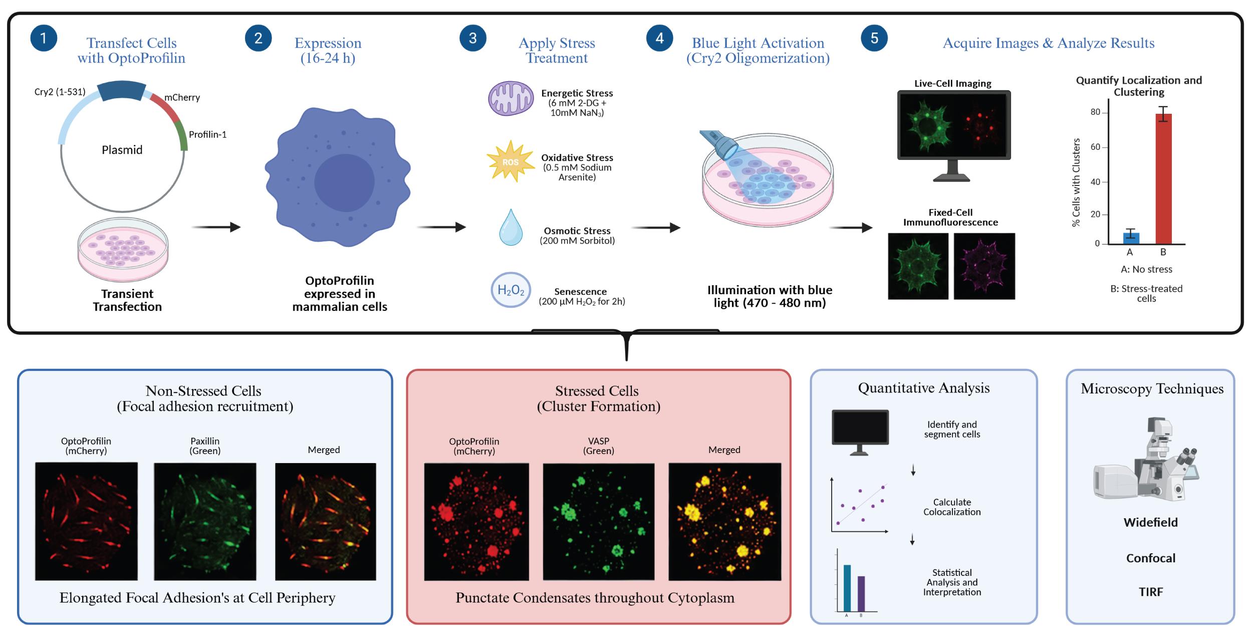
OptoProfilin applications in cells. Figure created using BioRender.
Background
The actin cytoskeleton is highly responsive to environmental and physiological stress. Stress-induced remodeling of actin-associated proteins contributes to processes including biomolecular condensate formation, stress granule assembly, senescence, neurodegeneration, and disease progression. Profilin-1 is a multifunctional actin-binding protein that regulates actin polymerization and interacts with numerous cytoskeletal regulatory proteins, including vasodilator-stimulated phosphoprotein (VASP).
OptoProfilin was developed by fusing mouse Profilin-1 to the photolyase homology region of Arabidopsis thaliana Cryptochrome 2 and mCherry [1]. Upon blue-light activation, Cry2 undergoes oligomerization, promoting localization of OptoProfilin to VASP-containing structures; the interaction is reversible in the absence of blue light. Under non-stress conditions, OptoProfilin localizes primarily to focal adhesions. Following cellular stress, OptoProfilin transitions into punctate condensates that serve as a visual indicator of stress-associated cytoskeletal remodeling. OptoProfilin was first investigated using a Leica DMi8 live cell imaging system; thus, this system and the associated LasX software are featured prominently in this protocol. However, these methods could reasonably be extended to other microscopy platforms.
Compared to multi-component biosensors, OptoProfilin requires expression of only a single construct, simplifying experimental implementation and increasing compatibility with multiplexed imaging approaches. This protocol is best suited to energetic (ATP depletion), oxidative, osmotic, and senescence-associated stress paradigms; it is not appropriate for detecting heat stress, as OptoProfilin does not form clusters under heat shock conditions. VASP (vasodilator-stimulated phosphoprotein) is an actin-associated protein and a known component of focal adhesions. Mutations to the VASP-binding region of Profilin-1 within OptoProfilin eliminate both its focal adhesion and stress cluster phenotypes. In cell lines with higher VASP expression (e.g., N2a), the assay may lose its ability to discriminate stressed from unstressed cells. In cell lines with lower endogenous VASP expression (e.g., HEK293T, NIH 3T3), only the stress-induced clustering phenotype is informative, whereas the dual-phenotype readout (focal adhesion recruitment versus stress-induced clustering) is most robust in HeLa cells.
Materials and reagents
Biological materials
1. OptoProfilin plasmids (Addgene plasmids, catalog numbers: 208286, 208287, 208288)
2. HeLa cells (ATCC, catalog number: CCL-2)
3. HEK293T cells (ATCC, catalog number: CRL-3216)
4. NIH 3T3 cells (ATCC, catalog number: CRL-1658)
5. Neuro-2a (N2a) cells (ATCC, catalog number: CCL-131)
6. Anti-VASP primary antibody (Cell Signaling, catalog number: 3132)
7. Anti-Paxillin primary antibody (Cell Signaling, catalog number: 12065)
8. Alexa Fluor 488-conjugated secondary antibody (Invitrogen, catalog number: A-11008)
Reagents
1. Dulbecco’s minimum essential medium (DMEM) with D-glucose and glutamine (Gibco, catalog number: 11965-092)
2. Fetalgro EX bovine growth serum (rmbio FGX-BBT) or fetal bovine serum (FBS) (Gibco, catalog number: A52568010)
3. Penicillin-streptomycin (Gibco, catalog number: 15140-122)
4. Trypsin-EDTA (0.25%), phenol red (Gibco, catalog number: 25200-056)
5. Calfectin transfection reagent (SignaGen, catalog number: SL100478)
6. Dulbecco’s phosphate-buffered saline (DPBS) containing Ca2+ and Mg2+ (Gibco, catalog number: 14040-133)
7. Sodium azide (NaN3) (Fisher Scientific, catalog number: BP9221)
8. 2-Deoxy-D-glucose (2-DG) (Sigma, catalog number: D8375)
9. Sodium (meta)arsenite (NaAsO2) (Sigma, catalog number: S7400)
10. Sorbitol (Fisher Scientific, catalog number: BP439)
11. Hydrogen peroxide (H2O2) (Sigma, catalog number: 216763)
12. Paraformaldehyde 16% (Fisher Scientific, Electron Microscopy Sciences, catalog number: 15710)
13. Triton X-100 (Acros, catalog number: 327372500)
14. Bovine serum albumin (BSA) (Fisher Scientific, catalog number: BP6711)
Solutions
1. ATP depletion medium (see Recipes)
2. Oxidative stress medium (see Recipes)
3. Osmotic stress medium (see Recipes)
4. Senescence induction medium (see Recipes)
5. Antibody dilution buffer (see Recipes)
6. 4% paraformaldehyde fixation solution (see Recipes)
Recipes
1. ATP depletion medium
| Reagent | Final concentration | Quantity or volume |
|---|---|---|
| DPBS (with Ca2+/Mg2+) | 1× | 9.84 mL |
| NaN3, 1 M stock | 10 mM | 100 μL |
| 2-DG, 1 M stock | 6 mM | 60 μL |
| Total | n/a | 10 mL |
2. Oxidative stress medium
| Reagent | Final concentration | Quantity or volume |
|---|---|---|
| NaAsO2 | 0.5 mM | 3.25 mg |
| DPBS | 1× | to 50 mL |
| Total | n/a | 50 mL |
3. Osmotic stress medium
| Reagent | Final concentration | Quantity or volume |
|---|---|---|
| D-sorbitol | 200 mM | 1.82 g |
| DPBS | 1× | to 50 mL |
| Total | n/a | 50 mL |
4. Senescence induction medium
| Reagent | Final concentration | Quantity or volume |
|---|---|---|
| H2O2, 9.79 M stock | 200 μM | 1.02 μL |
| DPBS | 1× | 49.999 mL |
| Total | n/a | 50 mL |
5. Antibody dilution buffer
| Reagent | Final concentration | Quantity or volume |
|---|---|---|
| 1× PBS | 1× | 9.97 mL |
| BSA | 1% (w/v) | 0.1 g |
| Triton X-100 | 0.3% (v/v) | 30 μL |
| Total | n/a | 10 mL |
6. 4% paraformaldehyde fixation solution
| Reagent | Final concentration | Quantity or volume |
|---|---|---|
| 16% paraformaldehyde | 4% (w/v) | 10 mL |
| DPBS | 0.75× | 30 mL |
| Total | n/a | 40 mL |
Laboratory supplies
1. 35 mm glass-bottom dishes (MatTek, catalog number: P35G-1.5-14-C)
2. Sterile microcentrifuge tubes (1.5 mL)
3. Sterile serological pipettes
Equipment
1. Tissue culture incubator (37 °C, 5% CO2)
2. Tissue culture microscope
3. Hemacytometer (Bright-LineTM Hemacytometer; Cambridge Instruments, Inc.)
4. Widefield fluorescence microscope: Leica DMi8 Live Cell Imaging System, equipped with an OKOLab stage-top live cell incubation system, Infinity TIRF module, Leica HCX PL APO 63×/1.40–0.60 n.a. oil objective, Leica HC PL APO 100×/1.47 oil TIRF objective, Lumencor LED light engine, CTRadvanced+ power supply, and a Leica DFC900 GT camera
5. Confocal microscope (Zeiss LSM 700 laser scanning microscope using ZEN Black 2012 software)
6. Blue LED illumination source (Sunlite LED Par30 Reflector, Item #80021, 4 W, 120 V)
Software and datasets
1. FIJI/ImageJ (ImageJ 1.54p 17 February 2025)
2. GraphPad Prism (10.6.1 (892) 31 October 2026)
3. ZEN Black 2012 or updated equivalent
4. LAS X software (3.7.6.25997)
Procedure
登录/注册后免费查看全文
文章信息
稿件历史记录
提交日期: Jul 6, 2026
接收日期: Sep 8, 2026
在线发布日期: Sep 20, 2026
出版日期: Oct 20, 2026
版权信息
© 2026 The Author(s); This is an open access article under the CC BY license (https://creativecommons.org/licenses/by/4.0/).
如何引用
Brown, C. J. and Hughes, R. M. (2026). Applications of OptoProfilin in Living Cells for the Imaging of Focal Adhesions and Stress-Associated Phenotypes. Bio-protocol 16(20): e5841. DOI: 10.21769/BioProtoc.5841.

 Download Citation in RIS Format
分类
细胞生物学 > 细胞成像 > 荧光
您对这篇实验方案有问题吗?
在此处发布您的问题,我们将邀请本文作者来回答。同时,我们会将您的问题发布到Bio-protocol Exchange,以便寻求社区成员的帮助。
 发布问题
发布问题


