


3D Iterative Immunofluorescence Imaging on Whole-Mount Samples

发布: 2026年10月05日第16卷第19期 DOI: 10.21769/BioProtoc.5838 浏览次数: 90
评审: Ivonne SehringRajesh D GunageAnonymous reviewer(s)
 PDF
PDF  Q&A
Q&A






Abstract
Multicellular organization relies on reciprocal interactions between molecular events, such as gene expression and protein state, and higher-scale properties, such as spatial patterning and tissue architecture. Understanding these processes requires methods that enable quantitative measurements at subcellular resolution, while maintaining the three-dimensional tissue organization. Conventional immunofluorescence imaging captures spatial information but is limited to the number of fluorescence markers that can be imaged simultaneously, whereas dissociation-based single-cell approaches can profile multimodal cellular states but lack positional information. Here, we describe 3D in toto iterative immunofluorescence imaging, termed 3D-4i, which enables up to ten-plex protein and protein state measurements in early zebrafish embryos. Leveraging sample immobilization on 96-well plates together with a gentle liquid handling system and high-content spinning disc confocal microscopy, this method comprises repeated rounds of antibody staining, optical clearing, confocal imaging, and antibody elution. Subsequent image analysis allows segmentation of nuclei and cells, extraction of quantitative single-cell features, and integration of molecular measurements with spatial context. Altogether, 3D-4i provides a scalable platform for investigating diverse biological processes in intact embryos, while maintaining both subcellular resolution and three-dimensional context.
Key features
• Enables volumetric, single-cell multiplexed protein imaging in intact early zebrafish embryos using iterative immunofluorescence imaging.
• Compatible with automated liquid-handling and high-throughput spinning disc confocal microscopy.
• Supports quantitative analysis of diverse biological processes at single-cell resolution.
Keywords: Fluorescence microscopy Multiplexed imaging
Antibody
High-throughput imaging
Volumetric imaging
Whole-mount samples
Automated liquid handling
Multiplexed imaging
Antibody
High-throughput imaging
Volumetric imaging
Whole-mount samples
Automated liquid handling
Graphical overview
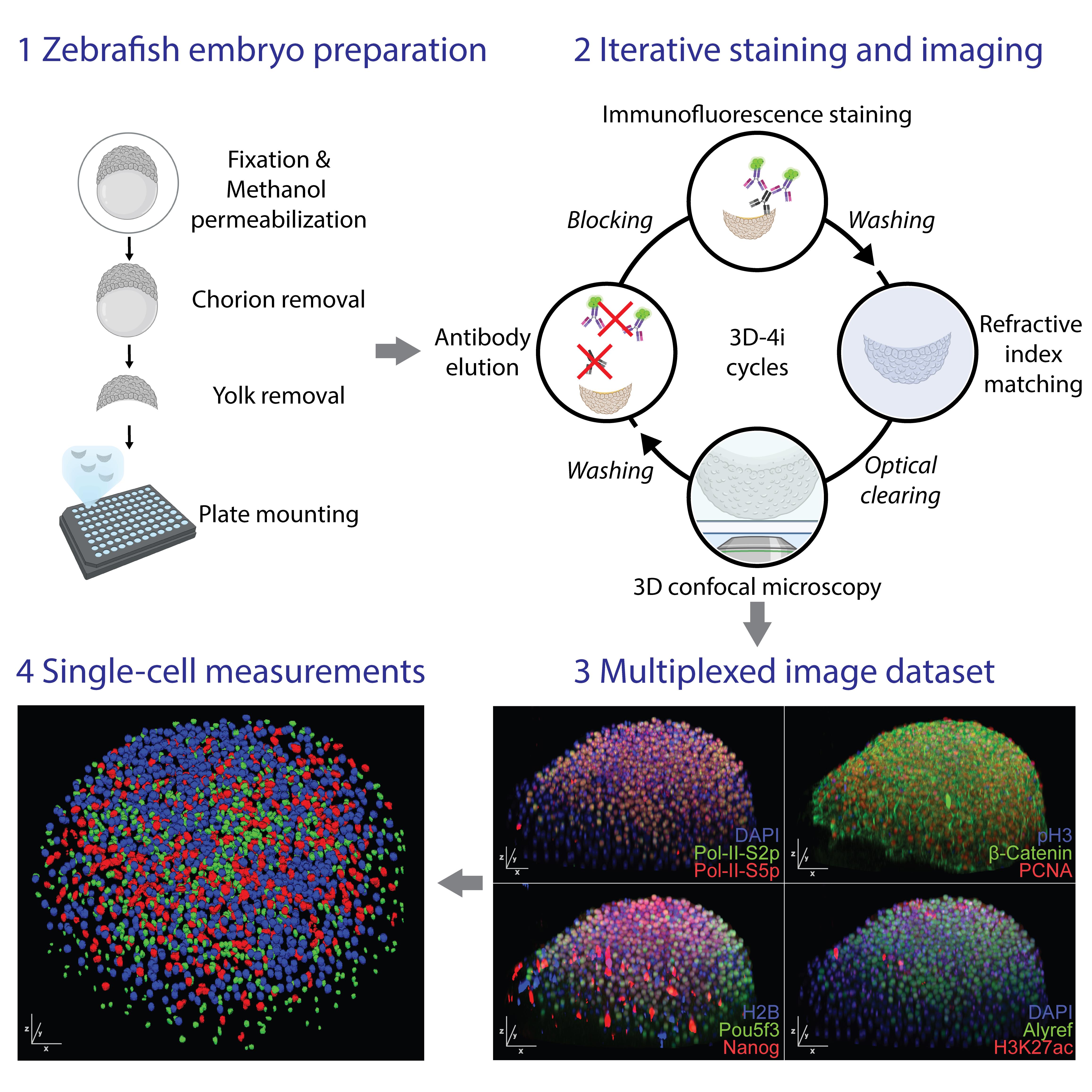
Workflow for multiplexed immunofluorescence in zebrafish embryos using the 3D-4i protocol and subsequent single cell measurements
Background
Multicellular development emerges from coordinated molecular, cellular, and tissue-scale processes. In embryos, proteins, chromatin states, signaling activities, cell-cycle progression, and cell–cell interactions are organized in three-dimensional space and change rapidly over developmental time. Methods that preserve this spatial organization while enabling quantitative measurements at single-cell or subcellular resolution are therefore essential for understanding how cellular states are coordinated within intact tissues. Immunofluorescence microscopy is widely used to visualize protein abundance and localization in fixed samples [1]. It provides direct spatial information and can resolve subcellular patterns, but conventional immunofluorescence is limited by spectral overlap between fluorophores and therefore typically allows only a small number of markers to be measured in the same specimen [2]. In contrast, dissociation-based single-cell approaches can profile many molecular features across large numbers of cells but generally remove cells from their native spatial context [3,4]. This loss of positional information is a major limitation when studying embryos and other organized three-dimensional systems, where cell state is strongly linked to tissue architecture, neighboring cells, and developmental position.
Cyclic immunofluorescence methods overcome part of this limitation by repeatedly staining, imaging, and removing or inactivating fluorescent signals from the same sample. Iterative immunofluorescence imaging uses repeated rounds of antibody staining, confocal imaging, and antibody elution to link multiplexed protein measurements with spatially resolved single-cell information [5–7]. Related cyclic imaging approaches, including CODEX and Immuno-SABER, have similarly enabled high-dimensional profiling of intact samples by increasing the number of measurable targets beyond the limits of standard fluorescence microscopy [8,9]. These methods have been particularly powerful in cultured cells and thin tissue sections, where samples are relatively accessible to antibodies, easy to immobilize, and compatible with repeated liquid exchange. Adapting cyclic immunofluorescence to intact three-dimensional developmental specimens remains technically challenging. In volumetric samples, antibodies must penetrate through many cell tiers, while maintaining specific and uniform staining. Repeated liquid exchange can disturb or detach fragile specimens, especially when samples are not naturally adherent. In addition, optical sectioning through thick samples introduces imaging challenges such as light scattering, signal attenuation, refractive index (RI) mismatch, and optical aberrations. Finally, multiplexed imaging requires that each staining and elution cycle be reproducible and that residual signal from previous cycles remains sufficiently low to avoid compromising later measurements.
The early zebrafish embryo is a useful system for developing and applying volumetric multiplexed imaging methods. Zebrafish embryos develop externally, are optically accessible, and can be collected in large numbers, making them well suited for high-content imaging approaches. However, early embryos are also challenging for cyclic imaging because they are spherical, non-adherent, and sensitive to handling. A successful protocol must therefore combine robust fixation and permeabilization, stable whole-mount immobilization, gentle liquid handling, efficient antibody elution, and high-throughput confocal imaging. Here, we describe 3D in toto iterative immunofluorescence imaging, termed 3D-4i, for multiplexed protein measurements in intact early zebrafish embryos [10]. The protocol immobilizes fixed and permeabilized embryos in coated 96-well plates and uses automated liquid handling to perform repeated rounds of antibody staining, washing, optical clearing, confocal imaging, and elution. High-content spinning disk confocal microscopy enables volumetric imaging of whole embryos, while downstream image analysis supports nuclear and cellular segmentation, extraction of quantitative features, and integration of protein measurements with three-dimensional spatial context.
Altogether, this protocol provides a scalable approach for multiplexed analysis of intact embryos. By preserving tissue organization while expanding the number of measurable protein markers, 3D-4i enables investigation of diverse developmental and cellular processes at single-cell resolution in their native three-dimensional context.
Materials and reagents
Biological materials
1. Zebrafish (Danio rerio) embryos, such as wild-type golden line or Tg(β-actin:H2A-mCherry), were used in this work
Note: This protocol has been rigorously tested for zebrafish embryos of stages 2.5–5 h post fertilization (hpf). Other biological samples, including tissues, whole animals, or embryos from other species, may also be compatible but will require optimization (see General notes).
Reagents
1. 10× DPBS (Thermo Fisher Scientific, catalog number: 14200075)
2. 1× DPBS (Thermo Fisher Scientific, catalog number: 14190144)
3. 16% paraformaldehyde (PFA) (Electron Microscopy Sciences, catalog number: 15710)
4. Bovine serum albumin (BSA) (Sigma-Aldrich, catalog number: B4287-5G)
5. CaCl2·2H2O (Merck Millipore, catalog number: 1023820250)
6. Diatrizoic acid (Sigma-Aldrich, catalog number: D9268)
7. Distilled water (Thermo Fisher Scientific, catalog number: 10977035)
8. Guanidine chloride (Sigma-Aldrich, catalog number: G4505)
9. HCl (e.g., 32%) (Sigma-Aldrich, catalog number: 1003191011)
10. Human fibronectin (1 mg/mL) (Sigma-Aldrich, catalog number: F0895)
11. Iodixanol (Sigma-Aldrich, catalog number: D1556)
12. KCl (Carl Roth, catalog number: 6781.1)
13. L-glycine (Sigma-Aldrich, catalog number: G7126)
14. Maleimide (Sigma-Aldrich, catalog number: 129585)
15. Methanol (Sigma-Aldrich, catalog number: 179957)
16. Methylene blue 1% (Thermo Fisher Scientific, catalog number: 042771.AE)
17. MgCl2·6H2O (Sigma-Aldrich, catalog number: M2670)
18. N-acetyl-cysteine (Sigma-Aldrich, catalog number: A9165)
19. N-methyl-d-glucamine (Sigma-Aldrich, catalog number: M2004)
20. NaCl (Carl Roth, catalog number: 3957.1)
21. NH4Cl (Sigma-Aldrich, catalog number: 254134)
22. Poly-D-lysine (0.1 mg/mL) (Thermo Fisher Scientific, catalog number: A3890401)
23. Sodium hydroxide (NaOH), 1N standard solution (Thermo Fisher Chemicals, catalog number: 124260010)
24. TCEP-HCl (Lucerna Chem, catalog number: BP-P1020)
25. Triton X-100 Surfact-AmpsTM detergent solution (10% solution) (Thermo Fisher Scientific, catalog number: 85112)
26. Tween20 (40% solution) (Sigma-Aldrich, catalog number: P9416)
27. Urea (Sigma-Aldrich, catalog number: U1250)
Solutions
1. E3 buffer (See Recipes)
2. 2× Fixation solution (FS) (see Recipes)
3. Quenching solution (QS) (see Recipes)
4. PBS-T washing solution (PBS-T) (see Recipes)
5. 2× conventional blocking buffer (cBB) (see Recipes)
6. Plate coating solution (see Recipes)
7. 4i Elution buffer (4i-EB) (see Recipes)
8. 2× 4i blocking buffer (sBB) (see Recipes)
9. PROTOS imaging buffer (PROTOS) (see Recipes)
Recipes
1. E3 buffer
60× stock:
| Reagent | Final concentration | Volume (2 L) |
|---|---|---|
| Water | - | 2 L |
| NaCl | 0.3 M | 34.8 g |
| KCl | 0.01 M | 1.6 g |
| CaCl2·2H2O | 0.02 M | 5.8 g |
| MgCl2·6H2O | 0.024 M | 9.78 g |
To prepare a 60× stock, dissolve the ingredients in H2O, to a final volume of 2 L. Adjust the pH to 7.2 with NaOH. Autoclave (https://cshprotocols.cshlp.org/content/2011/10/pdb.rec66449).
1× E3 buffer:
| Reagent | Final concentration | Volume (1 L) |
|---|---|---|
| 60× E3 buffer | 1× | 16 mL |
| Water | - | 984 mL |
| Methylene blue (1%) | 1 mg/L | 100 μL |
2. 2× Fixation solution (FS)
| Reagent | Final concentration | Volume (20 mL) |
|---|---|---|
| 10× DPBS | 2× | 4 mL |
| Water | - | 6 mL |
| 16% PFA | 8% | 10 mL |
First dilute the DPBS to 2× and then add the PFA for dilution. For use, add 1:1 to E3 buffer containing the sample to arrive at 1× DPBS and 4% PFA concentration.
Caution: PFA is a highly toxic chemical and should only be handled in a chemical safety cabinet.
Store the solution in the fridge and use it within a week; ideally, prepare fresh every time.
3. Quenching solution (QS)
| Reagent | Final concentration | Volume (10 mL) |
|---|---|---|
| 1× DPBS | 1× | 10 mL |
| NH4Cl | 100 mM | 53.5 mg |
Dissolve the NH4Cl powder in DPBS while shaking on a rocker.
4. PBS-T
| Reagent | Final concentration | Volume (50 mL) |
|---|---|---|
| 1× DPBS | 1× | 49.875 mL |
| Tween20 (40%) | 0.1% | 125 μL |
Add the Tween20 solution by pipetting up and down in the DPBS to make sure it is evenly distributed.
5. cBB
| Reagent | Final concentration | Volume (10 mL) |
|---|---|---|
| 1× DPBS | 1× | 9.75 mL |
| BSA | 1% | 100 mg |
| Triton-X-100 (10%) | 0.25% | 250 μL |
Dissolve the BSA powder in the DPBS by gently rocking the tube on a shaker. Store the solution in the fridge and use it within a month.
6. Plate coating solution
| Reagent | Final concentration | Volume (1 mL) |
|---|---|---|
| 1× DPBS | 1× | 400 μL |
| Poly-D-lysine (0.1 mg/mL) | 0.05 mg/mL | 500 μL |
| Human fibronectin (1 mg/mL) | 0.1 mg/mL | 100 μL |
Prepare the solution directly before use. Caution: Prolonged storage will reduce the coating material as it adsorbs to the container walls.
7. 4i Elution buffer (4i-EB)
Stock:
| Reagent | Final concentration | Volume (250 mL) |
|---|---|---|
| Water | - | To 250 mL final volume |
| L-glycine | 0.5 M | 9.38 g |
| Urea | 1.2 M | 18.02 g |
| Guanidine chloride | 3 M | 71.65 g |
Prepare the elution buffer stock containing water, L-glycine, urea, and guanidine chloride. Store at 4 °C for up to 6 months.
Ready-to-use solution:
| Reagent | Final concentration | Volume (5 mL) |
|---|---|---|
| 4i elution buffer stock | - | 5 mL |
| TCEP-HCl | 0.07 M | 100 mg |
| HCl (33%) | Adjust to pH 2.5 | ~77 μL |
Before use, add TCEP-HCl to the required volume and adjust the pH to 2.5 with HCl. Confirm the pH using a pH test strip.
8. 2× 4i blocking buffer (sBB)
| Reagent | Final concentration | Volume (10 mL) |
|---|---|---|
| 1× DPBS | 1× | 10 mL |
| BSA | 2% | 0.2 g |
| NH4Cl | 0.4 M | 0.2 g |
| Maleimide (just before use) | 0.6 M | 0.58 g |
The sBB can be prepared and stored in the fridge; however, maleimide is added just before use.
9. PROTOS imaging buffer (PROTOS)
| Reagent | Final concentration | Volume (100 mL) |
|---|---|---|
| Water | - | To 100 mL final volume |
| N-methyl-d-glucamine | 23.5% | 23.5 g |
| Diatrizoic acid | 29.4% | 29.4 g |
| Iodixanol | 32.4% | 32.4 g |
| N-acetyl-cysteine (add on the day before use) | 0.35 M | 5.7 g |
Add 50 mL of water to a beaker, followed by N-methyl-D-glucamine and diatrizoic acid. Stir until fully dissolved, then add Iodixanol and continue stirring until dissolved. Adjust the final volume to 100 mL with water. Store tightly sealed and protected from light to prevent evaporation and refractive index (RI) changes. Confirm an RI of approximately 1.45 using a refractometer. Add the required amount of N-acetyl-cysteine on the day of use.
Notes:
1. This refractive index matching solution is an adjusted version of the PROTOS clearing solution used in the Chung lab (https://docs.abcam.com/pdf/protocols/clarity-protocol.pdf).
2. Depending on the sample, the antibodies used, and the imaging platform used, other refractive index matching solutions can be used (e.g., CUBIC [11] or fructose-glycerol [12]).
Laboratory supplies
1. Glass Pasteur pipette (Hilgenberg, catalog number: 3150102)
2. Glass vials (e.g., Fisher Scientific, catalog number: 14-961-29)
3. Glass Petri dish (e.g., BRAND, catalog number: BR455717-10EA)
4. 5 mL tubes (e.g., Eppendorf, catalog number: 0030119401)
5. Imaging Spacer, 9 mm diameter × 0.12 mm depth (Sigma-Aldrich, catalog number: GBL654002-100EA)
6. Superfrost® ExcellTM slides (Menzel-Gläser, catalog number: J5800AMNZ)
7. 22 mm round cover slip glass (Menzel-Gläser, catalog number: DV40009)
8. 96-well imaging plate μCLEAR®, SCHWARZ, CELLSTAR® (Greiner Bio-One, catalog number: 655090)
9. pH testing strips (Macherey-Nagel, catalog number: 92110)
10. Nuclear counterstain dye (e.g., DAPI) (Invitrogen, catalog number: D1306)
11. Primary antibodies of choice [e.g., PCNA (D3H8P) rabbit monoclonal antibody; Cell Signaling Technology, catalog number: 13110]
12. Secondary antibodies of choice (e.g., Invitrogen, highly cross-adsorbed secondary antibody, Alexa FluorTM series)
13. (Optional) ZenonTM IgG Labeling kits (Invitrogen)
14. (Optional) Reagent reservoirs (BRAND, catalog number: BR701450)
15. (Optional) BRAVO compatible 70 μL tips in 384-unit box (Agilent Technologies, catalog number: 19133-142)
Note: Choice of primary antibody species and secondary detection antibodies must be compatible and tested with the sample. (see Supplementary Section C and General notes).
Equipment
1. Chemical safety cabinet/hood
2. Watchmaker's tweezers DUMONT Inox (DUMONT, model: Size 5)
3. Magnetic stirrer (e.g., IKA, model: RCT basic, catalog number: 0020129521)
4. Stereomicroscope (e.g., ZEISS, model: Stemi 508)
5. Orbital rocker-shaker (e.g., IKA, ROCKER 3D, catalog number: 0004001000)
6. FisherbrandTM UV crosslinker (Fisher Scientific, catalog number: 13-245-222)
7. Laboratory centrifuge with rotors compatible with 96-well plate inserts (e.g., Eppendorf, model: 5810R)
8. Spinning disk confocal microscope (e.g., Visitron, model: VisiScope CSU-X1)
9. pH meter (e.g., METTLER TOLEDO, model: SevenDirect series)
10. Pipetboy acu 2 (Integra, catalog number: 1550179)
11. Freezer (-20 °C)
12. Refrigerator (2–8 °C)
13. (Optional) Refractometer (e.g., KERN, model: analog refractometer)
14. (Optional) Liquid handling platform (e.g., Agilent Technologies, model: BRAVO)
Software and datasets
1. (Required) ImageJ/FIJI [13]
2. (Optional advanced analysis) All code used for the image analysis pipelines is available via GitHub at https://github.com/pelkmanslab/abbott/tree/v0.3.9.1, https://doi.org/10.5281/zenodo.22012790, https://github.com/pelkmanslab/abbott-features/tree/v0.1.5.2, https://doi.org/10.5281/zenodo.22012817, https://github.com/pelkmanslab/abbott-segmentation-tasks/tree/v0.4.2, https://doi.org/10.5281/zenodo.22012831, and https://github.com/MaksHess/manuscript-multiplexed-embryo-profiling, as also provided in [10,14].
Procedure
登录/注册后免费查看全文
文章信息
稿件历史记录
提交日期: Jul 2, 2026
接收日期: Aug 24, 2026
在线发布日期: Sep 15, 2026
出版日期: Oct 5, 2026
版权信息
© 2026 The Author(s); This is an open access article under the CC BY-NC license (https://creativecommons.org/licenses/by-nc/4.0/).
如何引用
Wyss, M. F., Hess, M., Shamipour, S. and Pelkmans, L. (2026). 3D Iterative Immunofluorescence Imaging on Whole-Mount Samples. Bio-protocol 16(19): e5838. DOI: 10.21769/BioProtoc.5838.

 Download Citation in RIS Format
分类
发育生物学
系统生物学
您对这篇实验方案有问题吗?
在此处发布您的问题,我们将邀请本文作者来回答。同时,我们会将您的问题发布到Bio-protocol Exchange,以便寻求社区成员的帮助。
 发布问题
发布问题


