


Disease Modeling in iPSC-Derived Human Bone Marrow Organoids

(*contributed equally to this work) 发布: 2026年10月20日第16卷第20期 DOI: 10.21769/BioProtoc.5837 浏览次数: 32
评审: Jessica DavisNeha SaxenaShanmugaPriyaa Madhukaran
 PDF
PDF  Q&A
Q&A
参见作者原研究论文
The authors used this protocol in:
Jan 2025





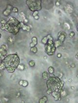
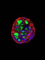
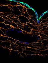
Abstract
Human bone marrow organoids provide a tractable three-dimensional platform for modeling hematopoiesis and hematologic disease in a human niche–like context. Here, we describe a stepwise protocol for utilizing human induced pluripotent stem cell (iPSC)-derived bone marrow organoids that support autonomous hematopoiesis for hematopoietic disease modeling, mouse xenograft hematopoiesis, and drug sensitivity testing. The workflow combines embryoid body formation, early mesoderm/angiogenic induction under hypoxia, hemogenic endothelial commitment, maturation within a collagen-containing hydrogel, and subsequent suspension culture as individual organoids. The resulting organoids contain endothelial, stromal, and hematopoietic components and reproduce key structural and cellular features of human marrow. We further describe procedures for engraftment of normal donor- or patient-derived CD34+ cells and implantation of mature organoids under the renal capsule of immunodeficient mice to assess in vivo hematopoietic maintenance. In prior applications of this platform, donor-derived CD34+ cells were shown to engraft within the organoid niche and undergo multilineage differentiation, enabling detection of selective erythroid defects caused by DDX41 deficiency and assessment of therapeutic suppression of JAK2V617F-mutant patient-derived hematopoietic cells in a human marrow–like microenvironment. This protocol, therefore, enables disease modeling, in vivo xenograft assessment, and ex vivo functional analysis of patient-derived hematopoietic cells using relatively small input samples.
Key features
• Stepwise differentiation of human iPSCs into bone marrow organoids containing endothelial, stromal, and hematopoietic compartments.
• Efficient engraftment of donor-derived CD34+ HSPCs into mature organoids using a Matrigel-assisted seeding method.
• Applicable to imaging, flow cytometry, cytogenetics, and downstream molecular analyses of organoid and donor-derived hematopoietic cells.
• Includes renal capsule implantation for in vivo xenograft assessment of organoid-supported hematopoiesis.
• Enables disease modeling and ex vivo functional studies of patient-derived hematopoietic cells, including analysis of erythroid and myeloid phenotypes and therapeutic responses.
Keywords: Bone marrow organoid iPSC-derived hematopoiesis
CD34
Myelodysplastic syndrome
Acute myeloid leukemia
Engraftment
JAK2
Myeloproliferative neoplasms
Erythropoiesis
DDX41
Drug sensitivity testing.
iPSC-derived hematopoiesis
CD34
Myelodysplastic syndrome
Acute myeloid leukemia
Engraftment
JAK2
Myeloproliferative neoplasms
Erythropoiesis
DDX41
Drug sensitivity testing.
Graphical overview
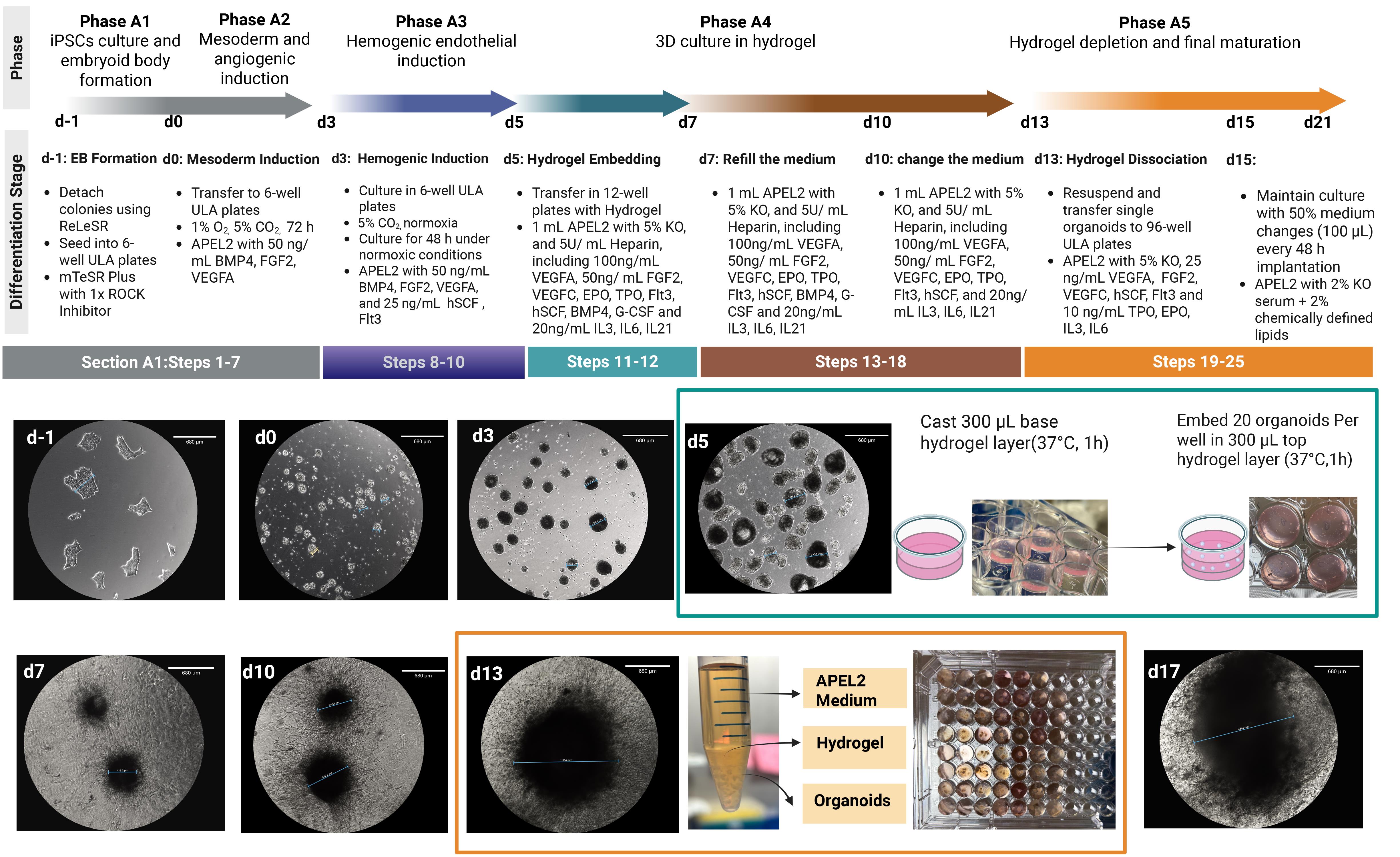
Overview of the bone marrow organoid differentiation in vitro protocol
Background
Human hematopoiesis is regulated by a highly organized bone marrow microenvironment composed of vascular, stromal, and hematopoietic cell populations that cooperate to control stem cell maintenance, lineage commitment, proliferation, and survival [1,2]. Conventional two-dimensional liquid culture systems do not adequately reproduce these multicellular and spatial interactions. Although murine xenograft models have been instrumental for studying human hematopoiesis, they remain limited by species-specific differences in niche-derived signals and by inconsistent engraftment of disease-derived human hematopoietic stem and progenitor cells (HSPCs), particularly in disorders such as myelodysplastic syndromes and other myeloid neoplasms.
Recent advances in human induced pluripotent stem cell (iPSC)-derived marrow organoid systems have established a framework for generating multilineage, vascularized bone marrow–like structures in vitro [3−5]. In particular, human iPSC-derived bone marrow organoids can recreate key endothelial, stromal, and hematopoietic components of the marrow niche in a three-dimensional architecture, providing a more physiologically relevant setting for modeling human hematopoiesis and hematologic disease than conventional suspension culture.
The protocol described here was adapted from that general framework and was used in our study demonstrating autonomous hematopoiesis and efficient engraftment of donor-derived CD34+ cells in human bone marrow organoids [5]. In this system, iPSC aggregates undergo cytokine-directed differentiation, hydrogel embedding, and maturation as individual suspension organoids through day 21, after which they can be engrafted with normal donor- or patient-derived CD34+ HSPCs or implanted under the renal capsule of immunodeficient mice. The resulting organoids support multilineage hematopoiesis and provide a marrow-like niche suitable for confocal Z-stack imaging, flow cytometric analysis, cytogenetic and genomic studies, disease modeling, and in vivo xenograft assessment.
A major advantage of the organoid engraftment platform is that it enables patient-derived hematopoietic cells to be studied within a structured human marrow–like microenvironment rather than in reductionist liquid culture [6]. This is particularly important for primary hematopoietic samples that depend on extracellular matrix, stromal interactions, vascular-associated cues, and short-range niche-derived survival signals. In this setting, donor-derived cells can be localized within the organoid niche and analyzed by imaging and flow cytometry, allowing lineage-specific phenotypes to be evaluated in a context that more closely resembles human marrow biology than standard suspension culture.
This platform has already been applied to multiple disease-focused studies. In one application, engrafted human bone marrow organoids were used to model DDX41-deficient hematopoiesis and revealed selective impairment of erythroid output from donor-derived cells, supporting the use of the system for studying lineage-specific disease phenotypes in a human marrow–like context [7]. In another application, the organoid engraftment model was used to study JAK2V617F-mutant patient-derived hematopoietic cells and to evaluate therapeutic perturbation, demonstrating suppression of donor-derived erythroid and myeloid output in response to cyclosporin A [8]. Together, these studies illustrate that the organoid platform is not only suitable for structural modeling of bone marrow development but also for functional interrogation of disease-relevant hematopoietic phenotypes and therapeutic responses using limited numbers of patient-derived cells.
Accordingly, this protocol includes procedures for organoid generation, donor-cell engraftment, renal capsule implantation, imaging and flow cytometric characterization, and downstream disease modeling applications using patient-derived hematopoietic cells. It provides a practical framework for studying human hematopoiesis, marrow niche interactions, and disease-associated hematopoietic defects in both ex vivo and in vivo settings.
Materials and reagents
Biological materials
1. Human induced pluripotent stem cells (iPSCs) (StemCell Technologies, SCTi003-A)
2. Human bone marrow aspirate samples from healthy donors or patients with myeloid disease, collected under approved institutional protocols
3. Primary normal human bone marrow CD34+ cells (StemCell Technologies, catalog number: 70002)
4. NSG mice (JAX lab, strain #005557)
Cell culture media and supplements
1. mTeSR Plus medium (StemCell Technologies, catalog number: 100-0276)
2. ReLeSR (StemCell Technologies, catalog number: 100-0483)
3. RevitaCell ROCK inhibitor (Thermo Fisher Scientific, catalog number: A2644501)
4. STEMdiff APEL2 medium (StemCell Technologies, catalog number: 05275)
5. StemPro-34 SFM (Thermo Fisher Scientific, catalog number: 10639011)
6. KnockOut serum replacement (Thermo Fisher Scientific, catalog number: 10828010)
7. Chemically defined lipids (Thermo Fisher Scientific, catalog number: 11905031)
8. CryoStor CSB medium (StemCell Technologies, catalog number: 100-1061)
9. Mycoplasma PCR Testing kit (Thermo Fisher Scientific, catalog number: J66117.AMJ)
10. Heparin (Sigma, catalog number: H0200000)
11. StemSpan CD34+ expansion supplement (StemCell Technologies, catalog number: 02691)
12. StemSpan SFEM medium (StemCell Technologies, catalog number: 09650)
13. Cyclosporin A (Sigma-Aldrich, catalog number: 30024-25MG)
14. DMEM/F12 (Thermo Fisher Scientific, catalog number: 11320033)
Cytokines and growth factors
1. BMP4 (Thermo Fisher Scientific, catalog number: PHC9534)
2. VEGFA (StemCell Technologies, catalog number: 78159.1)
3. VEGFC (StemCell Technologies, catalog number: 78202.1)
4. FGF2 (StemCell Technologies, catalog number: 78134.1)
5. IL-21 (StemCell Technologies, catalog number: 78193.1)
6. SCF (StemCell Technologies, catalog number: 78155.1)
7. FLT3 (StemCell Technologies, catalog number: 78137.1)
8. G-CSF (StemCell Technologies, catalog number: 78138.1)
9. TPO (StemCell Technologies, catalog number: 78210.1)
10. EPO (StemCell Technologies, catalog number: 78007)
11. IL-3 (StemCell Technologies, catalog number: 78194.1)
12. IL-6 (StemCell Technologies, catalog number: 78050)
Matrices and dissociation reagents
1. Matrigel for iPSC maintenance and engraftment overlay (Corning, catalog number: 354277)
2. GelTrex reduced-growth-factor basement-membrane matrix (Thermo Fisher Scientific, catalog number: A1413302)
3. VitroCol, Type I human collagen solution (Advanced BioMatrix, catalog number: 5007)
4. Type IV collagen powder (Advanced BioMatrix, catalog number: 5022)
5. Type I collagen powder (Advanced BioMatrix, catalog number: 5008)
6. Collagenase Type I (0.25%) (StemCell Technologies, catalog number: 07902)
General reagents
1. PBS (StemCell Technologies, catalog number: 07905)
2. BSA (Sigma-Aldrich, catalog number: A3294-50G)
3. EDTA (Sigma-Aldrich, catalog number: E9884-100G)
4. Ficoll-Paque density gradient medium (Sigma-Aldrich, catalog number: GE17-5442-02)
5. CD34 MicroBead kit (Miltenyi Biotec, catalog number: 130-046-702)
6. RBC lysis buffer (Thermo Fisher Scientific, catalog number: 00-4333-57)
7. MACS buffer (Miltenyi Biotec, catalog number: 130-091-221)
Cell labeling and viability assay reagents
1. CellTrace Far Red Cell Proliferation kit (Thermo Fisher Scientific, catalog number: C34564)
2. FITC-conjugated Annexin V (BD, catalog number: 556547)
3. DAPI (Thermo Fisher Scientific, catalog number: 41116113)
4. CellVue Far Red for cell membrane labeling (Sigma-Aldrich, catalog number: MIDCLARET-1KT)
Material and reagents for tissue clearing and confocal imaging
1. Fructose (Sigma-Aldrich, catalog number: F0127)
2. Glycerol (Sigma-Aldrich, catalog number: G5516)
3. Slides (Fisher Scientific, catalog number: NC9202659)
4. Coverslip (Sigma-Aldrich, catalog number: CLS285022)
5. Labeling tapes (Fisher Scientific, catalog number: 1590110B)
6. Goat serum (Thermo Fisher Scientific, catalog number: 50062Z)
7. Triton X-100 (Sigma-Aldrich, catalog number: T8787-50ML)
8. 16% paraformaldehyde (PFA) (Thermo Fisher Scientific, catalog number: 043368.9M)
9. Biotinylated UEA1 (Vector Laboratories, catalog number: B-1065-2)
Material and reagents for organoid implantation in mice
1. Mouse anesthesia system with heating pad
2. Ophthalmic ointment (Bausch&Lomb, catalog number: NDC24208-780-55)
3. Analgesic agents (Covetrus, catalog number: NDC11695-6936-1)
4. Absorbable suture (ETHICON, catalog number: J397)
5. Non-absorbable suture (ETHICON, catalog number: 8698)
6. Hair removal cream (Grainger, catalog number: 13Z999)
7. Betadine (Med Vet International, catalog number: PHVBETASCRUB16)
8. 70% ethanol (Thermo Fisher Scientific, catalog number: R40135)
9. Sterile dissecting scissors (Fine Science Tools, catalog number: 15003-08)
10. Fine forceps (Fine Science Tools, catalog number: 91115-10)
11. Sterile gauze (Dukal corporation, catalog number: 6208)
12. Sterile cotton swabs (PuritanTM, catalog number: 253206H 20MM)
13. Glass Pasteur pipette (Chemglass Life Sciences, catalog number: CGN3404102)
14. Saline (Cytiva, catalog number: SH30028.01)
Solutions
1. Collagen dissolving buffer (see Recipes)
2. Hydrogel dilution buffer (see Recipes)
3. Collagen solution (see Recipes)
4. Hydrogel working solution (see Recipes)
5. Mesoderm/angiogenic induction medium (see Recipes)
6. Hemogenic endothelial induction medium (see Recipes)
7. Hydrogel culture medium A (see Recipes)
8. Hydrogel culture medium B (see Recipes)
9. Organoid growth medium (see Recipes)
10. Engraftment-support medium (see Recipes)
11. Tissue clearing buffer (see Recipes)
Recipes
1. Collagen dissolving buffer
| Reagent | Quantity or volume | Final concentration |
|---|---|---|
| Glacial acetic acid | 0.25 mL | 0.25% (v/v) |
| Sodium acetate | 0.01 g | 0.01% (w/v) |
| H2O | 99.75 mL | - |
Note: Filter the prepared collagen dissolving buffer through a 0.22 μm filter.
2. Hydrogel dilution buffer
| Reagent | Quantity or volume | Final concentration |
|---|---|---|
| Sodium bicarbonate | 7.5 g | 0.075 g/mL |
| GlutaMax (200 mM) | 2 mL | 4 mM |
| Ham’s F12 | 69.6 mL | - |
| HEPES | 4.2 mL | - |
| 10× PBS | 20.86 mL | - |
| H2O | 3.34 mL | - |
| Total | 100 mL |
Note: Filter the prepared hydrogel dilution buffer through a 0.22 μm filter.
3. Collagen solution
| Reagent | Quantity or volume | Final concentration |
|---|---|---|
| Type I collagen powder | 15 mg | 3 mg/mL |
| Type IV collagen powder | 5 mg | 1 mg/mL |
| Collagen dissolving buffer | 5 mL | - |
Note: Mix Type I collagen powder with collagen dissolving buffer under sterile conditions in a biosafety cabinet. Agitate the mixture on a hula mixer at 4 °C for at least 12 h. Then, add the VitroCol solution to the collagen type IV powder and continue mixing on a hula mixer at 4 °C for an additional 12 h. During storage, keep the collagen solution at 4 °C with continuous agitation on a hula mixer.
4. Hydrogel working solution
| Reagent | Quantity or volume |
|---|---|
| VitroCol solution (3 mg/mL) | 0.2 mL |
| Hydrogel dilution buffer | 0.1 mL |
| GelTrex | 0.4 mL |
| Collagen solution | 0.3 mL |
| NaOH solution (1 M) | ~10 μL |
| Total | 1 mL |
Note: Prepare the hydrogel working solution on ice. Pre-cool the pipette tip with ice-cold PBS before aspirating Matrigel. Add 1 M sodium hydroxide in increments of less than 2 μL, allowing thorough mixing after each addition. Do not add excessive NaOH at once, as this may generate heat and cause a localized pH imbalance. After each addition, measure the pH using pH test paper. Prepare the hydrogel working solution in small volumes (≤1 mL), as pH adjustment with NaOH becomes difficult at larger volumes. The target pH is 7.0.
5. Mesoderm/angiogenic induction medium
| Reagent | Quantity or volume |
|---|---|
| APEL2 | 10 mL |
| BMP4 (100 ng/μL) | 5 μL |
| FGF2 (100 ng/μL) | 5 μL |
| VEGFA (100 ng/μL) | 5 μL |
Note: Prepare fresh before use.
6. Hemogenic endothelial induction medium
| Reagent | Quantity or volume |
|---|---|
| APEL2 | 10 mL |
| BMP4 (100 ng/μL) | 5 μL |
| FGF2 (100 ng/μL) | 5 μL |
| VEGFA (100 ng/μL) | 5 μL |
| hSCF (100 ng/μL) | 2.5 μL |
| Flt3 (100 ng/μL) | 2.5 μL |
Note: Prepare fresh before use.
7. Hydrogel culture medium A
| Reagent | Quantity or volume |
|---|---|
| APEL2 | 9.5 mL |
| KnockOut serum replacement | 0.5 mL |
| Heparin (1000 IU/mL) | 50 μL |
| VEGFA (100 ng/μL) | 10 μL |
| VEGFC (100 ng/μL) | 5 μL |
| Flt3 (100 ng/μL) | 5 μL |
| FGF2 (100 ng/μL) | 5 μL |
| hSCF (100 ng/μL) | 5 μL |
| EPO (100 ng/μL) | 5 μL |
| TPO (100 ng/μL) | 5 μL |
| IL3 (100 ng/μL) | 2.5 μL |
| IL6 (100 ng/μL) | 2.5 μL |
| IL21 (100 ng/μL) | 2.5 μL |
| BMP4 (100 ng/μL) | 5 μL |
| G-CSF (100 ng/μL) | 5 μL |
Note: Prepare fresh before use. Heparin is included to stabilize heparin-binding growth factors, particularly VEGF, and support their activity during vascular induction.
8. Hydrogel culture medium B
| Reagent | Quantity or volume |
|---|---|
| APEL2 | 9.5 mL |
| KnockOut serum replacement | 0.5 mL |
| Heparin (1000 IU/mL) | 50 μL |
| VEGFA (100 ng/μL) | 10 μL |
| VEGFC (100 ng/μL) | 5 μL |
| Flt3 (100 ng/μL) | 5 μL |
| FGF2 (100 ng/μL) | 5 μL |
| hSCF (100 ng/μL) | 5 μL |
| EPO (100 ng/μL) | 5 μL |
| TPO (100 ng/μL) | 5 μL |
| IL3 (100 ng/μL) | 2.5 μL |
| IL6 (100 ng/μL) | 2.5 μL |
| IL21 (100 ng/μL) | 2.5 μL |
Note: Prepare fresh before use.
9. Organoid growth medium
| Reagent | Quantity or volume |
|---|---|
| APEL2 | 9.5 mL |
| KnockOut serum replacement | 0.5 mL |
| Heparin (1000 IU/mL) | 50 μL |
| VEGFA (100 ng/μL) | 2.5 μL |
| VEGFC (100 ng/μL) | 2.5 μL |
| Flt3 (100 ng/μL) | 2.5 μL |
| FGF2 (100 ng/μL) | 2.5 μL |
| EPO (100 ng/μL) | 1 μL |
| TPO (100 ng/μL) | 1 μL |
| IL3 (100 ng/μL) | 1 μL |
| IL6 (100 ng/μL) | 1 μL |
| IL21 (100 ng/μL) | 1 μL |
Note: Prepare fresh before use.
10. Engraftment-support medium
| Reagent | Quantity or volume |
|---|---|
| StemPro-34 | 9.6 mL |
| KnockOut serum replacement | 0.2 mL |
| Chemically defined lipids | 0.2 mL |
| EPO (100 ng/μL) | 1 μL |
| TPO (100 ng/μL) | 1 μL |
| IL3 (100 ng/μL) | 1 μL |
| IL6 (100 ng/μL) | 1 μL |
Note: Prepare fresh before use.
11. Tissue clearing buffer
| Reagent | Quantity or volume |
|---|---|
| Glycerol | 330 mL |
| Fructose | 297.2 g |
| H2O | 70 mL |
Note: Stir the mixture in a magnetic stirrer overnight to fully dissolve the fructose. The final volume should reach approximately 660 mL after complete dissolution. Store the prepared solution at room temperature in a dry, light-protected area. To minimize water uptake, the solution is aliquoted into 10 mL portions in Falcon tubes, and the caps are sealed with Parafilm. Each aliquoted tube is discarded after use, and unopened sealed aliquots can be stored for up to 6 months under dry conditions.
Laboratory supplies
1. Ultra-low-attachment 6-well plates (Sigma-Aldrich, catalog number: CLS3471-24EA)
2. Ultra-low-attachment 96-well plate (Thermo Fisher Scientific, catalog number: 174925)
3. 6-well tissue culture plate (Sigma-Aldrich, catalog number: CLS3516-50EA)
4. 24-well tissue culture plate (Sigma-Aldrich, catalog number: CLS3524-100EA)
5. 100-μm cell strainers (Sigma-Aldrich, catalog number: CLS431752)
6. 40-μm cell strainers (Sigma-Aldrich, catalog number: 07-201-430)
Equipment
1. Biosafety cabinet (Thermo Fisher Scientific, catalog number: 1910179)
2. Normoxic CO2 incubator set to 37 °C and 5% CO2 (Thermo Fisher Scientific, catalog number: 51036157)
3. Hypoxia-capable incubator set to 37 °C, 1% O2, and 5% CO2 (Thermo Fisher Scientific, catalog number: 51036516)
4. Orbital shaker (Alkali Scientific, catalog number: RS7049)
5. Tube rotor (Thermo Fisher Scientific, catalog number: 88881001)
6. Inverted microscope (Echo Rebel)
Procedure
登录/注册后免费查看全文
文章信息
稿件历史记录
提交日期: Apr 20, 2026
接收日期: Sep 2, 2026
在线发布日期: Sep 17, 2026
出版日期: Oct 20, 2026
版权信息
© 2026 The Author(s); This is an open access article under the CC BY-NC license (https://creativecommons.org/licenses/by-nc/4.0/).
如何引用
Li, E., Ji, A., Zheng, A. and Ren, K. (2026). Disease Modeling in iPSC-Derived Human Bone Marrow Organoids. Bio-protocol 16(20): e5837. DOI: 10.21769/BioProtoc.5837.

 Download Citation in RIS Format
分类
干细胞 > 类器官培养
细胞生物学 > 细胞分离和培养 > 3D细胞培养
您对这篇实验方案有问题吗?
在此处发布您的问题,我们将邀请本文作者来回答。同时,我们会将您的问题发布到Bio-protocol Exchange,以便寻求社区成员的帮助。
 发布问题
发布问题


