


A SPAAC-Based Bioorthogonal Method for Verifying Protein Palmitoylation
基于 SPAAC 生物正交反应的蛋白质棕榈酰化验证方法

(*contributed equally to this work) 发布: 2026年09月20日第16卷第18期 DOI: 10.21769/BioProtoc.5835 浏览次数: 38
评审: Catherine HurdBhanu JagilinkiJoyce Chiu
 PDF
PDF  Q&A
Q&A
参见作者原研究论文
The authors used this protocol in:
May 2025






Abstract
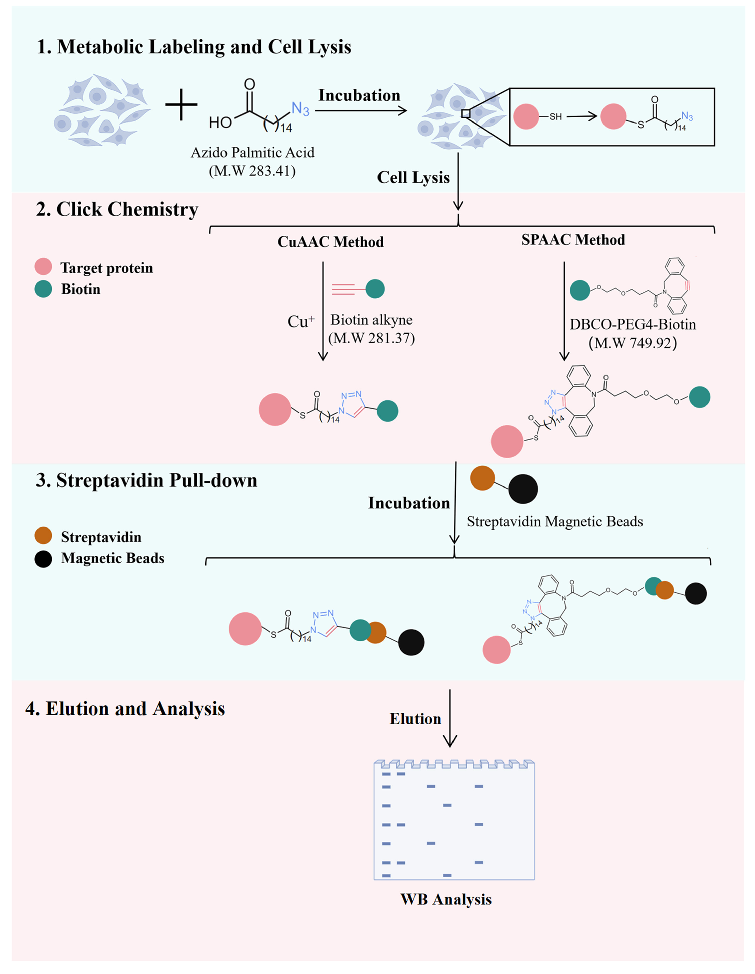
Palmitoylation is a crucial post-translational modification, and bioorthogonal chemistry based on azide-alkyne cycloaddition is typically used to verify protein palmitoylation. Traditional copper-catalyzed click chemistry (CuAAC) proceeds with fast kinetics and is widely used, but it requires a copper catalyst and suffers from copper-induced toxicity and nonspecific labeling. By contrast, strain-promoted click chemistry (SPAAC) has slower kinetics but is catalyst-free, offering high specificity, low cytotoxicity, and simple operation. However, SPAAC is mostly applied to live-cell labeling and imaging of known palmitoylated proteins in the field of palmitoylation, and its use in identifying novel palmitoylated proteins is still limited. Here, we present a SPAAC-based method for detecting endogenous protein palmitoylation. Compared with CuAAC, this method eliminates the need for copper catalysts and reducing agents, thereby simplifying the procedure and reducing reagent usage.
Key features
• A bioorthogonal method for identifying endogenous protein palmitoylation in cells.
• Metabolical labeling of palmitoylated proteins in cells using azido palmitic acid.
• The method requires simple steps and minimal reagents, reducing hands-on time and cost.
Keywords: Click chemistry (点击化学) CuAAC (CuAAC)
SPAAC (SPAAC)
Palmitoylation (棕榈酰化)
Protein tagging (蛋白质标记)
Post-translational modifications (翻译后修饰)
CuAAC (CuAAC)
SPAAC (SPAAC)
Palmitoylation (棕榈酰化)
Protein tagging (蛋白质标记)
Post-translational modifications (翻译后修饰)
Graphical overview
Background
Palmitoylation is a reversible post-translational modification that involves the covalent attachment of palmitic acid (C16:0) to cysteine residues via a thioester bond. It dynamically regulates protein membrane localization, stability, and signal transduction [1–3]. Aberrant palmitoylation is closely associated with cancer, neurodegenerative diseases, and infectious diseases [3–6]. Therefore, identifying and validating palmitoylation modifications is crucial for elucidating their biological functions and pathogenic mechanisms.
Methods for verifying palmitoylation are mainly divided into two categories: non-metabolic labeling and metabolic labeling [7]. The acyl-biotin exchange (ABE) method is a non-metabolic labeling strategy that offers direct applicability to tissue samples, high sensitivity, and good compatibility with downstream mass spectrometry analysis [8]. Nonetheless, it cannot readily distinguish between different lipid types, requires a complex procedure, and is susceptible to false-positive results [9,10]. Currently, metabolic labeling based on bioorthogonal chemistry is the most widely used strategy. In this approach, azide-bearing palmitic acid analogs (e.g., 15-azido pentadecanoic acid) are incorporated into palmitoylated proteins through cellular metabolism, followed by azide-alkyne cycloaddition to conjugate reporter groups (e.g., fluorophores or biotin) for detection, enrichment, and identification [10,11]. This strategy primarily includes copper-catalyzed click chemistry (CuAAC) and strain-promoted click chemistry (SPAAC). Traditional copper-catalyzed azide-alkyne cycloaddition (CuAAC) proceeds through the Cu(I)-mediated activation of a terminal alkyne to form a copper–acetylide intermediate, which then reacts with azides to yield a triazole. Although CuAAC offers fast reaction kinetics and is the most commonly used click reaction, it requires a copper ion catalyst, which can lead to cytotoxicity and nonspecific labeling [12]. This is partly because Cu(I) ions can coordinate with thiol groups of cysteine residues and imidazole groups of histidine residues in proteins and also generate reactive oxygen species (ROS), resulting in off-target modifications and cellular stress. In contrast, strain-promoted azide–alkyne cycloaddition (SPAAC) relies on the ring strain of cyclooctyne derivatives to drive a catalyst-free [3+2] cycloaddition with azides. Although SPAAC exhibits relatively slower reaction kinetics than CuAAC, it requires no catalyst and has become widely adopted owing to its high specificity, low cytotoxicity, and operational simplicity [13,14]. However, in the context of palmitoylation research, SPAAC is mainly applied to live-cell labeling and imaging for real-time tracking and localization of palmitoylated proteins, whereas few studies have applied it to the identification of previously unreported palmitoylated proteins.
Here, we describe a SPAAC-based protocol that eliminates copper catalyst usage, simplifies the workflow, and provides comparable sensitivity to CuAAC for endogenous palmitoylation detection. Moreover, it can also be extended to other applications, including the enrichment and mass spectrometry identification of palmitoylated proteins and the determination of palmitoylation sites.
Materials and reagents
Biological materials
1. HEK293T cell line
Reagents
1. 30% Acrylamide:Bisacrylamide (Acr/Bis) (29:1) (Solarbio, catalog number: A1010)
2. AEG-1 polyclonal antibody (Proteintech, catalog number: 13860-1-AP)
3. Ammonium persulfate (APS) (Aladdin, catalog number: A112450)
4. Azido palmitic acid (MCE, catalog number: HY-151656)
5. β-actin polyclonal antibody (Proteintech, catalog number: 20536-1-AP)
6. Biotin alkyne (MCE, catalog number: HY-138749)
7. Bovine serum albumin (BSA) (fatty acid free) (MCE, catalog number: HY-D0842A)
8. BCA Protein Assay kit (Biosharp, catalog number: 143179)
9. Chloroform (Aladdin, catalog number: C1506334)
10. Glycerol (Aladdin, catalog number: G116206)
11. CuSO4 (Aladdin, catalog number: C573445)
12. DBCO-PEG4-biotin (MCE, catalog number: HY-130809)
13. Dulbecco's modified Eagle medium (DMEM) (Vivacell, catalog number: C3130)
14. Dimethyl sulfoxide (DMSO) (Aladdin, catalog number: D103277)
15. Fetal bovine serum (FBS) (Vivacell, catalog number: C04001)
16. Fetal bovine serum (lipid depleted) (Vivacell, catalog number: C3840)
17. GPX4 polyclonal antibody (Proteintech, catalog number: 30388-1-AP)
18. Methanol (Aladdin, catalog number: M116115)
19. Non-fat milk (Solarbio, catalog number: D3840)
20. NaCl (Aladdin, catalog number: S433743)
21. NP-40 (Solarbio, catalog number: N8030)
22. Penicillin-streptomycin solution, 100× (Vivacell, catalog number: C3421)
23. Phosphate-buffered saline (PBS) (Biosharp, catalog number: BL601A)
24. Protease inhibitor cocktail (EDTA-free, 100×) (MCE, catalog number: HY-K0010)
25. PVDF membrane (Millipore, catalog number: IPVH00010)
26. Sodium dodecyl sulfate (SDS) (Aladdin, catalog number: S432158)
27. SDS-PAGE loading buffer (4×) (Proteintech, catalog number: PR20003)
28. Semi-dry membrane transfer solution (1×) (Biosharp, catalog number: BL1310A)
29. L-Ascorbic acid sodium salt (Aladdin, catalog number: S105026)
30. Streptavidin magnetic beads (Beyotime, catalog number: P2151)
31. N,N,N',N'-Tetramethylethylenediamine (TEMED) (Aladdin, catalog number: T105496)
32. Tris(3-hydroxypropyltriazolylmethyl)amine (THPTA) (Aladdin, catalog number: T405015)
33. Tris-buffered saline (1×) (Biosharp, catalog number: BL602A)
34. Tris-glycine-SDS running buffer (Biosharp, catalog number: BL603A)
35. Tris-HCl (1 M, pH 6.8) (Solarbio, catalog number: T1020)
36. Tris-HCl (1.5 M, pH 8.8) (Solarbio, catalog number: T1010)
37. Tween 20 (Solarbio, catalog number: T8820)
38. Trypsin EDTA solution (0.25%) (Vivacell, catalog number: C3530)
39. Ultrasensitive ECL Detection kit (Proteintech, catalog number: PK10003)
Solutions
1. DMEM medium (10% FBS) (see Recipes)
2. DMEM medium (5% lipid-depleted FBS) (see Recipes)
3. Fatty acid-free BSA solution (10%) (see Recipes)
4. Azido palmitic acid (50 mM) (see Recipes)
5. Azido-containing labeling medium (see Recipes)
6. NaCl solution (1.5 M) (see Recipes)
7. Cell lysis buffer (see Recipes)
8. Biotin alkyne (1 mM) (see Recipes)
9. DBCO-PEG4-biotin (10 mM) (see Recipes)
10. CuSO4 solution (20 mM) (see Recipes)
11. THPTA (100 mM) (see Recipes)
12. Sodium L-ascorbate solution (300 mM) (see Recipes)
13. TBST (see Recipes)
14. Washing buffer (see Recipes)
15. Spacer gel (see Recipes)
16. Stacking gel (see Recipes)
17. Blocking buffer (5% non-fat milk) (see Recipes)
Recipes
Note: In the following recipes, H2O is deionized water, and DMSO is cell culture grade.
1. DMEM medium (10% FBS)
| Reagent | Final concentration | Quantity or volume |
|---|---|---|
| DMEM | 89% | 44.5 mL |
| FBS | 10% | 5 mL |
| Penicillin-Streptomycin Solution, 100× | 1× | 500 μL |
| Total | n/a | 50 mL |
Store at 4 °C for a week.
2. DMEM medium (5% lipid-depleted FBS)
| Reagent | Final concentration | Quantity or volume |
|---|---|---|
| DMEM | 95% | 47.5 mL |
| Lipid-depleted FBS | 5% | 2.5 mL |
| Total | n/a | 50 mL |
Store at 4 °C for 4 weeks.
3. Fatty acid-free BSA solution (10%)
| Reagent | Final concentration | Quantity or volume |
|---|---|---|
| Fatty acid-free BSA | 10% | 1 g |
| PBS | n/a | 10 mL |
| Total | n/a | 10 mL |
Slowly stir with a magnetic stirrer at 4 °C until dissolved. Divide into 1 mL aliquots and store at -20 °C after filtering through a 0.22 μm filter in a biosafety cabinet.
4. Azido palmitic acid (50 mM)
| Reagent | Final concentration | Quantity or volume |
|---|---|---|
| Azido palmitic acid | 50 mM | 14.2 mg |
| DMSO | n/a | 1 mL |
| Total | n/a | 1 mL |
Divide into 200 μL aliquots in amber centrifuge tubes and store at -80 °C for 6 months.
5. Azido-containing labeling medium
| Reagent | Final concentration | Quantity or volume |
|---|---|---|
| Azido palmitic acid | 50 μM | 10 μL |
| 10% fatty acid-free BSA solution | 1% | 1 mL |
| DMEM medium (5% lipid-depleted FBS) | n/a | up to 10 mL |
| Total | n/a | 10 mL |
Prepare the reagents in the following order: first, combine azido palmitic acid with 10% fatty acid-free BSA solution in a sterile 15 mL centrifuge tube. Mix by inverting the tube or by gently pipetting up and down. Then, incubate the tube in a 37 °C water bath for 0.5–1 h. Finally, add DMEM medium (5% lipid-depleted FBS) and mix by pipetting. Use immediately after preparation. The volumes can be scaled up or down as needed.
Note: The purpose of this step is to allow azido palmitic acid to mix with fatty acid-free BSA, which facilitates cellular uptake of azido palmitic acid.
6. NaCl solution (1.5 M)
| Reagent | Final concentration | Quantity or volume |
|---|---|---|
| NaCl | 1.5 M | 8.8 g |
| H2O | n/a | 100 mL |
| Total | n/a | 100 mL |
7. Cell lysis buffer
| Reagent | Final concentration | Quantity or volume |
|---|---|---|
| NP-40 | 0.5% | 200 μL |
| Glycerol | 10% | 4 mL |
| Protease inhibitor cocktail (100×) | 1× | 400 μL |
| PBS, pH 7.4 | n/a | up to 40 mL |
| Total | n/a | 40 mL |
Aliquot into 10 mL aliquots and store at -20 °C.
Critical: The buffer must not contain Tris or EDTA, as they inhibit the click chemistry reaction [15].
8. Biotin alkyne (5 mM)
| Reagent | Final concentration | Quantity or volume |
|---|---|---|
| Biotin alkyne | 5 mM | 1.4 mg |
| DMSO | n/a | 1 mL |
| Total | n/a | 1 mL |
Aliquot into 500 μL aliquots in amber centrifuge tubes and store at -80 °C for 6 months.
9. DBCO-PEG4-biotin (10 mM)
| Reagent | Final concentration | Quantity or volume |
|---|---|---|
| DBCO-PEG4-biotin | 10 mM | 7.5 mg |
| DMSO | n/a | 1 mL |
| Total | n/a | 1 mL |
Divide into 200 μL aliquots in amber centrifuge tubes and store at -80 °C for 6 months.
10. CuSO4 solution (20 mM)
| Reagent | Final concentration | Quantity or volume |
|---|---|---|
| CuSO4 | 20 mM | 31.9 mg |
| H2O | n/a | 10 mL |
| Total | n/a | 10 mL |
Aliquot into 1 mL aliquots and store at -20 °C.
11. THPTA (100 mM)
| Reagent | Final concentration | Quantity or volume |
|---|---|---|
| THPTA | 100 mM | 43.5 mg |
| H2O | n/a | up to 1 mL |
| Total | n/a | 1 mL |
Divide into 200 μL aliquots in amber centrifuge tubes and store at -20 °C for 3 months.
12. Sodium L-ascorbate solution (300 mM)
| Reagent | Final concentration | Quantity or volume |
|---|---|---|
| L-ascorbate acid sodium salt | 300 mM | 594.3 mg |
| H2O | n/a | 10 mL |
| Total | n/a | 10 mL |
Aliquot into 1 mL aliquots and store at -20 °C protected from light.
13. TBST
| Reagent | Final concentration | Quantity or volume |
|---|---|---|
| Tween 20 | 0.05% | 500 μL |
| Tris-buffered saline (1×) | n/a | up to 1 L |
| Total | n/a | 1 L |
14. Washing buffer
| Reagent | Final concentration | Quantity or volume |
|---|---|---|
| Tween-20 | 0.05% | 50 μL |
| Fatty acid-free BSA solution (10%) | 0.1% | 1 mL |
| PBS | 98.95% | up to 100 mL |
| Total | n/a | 100 mL |
Use immediately after preparation.
15. Spacer gel
| Reagent | Final concentration | Quantity or volume |
|---|---|---|
| 30% Acr/Bis (29:1) | 10% | 3.3 mL |
| 1.5 M Tris-HCl, pH 8.8 | 375 mM | 2.5 mL |
| 10% SDS | 0.1% | 100 μL |
| 10% APS | 0.1% | 100 μL |
| TEMED | 0.2% | 20 μL |
| H2O | n/a | up to 10 mL |
| Total | n/a | 10 mL |
16. Stacking gel
| Reagent | Final concentration | Quantity or volume |
|---|---|---|
| 30% Acr/Bis (29:1) | 5% | 830 μL |
| 1 M Tris-HCl, pH 6.8 | 125 mM | 625 μL |
| 10% SDS | 0.1% | 50 μL |
| 10% APS | 0.15% | 75 μL |
| TEMED | 0.2% | 10 μL |
| H2O | n/a | up to 5 mL |
| Total | n/a | 5 mL |
17. Blocking buffer (5% non-fat milk)
| Reagent | Final concentration | Quantity or volume |
|---|---|---|
| Non-fat milk | 5% | 2 g |
| TBST | n/a | 40 mL |
| Total | n/a | 40 mL |
Laboratory supplies
1. 100 mm TC-treated culture dish (NEST, catalog number: 704202)
2. 1.5 mL microfuge tube (Biosharp, catalog number: BS-15-M)
3. 1.5 mL amber microfuge tube (Biosharp, catalog number: BS-15-A)
4. 2.0 mL microfuge tube (Biosharp, catalog number: BS-20-M)
5. 0.22 μm filters (Biosharp, catalog number: BS-NY13-22-S)
6. Cell scraper (Labselect, catalog number: 1601)
7. Pipette tips (Biosharp, catalog numbers: BS-10-T, BS-200-T, BS-1000-T)
Equipment
1. 4 °C and -20 °C freezer (Haier, model: BCD-390WGHC2B6W9U1)
2. -80 °C freezer (Haier, model: DW-86L486)
3. Automated chemiluminescence/fluorescence image analysis system (Tanon, model: 4600)
4. Biosafety cabinet (Thermo, model: MSC1)
5. CO2 cell culture incubator (Thermo, model: Heracell-150i)
6. Cell sonicator (Branson, model: S-450D)
7. Disc rotator (Scilogex, model: SCI-RD-E)
8. Inverted microscope (Leica, model: DM IL)
9. Magnetic stirrer (IKA, model: RET basic)
10. Metal bath heater (Yuejin, model: HDB1)
11. Orbital shaker (Scilogex, model: SLK-O3000-S)
12. Pipette (Thermo, catalog numbers: 4640110, 4640000, 4640030, 4640050, 4640060, 4640100)
13. Protein electrophoresis system (Bio-Rad, model: Mini-PROTEAN Tetra)
14. Refrigerated centrifuge (Thermo, model: ST16R)
15. Semi-dry transfer apparatus (Bio-Rad, model: Trans-Blot Turbo)
16. Vortex mixer (Scilogex, model: MX-S)
17. Water purification system (Neo Lab, model: Lab UP/RO)
Software and datasets
1. ImageJ (NIH, V14.3)
2. GraphPad Prism (GraphPad Software, V9.1)
Procedure
登录/注册后免费查看全文
文章信息
稿件历史记录
提交日期: Jun 5, 2026
接收日期: Sep 1, 2026
在线发布日期: Sep 15, 2026
出版日期: Sep 20, 2026
版权信息
© 2026 The Author(s); This is an open access article under the CC BY-NC license (https://creativecommons.org/licenses/by-nc/4.0/).
如何引用
Zhang, Y., Li, J., Pei, X., Ma, T., Wang, Y., Zhang, B. and Xu, C. (2026). A SPAAC-Based Bioorthogonal Method for Verifying Protein Palmitoylation. Bio-protocol 16(18): e5835. DOI: 10.21769/BioProtoc.5835.

 Download Citation in RIS Format
分类
生物化学 > 蛋白质 > 翻译后修饰
生物化学 > 蛋白质 > 标记
分子生物学 > 蛋白质 > 脱酰作用
您对这篇实验方案有问题吗?
在此处发布您的问题,我们将邀请本文作者来回答。同时,我们会将您的问题发布到Bio-protocol Exchange,以便寻求社区成员的帮助。
 发布问题
发布问题


