


Step-by-Step Protocol for Fluorescence-Based Analysis of Uptake in Transporter-Expressing Xenopus laevis Oocytes

发布: 2026年10月05日第16卷第19期 DOI: 10.21769/BioProtoc.5834 浏览次数: 20
评审: Prashanth N SuravajhalaSusmita SharmaAnonymous reviewer(s)
 PDF
PDF  Q&A
Q&A
参见作者原研究论文
The authors used this protocol in:

Jan 2023





Abstract
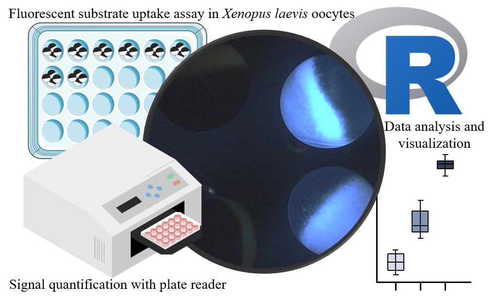
Xenopus laevis oocytes are widely used as a heterologous expression system for investigating the function of membrane proteins due to robust expression of heterologous protein and a low endogenous transport background. Traditionally, transporter activity in oocytes has been assessed using electrophysiology or radiolabeled uptake assays, approaches that are constrained by the requirement for electrogenicity of the transport process, availability of radiolabeled compounds, and instrumentation. Here, we describe a fluorescence-based uptake assay that enables direct and rapid quantification of transporter activity using a fluorescence plate reader. The protocol uses the Arabidopsis thaliana sucrose transporter 1 (SUC1) and its fluorescent substrate esculin as a case for how to set up the assay. The workflow includes optimizing assay conditions, sample preparation, fluorescent measurements, and downstream data analysis using R. This method can readily be adapted to other transporter-substrate pairs, and it supports applications such as transporter inhibitor screening, mutational analysis, characterization of kinetic properties, or indirect substrate specificity testing through competition assays. Overall, this protocol provides a simple and scalable alternative to traditional techniques, eliminating the need for radiolabeled compounds or electrophysiology while enabling easy quantitative assessment of transporter activity.
Key features
• Provides a simple, plate reader–based fluorescence uptake assay for quantifying transporter activity in Xenopus laevis oocytes.
• Adaptable to any transporter with a compatible fluorescent substrate, supporting rapid testing of substrate specificity indirectly through competition assays or mutant screening.
• Includes a complete workflow from assay setup and conduction to data analysis, suitable for both initial characterization and routine functional testing of membrane transporters.
Keywords: Xenopus laevis oocytes Heterologous expression
Fluorescence uptake assay
Plate reader
SUC1
Esculin
Transporter characterization
Heterologous expression
Fluorescence uptake assay
Plate reader
SUC1
Esculin
Transporter characterization
Graphical overview
Background
Xenopus laevis oocytes are a widely used heterologous expression system for the study of membrane proteins. Their large size, ease of manipulation, low endogenous transporter and channel background, and exceptional capacity for robust heterologous protein expression make them particularly well suited for functional characterization of membrane proteins [1–5]. Transporter and channel activity can be measured by two-electrode voltage clamp electrophysiology [6,7], scintillation counting of radiolabeled substrates [8], LC-MS/GC-MS detection [9–11], or fluorescent readouts [12–16].
When a fluorescent substrate for a target transporter is available, a fluorescent plate reader offers a rapid, safe, and experimentally accessible way to detect transporter activity in oocytes. Although their use depends on the availability of a suitable fluorescent substrate, recent advances in the generation of fluorescent analogs, including click chemistry–based labeling strategies [17–20], may help overcome this limitation and broaden the use of fluorescence-based transport measurements.
Fluorescence-based assays can detect transport either indirectly via fluorophore quenching of injected dyes upon binding to substrates imported by the target transporter [15,16] or directly by monitoring uptake of fluorescent substrates [12–14], both of which allow rapid quantification of transport activity. Fluorescence-based assays can be applied to uptake measurements, and can also be extended to inhibition, competition, or export assays.
In this protocol, we describe the fluorescence-based uptake assay using sucrose transporter 1 (SUC1), a high-affinity H+-sucrose symporter from Arabidopsis thaliana [21,22]. SUC1 has been functionally expressed in Xenopus laevis oocytes, where it mediates proton-coupled uptake of sucrose [22–24]. In yeast, SUC1 has additionally been shown to transport the fluorescent coumarin β-glucoside esculin [25], demonstrating that esculin is a suitable surrogate substrate for monitoring SUC1-dependent transport activity.
We previously applied a fluorescence-based analysis of uptake assay to plant glucosinolate transporters (GTR1/GTR2/GTR3) using artificial fluorescent glucosinolates [12]. This protocol establishes an accessible and generalizable workflow for SUC1 and esculin as a transporter–substrate pair, describing a complete workflow encompassing plate reader setup, initial assay optimization, oocyte assays, fluorescence measurement, and data analysis using R.
Materials and reagents
Biological materials
1. Xenopus laevis oocytes stage V–VI. Ovary lobes were kindly provided by Prof. Stephan Pless at the Department of Drug Design and Pharmacology, University of Copenhagen. Ovarian lobes were obtained shortly after surgical removal and transported to the laboratory in storage buffer supplemented with antibiotics. Transport of ovarian lobes to our lab was performed at ambient temperature and typically required 15–20 min, and collagenase treatment was commenced shortly after arrival. Defolliculated oocytes can be obtained commercially from Ecocyte Bioscience, Xenoocyte, or the European Xenopus Resource Centre (EXRC). Defolliculation and microinjection of stage V–VI oocytes followed the general procedures described in previous protocols [26–31].
Reagents
1. Esculin (Merck, Millipore, CAS: 66778-17-4, microbiology grade)
2. Sodium chloride (NaCl) (CAS: 7647-14-5, molecular biology grade)
3. Calcium chloride (CaCl2) (CAS: 10035-04-8, molecular biology grade)
4. Magnesium chloride (MgCl2) (CAS: 7791-18-6, molecular biology grade)
5. Potassium chloride (KCl) (CAS: 7447-40-7, molecular biology grade)
6. HEPES (CAS: 7365-45-9, molecular biology grade)
7. MES (CAS: 1266615-59-1, molecular biology grade)
8. DMSO (CAS: 67-68-5, ≥99.7%, Hybri-MaxTM grade); prepare 10% (v/v) DMSO and store at room temperature in a sealed container
9. Methanol (CAS: 67-56-1, ≥99.9%, HPLC grade); prepare 20% (v/v) methanol and store at room temperature in a sealed container
10. Ethanol (CAS: 64-17-5, 96% vol, molecular biology grade); prepare 70% (v/v) ethanol by dilution of 96% ethanol without correction; store at room temperature in a sealed container
11. MilliQ water
12. Amikacin disulfate salt (Thermo Scientific Chemicals, CAS no. 39831-55-5, catalog number: 455190050)
13. mMessage mMachine T7 Transcription kit (InVitrogen, Thermo Fisher Scientific, catalog number: AM1344)
14. Oocyte expression vector (e.g., pNB1 [32]) with gene of interest (SUC1 is used in the Validation of protocol section)
Solutions
1. Kulori buffer, pH 7.4 (see Recipes)
2. Kulori buffer, ph 4–6.5 (see Recipes)
3. 50 mM esculin in DMSO (see Recipes)
Recipes
1. Kulori buffer, pH 7.4
| Reagent | Final concentration | Quantity or volume |
| 5 M NaCl | 90 mM | 9 mL |
| 1 M CaCl2 | 1 mM | 500 μL |
| 1 M MgCl2 | 1 mM | 500 μL |
| 1 M KCl | 1 mM | 500 μL |
| HEPES (238.3 g/mol) | 5 mM | 0.596 g |
| Adjust to pH 7.4 with NaOH | ||
| MilliQ water | n/a | To 500 mL |
| Total | n/a | 500 mL |
2. Kulori buffer, pH 4–6.5
| Reagent | Final concentration | Quantity or volume |
| 5 M NaCl | 90 mM | 9 mL |
| 1 M CaCl2 | 1 mM | 500 μL |
| 1 M MgCl2 | 1 mM | 500 μL |
| 1 M KCl | 1 mM | 500 μL |
| MES (195.24 g/mol) | 5 mM | 0.488 g |
| Adjust pH with NaOH/HCl | ||
| MilliQ water | n/a | To 500 mL |
| Total | n/a | 500 mL |
Storage: Kulori solutions can be prepared in advance and stored at 4 °C. Under these conditions, the buffer is typically stable for several weeks, provided that no precipitation, turbidity, or microbial growth is observed. To minimize degradation of antibiotics, it is recommended to add antibiotics only to the volume required for immediate use rather than to the entire stock solution. Alternatively, a 10× Kulori stock can be prepared by increasing the concentration of all components tenfold and storing in 50-mL aliquots at -20 °C for several months. Prior to use, the stock should be diluted 1:10 with MilliQ water, and the pH verified and adjusted if necessary.
3. 50 mM esculin in DMSO
| Reagent | Final concentration | Quantity or volume |
| Esculin (367.31 g/mol) | 50 mM | 18.37 mg |
| DMSO | 100% | 1 mL |
| Total | n/a | 1 mL |
Esculin stock can be prepared in advance and stored at -20 °C. In our laboratory, esculin stocks stored under these conditions have been used successfully after at least 6 months of storage. To minimize repeated freeze/thaw cycles, we suggest preparing stock aliquots.
Laboratory supplies
1. Oocyte pipette (Pasteur pipettes with tips cut and smoothed) or plastic pipettes (e.g., BRANDTM, catalog number: 15202699 with FisherbrandTM Dropper Bulb, catalog number: 03-448-26; alternative: disposable plastic pipettes, e.g., Pastette, catalog number: LW4111)
2. Petri dishes, non-treated, 55 mm in diameter (any standard laboratory grade)
3. Petri dishes, non-treated, 90 mm in diameter (any standard laboratory grade)
4. 48-well plate (Sarstedt, catalog number: 83.3923 is used in this protocol, but any standard laboratory grade should work)
5. 1.5 mL microcentrifuge tubes (any standard laboratory grade)
6. P1000 pipette tips (any standard laboratory grade)
7. P200 pipette tips (any standard laboratory grade)
8. P10 pipette tips (any standard laboratory grade)
9. Ice
10. Black 96-well plate (Thermo ScientificTM, catalog number: 237108)
Equipment
1. Plate reader (BioTek, model: Synergy H1 Microplate reader)
2. Nanoject II (Drummond Scientific Company, Broomall, PA, USA)
3. Centrifuge for microcentrifuge tubes (HERMLE Labortechnic GmbH, Germany, model: Z216MK, and Thermo Scientific HERAEUS, Fresco 21, catalog number: 10651805; however, any centrifuge with a speed of >19,000× g can be used)
Procedure
登录/注册后免费查看全文
文章信息
稿件历史记录
提交日期: Jul 15, 2026
接收日期: Sep 1, 2026
在线发布日期: Sep 17, 2026
出版日期: Oct 5, 2026
版权信息
© 2026 The Author(s); This is an open access article under the CC BY-NC license (https://creativecommons.org/licenses/by-nc/4.0/).
如何引用
de Prado Parralejo, V., Nour-Eldin, H. H. and Kanstrup, C. (2026). Step-by-Step Protocol for Fluorescence-Based Analysis of Uptake in Transporter-Expressing Xenopus laevis Oocytes. Bio-protocol 16(19): e5834. DOI: 10.21769/BioProtoc.5834.

 Download Citation in RIS Format
分类
生物化学 > 蛋白质 > 活性
您对这篇实验方案有问题吗?
在此处发布您的问题,我们将邀请本文作者来回答。同时,我们会将您的问题发布到Bio-protocol Exchange,以便寻求社区成员的帮助。
 发布问题
发布问题


