


A NanoLuc-Based Protease Biosensor for Highly Sensitive Detection of Intracellular Protease Activity: Applications to Apoptosis and Coronavirus Infection
基于 NanoLuc 的高灵敏度细胞内蛋白酶活性生物传感器及其在细胞凋亡和冠状病毒感染研究中的应用

发布: 2026年09月20日第16卷第18期 DOI: 10.21769/BioProtoc.5833 浏览次数: 50
评审: Li HeDana Manuela SavulescuSébastien Gillotin
 PDF
PDF  Q&A
Q&A
参见作者原研究论文
The authors used this protocol in:

Jan 2023






Abstract
FlipNanoLuc is a highly sensitive protease biosensor based on the β-strand-flipping principle of NanoLuc luciferase, which is derived from Oplophorus gracilirostris. In the inactive configuration, one β-strand of NanoLuc is repositioned, thereby suppressing luciferase activity. Upon cleavage of the embedded protease recognition sequence by a target protease, the flipped β-strand is released, and luciferase activity is reconstituted. Incorporation of the LgBiT fragment (NanoBiT technology) yields strong luminescent output once the reporter is reconstituted, whereas the CL1-PEST1 degradation tag lowers background luminescence by promoting the degradation of the uncleaved, inactive form; together, these two modifications widen the dynamic range. A firefly luciferase normalization cassette connected via a P2A self-cleaving peptide is encoded in the same reporter plasmid, thereby eliminating the need for separate co-transfection. Because the readout directly reports intracellular protease activity in living cells, the system is suitable for detecting protease activation during apoptosis or viral infection and, in principle, for evaluating protease inhibitors and antiviral compounds. This protocol describes the following: (1) generation of HEK293T cells stably expressing FlipNanoLuc by retroviral transduction; (2) validation of reporter activity by protease overexpression; (3) detection of apoptosis using staurosporine; (4) detection of human coronavirus OC43 infection; and (5) detection of SARS-CoV-2 infection in BHK cells stably expressing hACE2. All luminescence assays employ dual-luciferase normalization and are compatible with standard 384-well plate readers.
Key features
• Highly sensitive detection of intracellular protease activity using a β-strand-flipping NanoLuc biosensor with dual-luciferase normalization encoded in a single plasmid.
• Detects protease activity in living cells, including caspase-3 during apoptosis and 3CLpro during authentic OC43 and SARS-CoV-2 infection.
• Quantitative luminescence readout in a 384-well plate format with a standard plate reader.
• Adaptable to other proteases and to inhibitor or antiviral screening by exchanging the embedded recognition sequence.
Keywords: FlipNanoLuc (FlipNanoLuc) Protease sensor (蛋白酶传感器)
Coronavirus (冠状病毒)
SARS-CoV-2 (SARS-CoV-2)
3CL protease (3CL 蛋白酶)
NanoLuc (NanoLuc)
Apoptosis (细胞凋亡)
Protease activity reporter (蛋白酶活性报告系统)
Protease sensor (蛋白酶传感器)
Coronavirus (冠状病毒)
SARS-CoV-2 (SARS-CoV-2)
3CL protease (3CL 蛋白酶)
NanoLuc (NanoLuc)
Apoptosis (细胞凋亡)
Protease activity reporter (蛋白酶活性报告系统)
Graphical overview
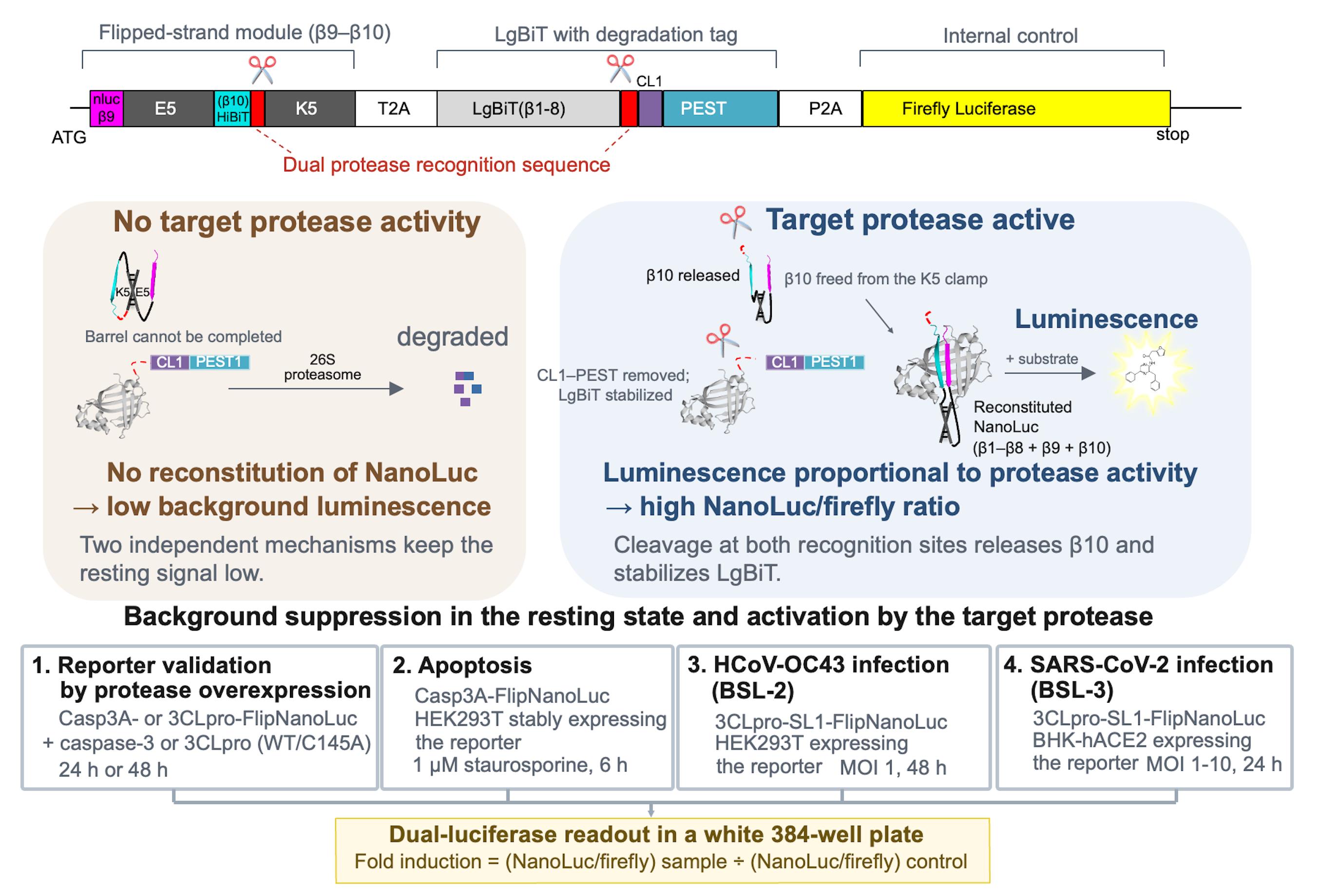
Principle and applications of the NanoLuc-based protease biosensor FlipNanoLuc. Architecture of the reporter, which is encoded as a single transcript (top panel). Suppression of the resting signal and activation of the reporter by the target protease (middle panels). The four applications described in this protocol are all read out by dual-luciferase measurement in a 384-well format (bottom panels).
Background
Protease activity is a central regulatory event in diverse biological processes, including apoptosis, viral replication, and innate immune signaling. Therefore, sensitive and quantitative detection of protease activity in living cells is of broad utility in both basic research and antiviral drug discovery.
Bioluminescent reporters based on NanoLuc luciferase (NLuc) offer high sensitivity and a wide dynamic range compared with conventional firefly or Renilla luciferase systems. The NanoBiT complementation system [1] splits NLuc into a large fragment (LgBiT, 18 kDa) and a small peptide (SmBiT or HiBiT), thereby enabling the reconstitution-based detection of protein–protein interactions. Splitting NLuc in this way is advantageous for the design of activity reporters: the separated fragments are essentially non-luminescent on their own, so reconstitution can be made strictly dependent on a designed molecular event, whereas the high affinity of the LgBiT–HiBiT pair restores strong luminescence once reconstitution is permitted. Because HiBiT is a short peptide, it can also be repositioned within an engineered polypeptide with little steric penalty, which is not feasible with intact NLuc or with the substantially larger firefly and Renilla luciferases [1,2].
FlipNanoLuc is encoded as a single transcript that yields three products: a flipped-strand module, the LgBiT fragment of NLuc carrying a CL1-PEST1 destabilization tag, and firefly luciferase, which are separated by T2A and P2A self-cleaving peptides (Graphical overview, top panel). In the flipped-strand module, the β10 strand of NLuc (HiBiT) is held by an E5/K5 coiled-coil pair in a flipped, parallel orientation relative to β9, so that the NLuc barrel cannot be completed, and catalytic activity is abolished. A protease recognition sequence is placed within the flexible linkers of this module and of the LgBiT fragment. Cleavage by the target protease releases β10 from the coiled-coil constraint and removes the CL1-PEST1 tag from LgBiT, allowing the strand to re-engage the barrel, reconstituting luminescence (Graphical overview, middle panels). Conversely, in the absence of protease activity, the reporter remains suppressed by two independent mechanisms: LgBiT requires HiBiT for full activity, and the CL1-PEST1 tag accelerates the degradation of the uncleaved form before it can fold spontaneously. Together, these features yield a reporter with a low baseline signal and high fold induction upon protease activation [3].
Because reporter cleavage depends on the catalytic activity of the target protease, FlipNanoLuc provides a quantitative, live-cell activity readout that extends beyond simple detection. The embedded recognition sequence is modular and can be exchanged to monitor other cellular or viral proteases. The same readout can, in principle, be applied to the evaluation of protease inhibitors and antiviral compounds, as the inhibition of the target protease is reflected by reduced reporter induction (for example, 3CLpro inhibition during coronavirus infection).
This protocol details the complete workflow, from stable cell line generation to luminescence data acquisition and normalization, covering four experimental applications: protease overexpression validation, staurosporine-induced apoptosis, OC43 coronavirus infection, and SARS-CoV-2 infection.
Materials and reagents
Biological materials
1. HEK293T cells (ATCC, catalog number: CRL-3216)
2. BHK cells stably expressing hACE2 (parental BHK cells; JCRB Cell Bank, catalog number: JCRB9020); generated in-house using the PiggyBac transposon system; see General notes
3. SARS-CoV-2 (BSL-3); the virus stock is the same as that described in Yoshida et al. [4] and must be obtained under appropriate institutional and regulatory approval
4. Human coronavirus OC43 (BSL-2) (ATCC, catalog number: VR-1558)
5. FlipNanoLuc reporter plasmids (available via Addgene; see Table 1 for the plasmid list)
Table 1. FlipNanoLuc reporter and protease expression plasmids used in this protocol, with their Addgene accession numbers.
| Construct (used in this protocol) | Addgene plasmid name | Addgene ID |
|---|---|---|
| 3CLpro-FlipNanoLuc reporter, CoVA WT | pQC.Flip-nluc(LgBiT1-8)CP[CoVA]-Fluc | 200125 |
| 3CLpro-SL1-FlipNanoLuc reporter, CoVA WT (used for SARS-CoV-2/OC43 infection assays) | pQC.SARS2_5′UTR(SL1)-Flip-nluc(LgBiT1-8)CP[CoVA]-Fluc | 200126 |
| 3CLpro-SL1-FlipNanoLuc reporter, CoVA Q5A (cleavage-site mutant, negative control) | pQC.SARS2_5′UTR(SL1)-Flip-nluc(LgBiT1-8)CP[CoVA(Q5A)]-Fluc | 200127 |
| 3CLpro-SL1-FlipNanoLuc reporter, CoVA SA (cleavage-site mutant, negative control) | pQC.SARS2_5′UTR(SL1)-Flip-nluc(LgBiT1-8)CP[CoVA(SA)]-Fluc | 200128 |
| Casp3A-FlipNanoLuc reporter, WT (apoptosis assay) | pQC.Flip-nluc(LgBiT1-8)CP[Casp3A]-Fluc | 200123 |
| Casp3A-FlipNanoLuc reporter, D5A (cleavage-site mutant, negative control) | pQC.Flip-nluc(LgBiT1-8)CP[Casp3A(D5A)]-Fluc | 200124 |
| Caspase-3 (WT) protease expression plasmid (apoptosis/reporter validation) | pCAG.CASP3-Myc | 200119 |
Note: Construct names follow the Addgene depositor records. CoVA Q5A and SA, and Casp3A D5A, are cleavage-site mutants used as negative controls. pCAG.CASP3-Myc encodes the caspase-3 protease (not a FlipNanoLuc reporter) and is used to validate the Casp3A reporter. The Q5A and SA cleavage-site mutants are available only in the SL1-containing backbone; see the note in Part 2, section B.
6. Gag-pol expression plasmid for retroviral particle production, as described in Morita et al. [5]
7. VSV-G expression plasmid for retroviral particle production, as described in Naldini et al. [6]
8. Protease expression plasmids: SARS-CoV-2 3CLpro, wild-type (pCAG.Myc-SARS2_3CL; Addgene, plasmid number: 200120) and the catalytically inactive C145A mutant [pCAG.Myc-SARS2_3CL(C145A); Addgene, plasmid number: 200121]; caspase-3, wild-type (pCAG.CASP3-Myc; Addgene, plasmid number: 200119)
9. PiggyBac transposon vector encoding hACE2, constructed in PB-CMV-MCS-EF1α-RedPuro (System Biosciences, catalog number: PB514B-2)
10. Super PiggyBac transposase expression vector (System Biosciences, catalog number: PB200A-1; currently supplied as catalog number: PB210PA-1)
11. Empty vector (pCAG-empty), constructed in-house; the pCAG backbone without an insert was used as the mock control in Tables 2 and 3
Table 2. Plasmid combinations for the 3CLpro (SARS-CoV-2) overexpression assay.
In every condition, 0.5 μg of reporter plasmid and 0.5 μg of co-transfected plasmid are used per well (1:1, w/w; 1 μg of total DNA).
| Condition | Reporter plasmid | Reporter Addgene ID | Co-transfected plasmid | Purpose/expected result |
|---|---|---|---|---|
| Test | 3CLpro-FlipNanoLuc (CoVA WT) | 200125 | SARS-CoV-2 3CLpro (WT) | Active protease cleaves the reporter → high NanoLuc signal |
| Protease-inactive control | 3CLpro-FlipNanoLuc (CoVA WT) | 200125 | SARS-CoV-2 3CLpro (C145A) | Catalytically dead protease → no signal; confirms dependence on protease activity |
| Cleavage-site specificity reference | 3CLpro-SL1-FlipNanoLuc (CoVA WT) | 200126 | SARS-CoV-2 3CLpro (WT) | Reference for the cleavage-site mutant control below |
| Reporter cleavage-site mutant control | 3CLpro-SL1-FlipNanoLuc (CoVA Q5A or SA) | 200127 or 200128 | SARS-CoV-2 3CLpro (WT) | Non-cleavable recognition site → no signal; confirms sequence specificity |
| Mock | 3CLpro-FlipNanoLuc (CoVA WT) | 200125 | Empty vector (pCAG-empty) | No protease → background reference |
Note: The Q5A and SA cleavage-site mutants exist only in the SL1-containing backbone. For cleavage-site specificity experiments, therefore, use the SL1-containing WT reporter (number: 200126) as the reference and compare it with the SL1-containing Q5A and SA reporters (numbers: 200127 and 200128), so that the constructs differ only in the protease recognition sequence. Do not compare the mutant reporters directly with the reporter used in the other conditions (number: 200125), which lacks SL1, because the two constructs differ in their 5′UTR and therefore in the level of reporter expression.
Table 3. Plasmid combinations for the caspase-3 (apoptosis) overexpression assay.
In every condition, 0.5 μg of reporter plasmid and 0.5 μg of co-transfected plasmid are used per well (1:1, w/w; 1 μg of total DNA).
| Condition | Reporter plasmid | Reporter Addgene ID | Co-transfected plasmid | Purpose/expected result |
|---|---|---|---|---|
| Test | Casp3A-FlipNanoLuc (WT) | 200123 | Caspase-3 (WT) | Active caspase-3 cleaves the reporter → high NanoLuc signal |
| Reporter cleavage-site mutant control | Casp3A-FlipNanoLuc (D5A) | 200124 | Caspase-3 (WT) | Non-cleavable recognition site → no signal; confirms sequence specificity |
| Mock | Casp3A-FlipNanoLuc (WT) | 200123 | Empty vector (pCAG-empty) | No protease → background reference |
Cell culture reagents
1. Dulbecco's modified Eagle medium (DMEM), high glucose (Nacalai Tesque, catalog number: 08458-16)
2. Minimum essential medium (MEM) (Sigma-Aldrich, catalog number: M4655)
3. Fetal bovine serum (FBS) (Gibco, Thermo Fisher Scientific, catalog number: 10270-106); heat-inactivate before use (see Recipes)
Note: This catalog number is currently listed as unavailable by the supplier; an equivalent qualified FBS may be used.
4. Benzylpenicillin potassium (Fujifilm Wako Pure Chemical Corporation, catalog number: 021-07732)
5. Streptomycin sulfate (Tokyo Chemical Industry, catalog number: S0585)
6. Puromycin dihydrochloride (InvivoGen, catalog number: ant-pr-1)
7. Opti-MEM I reduced serum medium (Thermo Fisher Scientific, catalog number: 31985070)
8. Phosphate-buffered saline (PBS), without calcium and magnesium (Nacalai Tesque, catalog number: 14249-24)
9. Trypsin-EDTA solution (0.25%) (Nacalai Tesque, catalog number: 32777-15)
Transfection reagents
1. Polyethylenimine (PEI MAX, MW 40,000; 1 mg/mL stock in water) (Polysciences, catalog number: 24765-1)
Luminescence assay reagents
1. Nano-Glo® luciferase assay system (Promega, catalog number: N1120)
2. Bright-GloTM luciferase assay system (Promega, catalog number: E2620)
Lysis buffer components
1. NaCl (Nacalai Tesque, catalog number: 31320-05)
2. Tris (Tris[hydroxymethyl]aminomethane) (Nacalai Tesque, catalog number: 35406-91)
3. Triton X-100 (Nacalai Tesque, catalog number: 35501-15)
4. cOmpleteTM Mini protease inhibitor cocktail tablets (Roche, catalog number: 11836153001)
5. Hydrochloric acid (HCl), for pH adjustment
6. Distilled water (used for the preparation of buffers and stock solutions)
Apoptosis assay
1. Staurosporine (AdipoGen Life Sciences, catalog number: AG-CN2-0022; CAS 62996-74-1); prepare 200 μM stock in DMSO
2. Dimethyl sulfoxide (DMSO) (Nacalai Tesque, catalog number: 13406-55)
Solutions
1. 1× lysis buffer (see Recipes)
2. 100× penicillin G + streptomycin stock solution (see Recipes)
3. Complete culture medium (see Recipes)
Recipes
1. 1× lysis buffer
| Component | Final concentration |
|---|---|
| NaCl | 150 mM |
| Tris-HCl, pH 7.4 | 50 mM |
| Triton X-100 | 1% (v/v) |
| Protease inhibitor cocktail | 1× |
Dissolve NaCl and Tris-HCl in approximately 80% of the final volume of distilled water. Add Triton X-100 and mix until dissolved. Adjust the pH to 7.4 using HCl. Bring to the final volume. Store at 4 °C for up to one month.
Note: Add the protease inhibitor cocktail immediately before use, at one tablet per 10 mL of lysis buffer (1×).
2. 100× penicillin G + streptomycin stock solution
Dissolve 0.626 g of benzylpenicillin potassium and 1 g of streptomycin sulfate in PBS to a final volume of 100 mL. Sterilize through a 0.22 μm filter and store at 4 °C for up to 6 months; for longer storage, dispense into aliquots and keep at -20 °C. This stock is used at 1× (1:100 dilution) in the respective complete culture medium (DMEM for HEK293T cells and MEM for BHK cells stably expressing hACE2).
Note: In this protocol, Pen/Strep refers to the homemade 100× penicillin G + streptomycin stock solution described in this recipe. A commercial penicillin-streptomycin solution may be substituted at the manufacturer’s recommended working concentration.
3. Complete culture medium
Combine 500 mL of basal medium (DMEM for HEK293T cells and MEM for BHK cells stably expressing hACE2), 50 mL (final 10%) of FBS that has been heat-inactivated (56 °C, 45 min), and 5 mL (final 1×) of 100× penicillin G + streptomycin stock solution. Mix gently. Store at 4 °C for up to 2 months. For the maintenance medium of BHK cells stably expressing hACE2, additionally supplement with puromycin dihydrochloride to a final concentration of 3 μg/mL.
Laboratory supplies
1. White 384-well plates (Greiner Bio-One, catalog number: 784075)
2. 24-well cell culture plates (Violamo, catalog number: 2-8588-03)
3. 96-well cell culture plates (Violamo, catalog number: 2-8588-05)
4. 3.5 cm (35 mm) cell culture dishes (Thermo Scientific, catalog number: 130180)
5. Microcentrifuge tubes, 1.5 mL (Watson, catalog number: 131-815C)
6. Syringe filters, 0.22 μm (AS ONE, catalog number: 033022SO-SFCA)
Equipment
1. Varioskan LUX multimode plate reader (Thermo Fisher Scientific, catalog number: VLBL00D1) or an equivalent luminometer capable of luminescence detection in 384-well format
2. Multichannel pipette (suitable for 10 μL) (Thermo Scientific, catalog number: 4661040N)
3. Refrigerated microcentrifuge capable of 500× g at 4 °C (Thermo Scientific, catalog number: 75002559)
4. BSL-2 and BSL-3 certified biosafety cabinets (as appropriate) (HITACHI, catalog number: SCV-1303ECIIB)
5. 37 °C incubator with 5% CO2 (PHC, catalog number: MCO-170AIC-PJ)
6. Inverted phase-contrast microscope (OLYMPUS, model: CKX53)
7. Water bath capable of 56 °C (Yamato Scientific, catalog number: BF200)
Software and datasets
1. GraphPad Prism (version 11.0.0 for macOS) (GraphPad Software, https://www.graphpad.com)
2. SkanIt Software for microplate readers (Thermo Fisher Scientific), used for data acquisition on the Varioskan LUX plate reader v. 6.0.2.3
Procedure
登录/注册后免费查看全文
文章信息
稿件历史记录
提交日期: Jun 25, 2026
接收日期: Aug 31, 2026
在线发布日期: Sep 14, 2026
出版日期: Sep 20, 2026
版权信息
© 2026 The Author(s); This is an open access article under the CC BY license (https://creativecommons.org/licenses/by/4.0/).
如何引用
Arakawa, M. and Morita, E. (2026). A NanoLuc-Based Protease Biosensor for Highly Sensitive Detection of Intracellular Protease Activity: Applications to Apoptosis and Coronavirus Infection. Bio-protocol 16(18): e5833. DOI: 10.21769/BioProtoc.5833.

 Download Citation in RIS Format
分类
微生物学 > 病原体检测 > 生物传感器
细胞生物学 > 基于细胞的分析方法 > 病毒性感染
细胞生物学 > 基于细胞的分析方法 > 酶学测定
您对这篇实验方案有问题吗?
在此处发布您的问题,我们将邀请本文作者来回答。同时,我们会将您的问题发布到Bio-protocol Exchange,以便寻求社区成员的帮助。
 发布问题
发布问题


