


Isolation of Human Umbilical Cord Blood Hematopoietic Stem Cells and Directed Differentiation Into Megakaryocytes
人脐带血造血干细胞的分离及向巨核细胞定向分化

发布: 2026年09月20日第16卷第18期 DOI: 10.21769/BioProtoc.5822 浏览次数: 348
评审: Jessica DavisXinyuan ZhangAnonymous reviewer(s)
 PDF
PDF  Q&A
Q&A
参见作者原研究论文
The authors used this protocol in:
Oct 2025





Abstract
Platelets originate from megakaryocytes, whose generation involves a series of biological processes including directed differentiation, proliferation, polyploidization, and maturation of hematopoietic stem cells. Abnormalities in megakaryocyte development and maturation can lead to quantitative and functional defects in platelets, thereby contributing to hemostatic or thrombotic disorders as well as the development of malignancies. Investigating megakaryocyte development and maturation and platelet production can provide important theoretical foundations for the diagnosis and treatment of thrombocytopenia, thrombotic diseases, and myeloproliferative neoplasms. Currently, there are three main clinical sources of hematopoietic stem cells (HSCs): bone marrow (BM), peripheral blood (PBSC), and umbilical cord blood (UCB). Among these, umbilical cord blood (UCB)-derived HSCs, due to their higher differentiation efficiency and stronger proliferative capacity, are the preferred starting cell source for studying megakaryocyte (MK) development and maturation and the mechanisms of platelet production. This article describes a detailed protocol covering all necessary steps for isolating CD34+ hematopoietic stem cells from umbilical cord blood, followed by in vitro induction culture with stem cell factor (SCF) and thrombopoietin (TPO) to generate mature megakaryocytes that highly express early megakaryocyte markers (CD41a, CD61) and late maturation markers (CD42a, CD42b). This protocol provides an effective tool for studying megakaryocyte development and platelet production and holds potential value for application in research on megakaryocyte-related diseases.
Key features
• Enables efficient isolation of highly pure and viable CD34+ hematopoietic stem cells from human umbilical cord blood using magnetic bead enrichment.
• Establishes a serum-free, chemically defined culture system that minimizes batch-to-batch variability and ensures reproducible megakaryocyte differentiation.
• Uses two key cytokines (SCF and TPO) to effectively induce directed differentiation of CD34+ cells into mature megakaryocytes.
• This protocol is compatible with downstream analyses, including flow cytometry, qPCR, and platelet morphology, and can be used for disease modeling and drug screening.
Keywords: Umbilical cord blood (脐带血) Hematopoietic stem cells (造血干细胞)
Megakaryocyte differentiation (巨核细胞分化)
Thrombopoiesis (血小板生成)
CD34+ (CD34+)
SCF (SCF)
TPO (TPO)
Hematopoietic stem cells (造血干细胞)
Megakaryocyte differentiation (巨核细胞分化)
Thrombopoiesis (血小板生成)
CD34+ (CD34+)
SCF (SCF)
TPO (TPO)
Graphical overview
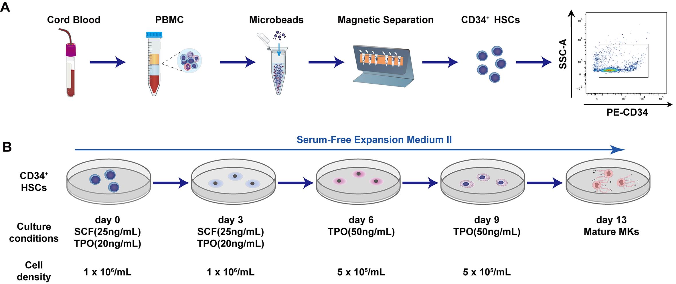
Schematic of human umbilical cord blood–derived CD34+ hematopoietic stem cell (HSC) isolation (A) and directed differentiation into mature megakaryocytes (B)
Background
Megakaryocytes are the precursor cells of platelets. Platelets are indispensable for hemostasis and tissue repair, and their clinical demand is enormous. However, reliance on donor-derived platelets faces challenges such as supply shortages and short storage periods. Therefore, in vitro generation of megakaryocytes and platelets has become a key direction in transfusion medicine and regenerative medicine [1]. Common sources of hematopoietic stem cells include bone marrow, peripheral blood, and umbilical cord blood. Bone marrow is a classic source, but its collection is invasive, and the quality of cells is influenced by donor age and health status. Under homeostatic conditions, only a very small number of stem cells circulate in peripheral blood [2]. Hematopoietic stem cells (HSCs) reside within the bone-marrow niche, where they are regulated by a complex network of cellular components [3]. Meanwhile, hematopoietic stem cells are capable of trafficking between bone marrow and peripheral blood. Accordingly, stem cell mobilization can be achieved via specific pharmacological agents or interventions, which displace hematopoietic stem and progenitor cells (HSPCs) from the bone-marrow niche into the peripheral circulation. Sufficient CD34+ cells can then be collected by leukapheresis [4]. Clinically, the most frequently applied mobilization regimens include granulocyte colony-stimulating factor (G-CSF) monotherapy or its combination with plerixafor (AMD3100) [4]. In addition, cyclophosphamide (CY)-based chemotherapy-assisted mobilization represents a classic strategy: cyclophosphamide is administered on day 1, followed by G-CSF treatment from day 2 to day 11, and leukapheresis is initiated on day 12. Nevertheless, multiple factors may affect CD34+ cell collection efficiency, including donor demographics (age, sex, body weight), baseline blood cell counts, and the selection of the mobilization regimen. In contrast, umbilical cord blood can be collected non-invasively and is rich in CD34+ cells, making it an ideal starting material for in vitro megakaryocytic differentiation [5].
This protocol describes in detail the isolation of CD34+ hematopoietic stem cells from human cord blood using magnetic bead separation technology and the induction of these hematopoietic stem cells into mature megakaryocytes [1,6,7]. Using magnetic bead enrichment, high-purity CD34+ cells are efficiently obtained from cord blood mononuclear cells and then directed toward megakaryocytic differentiation in serum-free medium supplemented with only thrombopoietin (TPO) and stem cell factor (SCF). As core cytokines governing hematopoiesis, SCF and TPO exert complementary and synergistic pivotal functions in the HSC-to-MK induction system. SCF delivers basal signals supporting the proliferation and self-renewal of hematopoietic stem and progenitor cells, whereas TPO drives lineage commitment and maturation toward the megakaryocytic lineage. At the signaling level, their synergistic effect manifests as enhanced and prolonged activation of pathways such as JAK2-STAT5. Functionally, this synergy drives massive expansion of megakaryocyte progenitors for subsequent megakaryocyte differentiation [8–10]. Compared with existing methods, this protocol provides more detailed operational steps and offers compatibility with various downstream analyses, including flow cytometry, qPCR, and platelet morphology. At the application level, this protocol can be used not only for basic research but also to provide an alternative source for platelet transfusion. Clinical trials based on cord blood–derived megakaryocyte progenitors have already shown good tolerability [5,11–12]. Furthermore, it can be used to construct models of inherited platelet disorders or serve as a platform for screening candidate drugs for hematological toxicity.
To address the technical limitations of previously reported megakaryocyte induction systems, we established an optimized two-stage gradient cytokine differentiation protocol modified from existing culture workflows. Published differentiation protocols either employ complex multi-cytokine cocktails that increase experimental batch variability or rely solely on TPO, which restricts early HSPC expansion. Our simplified SCF/TPO dual-factor culture system retains robust megakaryocyte terminal maturation efficiency while minimizing reagent-induced variation. More importantly, we established a standardized double-round MACS enrichment workflow; this purification strategy consistently yields CD34+ cell purity above 90%, which markedly outperforms the 60%–70% purity obtained by single-column separation in reference protocols [13]. Beyond cytokine and enrichment optimizations, continuous multi-time-point sampling for dynamic differentiation tracking and standardized post-staining antibody washing steps are integrated into our complete workflow to resolve the deficiencies of prior protocols, including insufficient CD34 purity control and non-standardized flow cytometry staining procedures [14–16].
Materials and reagents
Biological materials
1. Human umbilical cord blood (UCB): Fresh human umbilical cord blood collected from full-term deliveries
Reagents
1. Ficoll-PaqueTM PLUS (Cytiva, catalog number: 17144002)
2. AutoMACS rinsing solution (Miltenyi Biotec, catalog number: 130-091-222)
3. MACS BSA stock solution (Miltenyi Biotec, catalog number: 130-091-376)
4. CD34 MicroBead kit, human (Miltenyi Biotec, catalog number: 130-046-702)
5. Penicillin/streptomycin (Invitrogen, catalog number: 15140-122)
6. 10 μg recombinant human TPO (Peprotech, catalog number: 300-18)
7. 10 μg recombinant human SCF (Peprotech, catalog number: 300-07)
8. Phosphate-buffered saline (PBS) (without Ca2+ and Mg2+) (VivaCell BIOSCIENCES, catalog number: C3580-0500)
9. Fetal bovine serum (FBS) (Gibco, catalog number: A3161001C)
10. FcR blocking reagent, human (Miltenyi Biotec, catalog number: 130-059-901)
11. Dimethyl sulfoxide (DMSO) (Sigma-Aldrich, catalog number: D2650)
12. DMEM (Gibco, catalog number: C11995500BT)
13. EDTA, 0.5 M, pH 8.0 (Beyotime, catalog number: ST066)
14. APC mouse anti-human CD41a antibody (clone: HIP8) (BD Biosciences, catalog number: 559777)
15. PE mouse anti-human CD61 antibody (clone: VI-PL2) (BD Biosciences, catalog number: 555754)
16. PE mouse anti-human CD42a antibody (clone: ALMA.16) (BD Biosciences, catalog number: 558819)
17. PE mouse anti-human CD42b antibody (clone: HIP1) (BD Biosciences, catalog number: 555473)
18. APC mouse IgG1 κ isotype control (clone: MOPC-21) (BD Biosciences, catalog number: 555751)
19. PE mouse IgG1 κ isotype control (clone: MOPC-21) (BD Biosciences, catalog number: 555749)
20. PE mouse anti-human CD34 antibody (clone: 581) (BD Biosciences, catalog number: 555822)
21. Cell staining buffer (Biolegend, catalog number: 420201)
22. Trypan Blue stain, 0.4% (Gibco, catalog number: 15250-061)
23. 75% ethanol (LIRCON, 500 mL)
24. StemSpanTM SFEM II (Stem Cell Technologies, catalog number: 09605)
Solutions
1. Buffer 1 (see Recipes)
2. Buffer 2 (see Recipes)
3. Cell cryopreservation medium (see Recipes)
4. DMEM + 10% FBS (see Recipes)
5. 25 μg/mL SCF (see Recipes)
6. 20 μg/mL TPO (see Recipes)
7. SFEM (see Recipes)
Recipes
1. Buffer 1
| Reagent | Final concentration | Volume |
|---|---|---|
| PBS | n/a | 488 mL |
| FBS | 2% | 10 mL |
| EDTA | 2 mM | 2 mL |
| Total | n/a | 500 mL |
After buffer 1 is prepared, store it at 4 °C.
2. Buffer 2
| Reagent | Final concentration | Quantity or volume |
|---|---|---|
| AutoMACS rinsing solution | n/a | 100 mL |
| MACS BSA stock solution | n/a | 5 mL |
| Total | n/a | 105 mL |
After Buffer 2 is prepared, filter through a 0.22 μm membrane, store at 4 °C protected from light, and do not warm to room temperature (RT) before use.
3. Cell cryopreservation medium
| Reagent | Final concentration | Quantity or volume |
|---|---|---|
| DMEM | 75% | 750 μL |
| FBS | 15% | 150 μL |
| DMSO | 10% | 100 μL |
| Total | n/a | 1 mL |
For every 5 × 10⁵ cells, use 1 mL of cryopreservation medium (prepare fresh and use immediately).
4. DMEM + 10% FBS
| Reagent | Final concentration | Quantity or volume |
|---|---|---|
| DMEM | 89% | 89 mL |
| FBS | 10% | 10 mL |
| Penicillin-streptomycin | 1% | 1 mL |
| Total | n/a | 100 mL |
Store the prepared complete medium at 4 °C and use within 2–4 weeks. Allow the medium to equilibrate to RT prior to experimental use.
5. 25 μg/mL SCF
| Reagent | Final concentration | Quantity or volume |
|---|---|---|
| SCF | 25 μg/mL | 10 μg |
| ddH2O | n/a | 0.4 mL |
| Total | n/a | 0.4 mL |
Prepare single-use 10 μL aliquots of working stocks. Store at -80 °C, avoid repeated freeze-thaw cycles, and use within 6 months after preparation.
6. 20 μg/mL TPO
| Reagent | Final concentration | Quantity or volume |
|---|---|---|
| TPO | 20 μg/mL | 10 μg |
| ddH2O | n/a | 0.5 mL |
| Total | n/a | 0.5 mL |
Prepare single-use 10 μL aliquots of working stocks. Store at -80 °C, avoid repeated freeze-thaw cycles, and use within 6 months after preparation.
7. SFEM
| Reagent | Final concentration | Quantity or volume |
|---|---|---|
| StemSpanTM SFEM II | 90% | 99 mL |
| Penicillin-streptomycin | 1% | 1 mL |
| Total | n/a | 100 mL |
Store the prepared medium at 4 °C and use within 2–4 weeks. Allow the medium to equilibrate to RT prior to experimental use.
Note: All commercial reagents shall be stored according to the manufacturer’s instructions and used before the manufacturer-indicated expiry date. For opened commercial reagents and laboratory-prepared cytokine working stocks, an additional in-house recommended shelf-life is specified. Do not use any reagent beyond its expiry or recommended usage period.
Laboratory supplies
1. 3 mL Pasteur pipette (NEST, catalog number: 318314)
2. 12-well cell culture plate (NEST, catalog number: 712001)
3. 24-well cell culture plate (NEST, catalog number: 702001)
4. 15-mL centrifuge tube (NEST, catalog number: 601052)
5. 50-mL centrifuge tube (NEST, catalog number: 602052)
6. FACS tubes 5 mL (Falcon, catalog number: 352054)
7. 1.5 mL Eppendorf tubes (Axygen, catalog number: MCT-150-CS)
8. 0.22 μm filter (Millipore, catalog number: 431229)
9. Hemocytometer (Countstar, catalog number: C0010101)
10. 2 mL cryogenic vials (Corning, catalog number: 430659)
11. 25 mL pipette (BeyoGold, catalog number: FPIP125)
12. μ-dish 35 mm, high (ibidi, catalog number: 81156)
13. 10 mL BD Vacutainer® K2EDTA blood collection tubes (BD, catalog number: 367525)
14. MS columns (Miltenyi Biotec, catalog number: 130-042-201)
15. LS columns (Miltenyi Biotec, catalog number: 130-042-401)
16. Cell strainers (BeyoGold, catalog number: FSTR070)
17. RNase-free pipette tips (10 μL, 200 μL, 1 mL) (Axygen, catalog numbers: AXY-TF-300-R-S, TF-200, TF-1000)
Equipment
1. Biosafety cabinet, BL2 level (Haier, model: HR1200-IIA2)
2. CO2 cell incubator (37 °C, 5% CO2) (Thermo Fisher Scientific, model: 3111)
3. Benchtop centrifuge (Xiangyi, model: L5-35R)
4. Automated cell counter (Countstar, model: IC1000)
5. 4 °C refrigerator (Haier, model: HYC-940C)
6. -80 °C laboratory freezer (Thermo Fisher Scientific, model: 994)
7. Liquid nitrogen storage tank (Haier, model: YDS-115-216-FZ)
8. Inverted microscope (Olympus, model: IX73)
9. Flow cytometer (BD, model: FACSCantoII)
10. Motorized pipette filler (Thermo Fisher Scientific, model: S1)
11. Water bath (Bluepard, model: HWS-12)
12. MiniMACS separator (Miltenyi Biotec, catalog number: 130-042-102/120-060-068)
13. Adjustable-volume pipette (1–10 μL, 20–200 μL, 100–1,000 μL) (Thermo ScientificTM FinnpipetteTM F1, model: 4641040N, 4641080N, 4641100N)
14. Cell freezing container (Corning, model: 432002)
Procedure
登录/注册后免费查看全文
文章信息
稿件历史记录
提交日期: Jul 2, 2026
接收日期: Aug 18, 2026
在线发布日期: Aug 31, 2026
出版日期: Sep 20, 2026
版权信息
© 2026 The Author(s); This is an open access article under the CC BY-NC license (https://creativecommons.org/licenses/by-nc/4.0/).
如何引用
Xia, W., Miao, Z., Zhang, W., Liu, Z., Wu, H. and Fan, Y. (2026). Isolation of Human Umbilical Cord Blood Hematopoietic Stem Cells and Directed Differentiation Into Megakaryocytes. Bio-protocol 16(18): e5822. DOI: 10.21769/BioProtoc.5822.

 Download Citation in RIS Format
分类
干细胞 > 多能干细胞 > 细胞分化
细胞生物学 > 细胞分离和培养 > 细胞分化
免疫学 > 免疫细胞分化 > 造血干细胞和祖细胞
您对这篇实验方案有问题吗?
在此处发布您的问题,我们将邀请本文作者来回答。同时,我们会将您的问题发布到Bio-protocol Exchange,以便寻求社区成员的帮助。
 发布问题
发布问题


