


How to Perform a Tracer Displacement BRET Assay for the TRPML1 Ion Channel
TRPML1 离子通道示踪剂置换 BRET 检测方法

(*contributed equally to this work, § Technical contact) 发布: 2026年09月20日第16卷第18期 DOI: 10.21769/BioProtoc.5820 浏览次数: 692
评审: Elena A. OstrakhovitchAnonymous reviewer(s)
 PDF
PDF  Q&A
Q&A
参见作者原研究论文
The authors used this protocol in:
Jun 2023





Abstract
The transient receptor mucolipin subtype 1 (TRPML1) is a ubiquitously expressed ion channel involved in lysosomal homeostasis. Recent pharmaceutical interest in developing agonist ligands has emerged due to beneficial effects in neurodegenerative diseases. The major high-throughput screening techniques to investigate this ion channel involve fluorescent calcium imaging and electrophysiology. Despite their high capacity for screening compounds, it is well known that both methods face hurdles, such as the need for expensive, specialized equipment. Here, we present a novel technique to screen for ligands of TRPML1 using a bioluminescence resonance energy transfer (BRET) assay. This assay consists of a target engagement assay in live cells, which permits the determination of binding constants between ligands and the target of interest in equilibrium or time-dependently. We employ a full-length TRPML1 C-terminally tagged with the small bioluminescent protein nanoluciferase. This ensures the correct localization of the ion channel in the lysosomal membrane and an optimal placement of the luciferase in the cytoplasm. We also developed a cell- and lysosome-permeable fluorescent BRET tracer that gives a BRET signal only when bound to the ion channel. This new protocol allows researchers worldwide to screen compounds that would interact with TRPML1 by using any plate reader with luminescent and fluorescence filters.
Key features
• This assay enables high-throughput screening of ligands for the TRPML1 ion channel without the need for a kinetic plate reader.
• With the tracer displacement assay, it is possible to derive the apparent ligand affinity (KdA) for TRPML1.
• The assay uses full-length TRPML1, preserving the physiological context of lysosomal TRPML1.
Keywords: TRPML1 (TRPML1) BRET (BRET)
Tracer (示踪剂)
ML-SA1 (ML-SA1)
Cell-based assay (细胞检测)
Biophysical assay (生物物理检测)
BRET (BRET)
Tracer (示踪剂)
ML-SA1 (ML-SA1)
Cell-based assay (细胞检测)
Biophysical assay (生物物理检测)
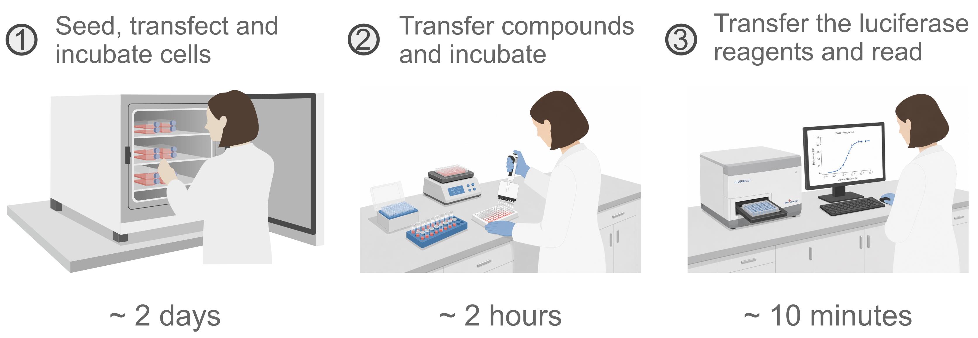
Graphical overview
Background
The transient receptor mucolipin subtype 1 (TRPML1) is a lysosomal ion channel expressed in every cell type [1]. Its deregulation has been implicated in many diseases, including genetic diseases (e.g., Mucolipidosis type IV [2], Niemann–Pick [3], and Duchenne muscular dystrophy [4]), bacterial [5] and viral infections [6], and cancer [7–14]. Recent pharmaceutical interest in developing agonist ligands has emerged due to beneficial effects in neurodegenerative diseases [15]. The identification of cell-permeable modulators for TRPML1 has been challenging due to the dynamic nature of these proteins and the inability to express and purify them in bacteria because of their complex structure. As a result, in vitro biochemical assays are largely unavailable. To overcome these issues, researchers have essentially relied on compound screening in cells overexpressing a cytoplasmic version of TRPML1 rather than the endolysosomal form [16–19]. This allows the apical application of compounds and the direct measurement of channel activity by calcium (Ca2+) imaging assay or by electrophysiology. However, these assays represent a non-physiological condition for drug screening because the compounds do not target the receptor in its native location [20,21]. More recently, the use of genetically encoded calcium indicators (GECI, such as GCaMP’s), specific intracellular calcium-sensitive dyes (such as Fura-2 and Fluo-4), and lysosomal preparations for patch-clamping has changed how we investigate compound binding to wild-type TRPML1 [22,23]. Despite these advancements, drug-screening campaigns remain extremely work-intensive due to successive washing steps and special handling of cells, and, more importantly, the use of expensive equipment for the functional assays.
Target engagement (TE) assays have emerged as an essential confirmatory assay for on-target activity in live cells, which accounts for compound permeability, biological target localization, and competition with intracellular constituents [24]. Among the different assay modalities, the bioluminescent resonance energy transfer (BRET) technology has been widely used to investigate compound binding to important targets such as kinases, GPCRs, and others [25]. This assay depends on the intracellular tracer that emits fluorescence once bound to the target protein fused to a luciferase [26]. The tracer (also called a BRET probe) is composed of a small molecule with experimental data on potency and mode of action in the target of interest, by either cryo-electron microscopy (cryo-EM), X-ray diffraction, or other structural biology techniques. This compound is rationally modified to covalently incorporate a fluorescent moiety in a position that would not interfere with binding to the protein target. This compound must also be cell-permeable in live cells and must bind to the target in a pocket that is placed at an optimal distance from the luciferase [27]. Once these parameters are achieved, one can perform the competition assay between a known concentration of the tracer and non-fluorescent test compounds, referred to as the tracer displacement (TD) assay. It is important to note that if the parent molecule that originated the tracer is promiscuous within a protein family, it allows the researchers to use the same tracer to investigate many proteins of the family [28]. Finally, it is worth mentioning that the BRET assay will only give data for compounds that bind to the same region of the tracer. Therefore, one of the major challenges to establishing a functional BRET assay is to develop a validated BRET tracer for the intended target.
Given the need for new screening assays for the TRPML1 ion channel and the ability of TE assays to investigate targets in their native environment, we set out to develop a BRET assay for TRPML1 using a new validated tracer called MRC087. The tracer was designed using a molecular docking approach to incorporate a fluorescent BODIPY-based compound into the mucolipin synthetic agonist-1 (referred to as ML-SA1). ML-SA1 was chosen due to its wide use in the scientific community as a TRPML1 agonist with a defined mode of action by cryo-EM [1,29,30] and reported structure-activity relationships [16].
Materials and reagents
Biological materials
1. Human embryonic kidney 293T cells (American Type Culture Collection, CRL-3216)
Reagents
1. Flexi® cloning system (Promega, catalog number: C8640)
2. hTRPML1 fused to YFP plasmid (Addgene, catalog number: 18826)
3. Plasmid constructs: The gene encoding the full-length human TRPML1 (Uniprot: Q9GZU1) was cloned in frame with nanoluciferase in plasmid pFC32K, part of the Flexi® cloning system. The coding sequence for hTRPML1 was PCR-amplified from hTRPML1 fused to YFP plasmid using primers hTRPML1-fb003 (GGCTGCGATCGCCATGACAGCCCCGGCGGGTCC) and hTRPML1-rb003 (ATGGGTTTAAACATTCACCAGCAGCGAATGCTCCTCCG). Correct cloning was confirmed by sequencing.
4. Fetal bovine serum (FBS) (Gibco, catalog number: A5256701)
5. FuGENE® HD transfection reagent (Promega, catalog number: E2311)
6. Nanoluciferase substrate/inhibitor (Promega, catalog number: N2162)
Note: The nanoluciferase substrate (furimazine-based) [31] and inhibitor [32] are both proprietary of Promega Corp, thus the exact composition of the kit is not fully disclosed.
7. Tracer dilution buffer (Promega, catalog number: N2191)
8. MRC087, 10 mM stock in DMSO (self-made, see [33]); validated and deposited in tracerDB (ID T000049; tracerdb.org) [34]
9. Dimethyl sulfoxide (DMSO), molecular biology grade (Sigma-Aldrich, catalog number: D2650-100ML)
10. NucleoBond Xtra Maxi Plus kit (Macherey Nagel, catalog number: 740416.50)
11. DMEM, high glucose (Gibco, catalog number: 11965092)
12. Penicillin-streptomycin (10,000 U/mL) (Gibco, catalog number: 15140122)
13. Dulbecco’s phosphate-buffered saline (PBS) (Sigma-Aldrich, catalog number: D8537-500ML)
14. 0.25% trypsin-EDTA (Gibco, catalog number: 25200072)
15. Trypan Blue solution, 0.4% (Gibco, catalog number: 15250061)
16. Opti-MEM serum-free without phenol red and antibiotics (Gibco, catalog number: 11058021)
Laboratory supplies
1. Cell culture flask, 250 mL, 75 cm2 (Greiner, catalog number: 658175)
2. 15 mL centrifuge tube (Corning, catalog number: 430766)
3. 96-well, white-walled/opaque bottom plate (Greiner Bio-One, catalog number: 655098)
4. 125 μL tips, sterile, with filter (Integra, catalog number: 4425)
5. 12.5 μL tips, sterile, with filter (Integra, catalog number: 4405)
6. Neubauer chamber (Cral, catalog number: C1010)
7. Reagent reservoir, 25 mL (Thermo, catalog number: 809311)
8. Aluminum foil
9. 10 mL sterile serological pipette (Sarstedt, catalog number: 86.1254.001)
Equipment
1. Inverted microscope (Leica, model: DMi8)
2. Centrifuge (Eppendorf, model: 5810R) with S-4-104 rotor
3. Plate reader (BMG LABTECH CLARIOstar)
4. Pipette controller (USA Scientific Inc, model: ErgoOne FAST)
5. Multichannel pipette 100 μL (Thermo Fisher Scientific, model: Finnpipette F1)
6. Orbital shaker (Heidolph, model: Titramax 101)
7. Biosafety cabinet (ESCO, model: Airstream AC2-4S8)
Software and datasets
1. GraphPad Prism (GraphPad, Version 11.0.2)
2. tracerDB [34] (tracerdb.org): a crowdsourced database of experimentally validated fluorescent tracers for target engagement assays (last accessed July 2026)
Procedure
登录/注册后免费查看全文
文章信息
稿件历史记录
提交日期: Jul 10, 2026
接收日期: Aug 10, 2026
在线发布日期: Aug 28, 2026
出版日期: Sep 20, 2026
版权信息
© 2026 The Author(s); This is an open access article under the CC BY-NC license (https://creativecommons.org/licenses/by-nc/4.0/).
如何引用
Readers should cite both the Bio-protocol article and the original research article where this protocol was used:
- Cunha, M. R., Catta-Preta, C. M. C., Massirer, K. B. and Couñago, R. M. (2026). How to Perform a Tracer Displacement BRET Assay for the TRPML1 Ion Channel. Bio-protocol 16(18): e5820. DOI: 10.21769/BioProtoc.5820.
- Cunha, M. R., Catta-Preta, C. M., Takarada, J. E., Moreira, G. A., Massirer, K. B. and Couñago, R. M. (2023). A novel BRET-based assay to investigate binding and residence time of unmodified ligands to the human lysosomal ion channel TRPML1 in intact cells. J Biol Chem. 299(6): 104807. https://doi.org/10.1016/j.jbc.2023.104807

 Download Citation in RIS Format
分类
生物化学 > 蛋白质 > 相互作用 > 蛋白质-蛋白质相互作用
细胞生物学 > 基于细胞的分析方法 > 药物筛选
分子生物学 > 蛋白质 > 离子通道信号转导
您对这篇实验方案有问题吗?
在此处发布您的问题,我们将邀请本文作者来回答。同时,我们会将您的问题发布到Bio-protocol Exchange,以便寻求社区成员的帮助。
 发布问题
发布问题


