


Stereotaxic Injection of Lysophosphatidylcholine Into Mouse Corpus Callosum for Establishment of a Focal Demyelination Model
通过向小鼠胼胝体立体定位注射溶血磷脂酰胆碱建立局灶性脱髓鞘模型

(*contributed equally to this work) 发布: 2026年10月20日第16卷第20期 DOI: 10.21769/BioProtoc.5818 浏览次数: 184
评审: Anonymous reviewer(s)
 PDF
PDF  Q&A
Q&A





Abstract
Multiple sclerosis (MS) is a chronic autoimmune disease characterized primarily by inflammatory demyelination of the central nervous system and is one of the leading causes of non-traumatic neurological disability in young and middle-aged adults worldwide. Myelin loss leads to impaired neural conduction, while progressive axonal degeneration resulting from failed remyelination constitutes a major pathological basis for irreversible disability in patients. Among currently approved treatments for MS, effective therapies that directly promote remyelination are still lacking; therefore, establishing animal models that can precisely recapitulate the myelin injury-repair process is essential for elucidating the mechanisms of remyelination and screening remyelination-promoting drugs. Focal demyelination models are important tools for investigating the mechanisms of remyelination and for developing therapeutic strategies for demyelinating diseases such as multiple sclerosis. Unlike the inflammation-driven injury of the experimental autoimmune encephalomyelitis (EAE) model and the systemic metabolic toxicity-induced demyelination of the cuprizone model, the lysophosphatidylcholine (LPC) injection model directly disrupts myelin in the corpus callosum through local injection of a membrane-solubilizing lipid, inducing focal demyelinating lesions and enabling investigators to study, in a controlled manner, the recruitment and differentiation of oligodendrocyte progenitor cells as well as the dynamic process of remyelination. This protocol describes the complete workflow for establishing focal demyelinating lesions by stereotaxic injection of LPC into the mouse corpus callosum, covering surgical preparation, coordinate localization, controlled injection, and postoperative care. Compared with existing methods, its main advantages lie in the precise control of the lesion and the synchronization of the post-injury repair phase, making it highly suitable for quantitative comparisons. Beyond the corpus callosum, this method is also broadly applicable to focal demyelination studies in other white matter tracts (including the spinal cord, optic nerve, and others), serving as a versatile platform for investigating region-specific myelin injury and repair.
Key features
• Precise injury that better enables quantitative analysis of demyelination and remyelination dynamics within defined time courses compared with existing techniques.
• Compatible with multiple downstream analyses, including immunohistochemistry, electron microscopy, and molecular profiling.
• Incorporates requirements for surgical technique, sham surgery controls, and predefined exclusion criteria for mistargeting, enabling rigorous quantitative analysis of demyelinating lesions.
• Practical validation timeline for myelin injury or remyelination, spanning the demyelination phase at 3–7 dpi through the remyelination phase at ~14–28 dpi.
Keywords: LPC (LPC) Remyelination (再髓鞘化)
White matter injury (白质损伤)
Oligodendrocyte lineage (少突胶质细胞谱系)
Histological validation (组织学验证)
Image quantification (图像定量)
Remyelination (再髓鞘化)
White matter injury (白质损伤)
Oligodendrocyte lineage (少突胶质细胞谱系)
Histological validation (组织学验证)
Image quantification (图像定量)
Graphical overview
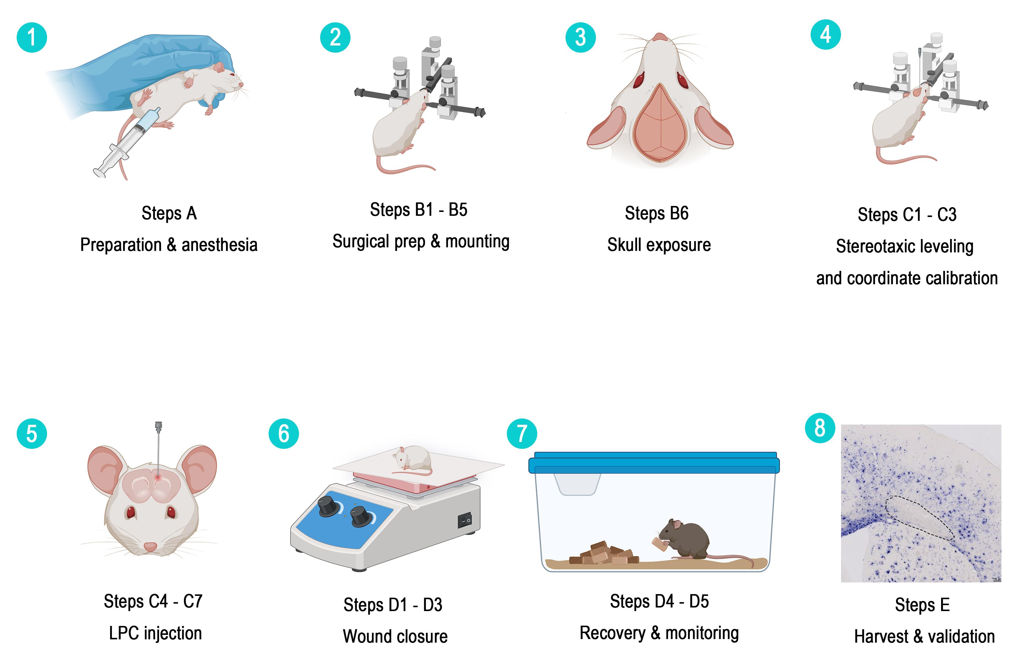
Step-by-step numbered schematic of this protocol. 1) Preparation and anesthesia (section A). 2) Surgical preparation and stereotaxic fixation (steps B1–5). 3) Skull exposure (step B6). 4) Stereotaxic leveling and coordinate calibration (steps C1–3). 5) Controlled LPC injection (steps C4–7). 6) Wound closure (steps D1–3). 7) Recovery and monitoring (steps D4–5). 8) Tissue collection and histological validation (section E).
Background
Demyelinating diseases such as multiple sclerosis (MS) involve damage to the myelin sheath surrounding axons in the central nervous system. Focal demyelination models allow investigators to study the cellular and molecular mechanisms of demyelination and remyelination in a spatiotemporally controlled manner. Current animal models for studying demyelination and remyelination fall mainly into three categories: the experimental autoimmune encephalomyelitis (EAE) model, the cuprizone model, and focal toxin-injection models [such as lysophosphatidylcholine (LPC) and ethidium bromide (EB) injection]. Because no single animal model recapitulates all features of human demyelinating disease, the choice of model should depend on the biological question being addressed. Experimental autoimmune encephalomyelitis (EAE) is suitable for studying autoimmune inflammation, but its lesions are temporally asynchronous, and spatial control is limited. Cuprizone intoxication produces pronounced corpus callosum demyelination after several weeks of systemic feeding, but the lesions are diffuse rather than experimentally targeted. Ethidium bromide can produce focal lesions but is broadly cytotoxic, damaging astrocytes and oligodendrocyte lineage cells in addition to myelin. By contrast, LPC is a membrane-disrupting lysophospholipid that produces spatially confined lesions, relatively spares axons, and exhibits a predictable demyelination–spontaneous remyelination sequence [1–9]. The LPC injection model is widely used because it induces focal demyelination without damaging axons, making it an ideal system for studying remyelination. Stereotaxic injection of LPC into the corpus callosum targets an anatomically well-defined and functionally important white matter tract, which is highly suitable for studying the recruitment and differentiation of oligodendrocyte progenitor cells. Compared with systemic demyelination models (such as the cuprizone model), this protocol allows precise control of lesion location and size, enables paired comparisons with the contralateral hemisphere, and facilitates the correlation of lesions with specific behavioral deficits [10–12]. Based on these advantages, this protocol describes in detail a standardized procedure for establishing a focal demyelination model by stereotaxic injection of LPC into the mouse corpus callosum and provides a complete histological validation method, with the aim of offering researchers in this field—especially beginners—a reproducible and easy-to-perform experimental reference (Table 1).
Table 1. Comparison of commonly used demyelination models
| Model | Primary mechanism | Advantages | Limitations | Representative references |
|---|---|---|---|---|
| EAE | Autoimmune inflammation | Clinically relevant immune component; suitable for immunotherapy studies | Lesions are temporally asynchronous; limited spatial control; predominantly spinal cord involvement in many paradigms | [3,4] |
| Cuprizone [bis(cyclohexanone)oxaldihydrazone] | Systemic oligodendrocyte mitochondrial toxicity | Pronounced corpus callosum demyelination; spontaneous remyelination after withdrawal | Diffuse lesions; requires several weeks of feeding; targeted location cannot be controlled | [4,5] |
| Ethidium bromide | Focal DNA intercalation and cytotoxicity | Large lesions with clear spatial boundaries | Broad glial cytotoxicity; under certain conditions, repair is slower and less complete than with LPC | [5,8] |
| LPC | Focal disruption of myelin lipids | Precise targeting, rapid onset, high reproducibility, relative axonal sparing, synchronized remyelination | Injection-related tissue damage; lack of autoimmune inflammation; local inflammatory responses still occur | [8,9] |
The LPC paradigm is not restricted to the corpus callosum. Focal LPC lesions have been established in spinal cord white matter, the caudal cerebellar peduncle, the internal capsule, the optic nerve, and the optic chiasm [5,13–19]. Therefore, this method is broadly applicable to region-specific questions of myelin injury and repair. However, coordinates, injection volumes, needle gauges, and tissue collection endpoints must be re-optimized for each target structure and species.
In the corpus callosum, demyelination can be detected within the first few days after LPC injection, typically peaking at around 3–7 dpi, followed by recruitment and differentiation of oligodendrocyte progenitor cells and remyelination at approximately 14–21 dpi. The exact time course depends on species, strain, age, injection volume, concentration, and target site [4,12,14,19,20]. Published volumetric analyses using defined mouse corpus callosum protocols report demyelination volumes of approximately 0.2–0.3 mm3 at 4 dpi and 0.05–0.15 mm3 at 14 dpi; these values are protocol-specific and should not be regarded as universal constants [16].
Adult C57BL/6 mice aged 8–10 weeks are commonly used because of their robust baseline remyelination capacity and well-defined time course. Aged mice may also be used when the experimental question concerns age-dependent repair; however, remyelination in aged animals is slower and less efficient, so age-matched controls should be included and tissue collection times adjusted to account for the slower remyelination in aged animals [15,21–23].
The target coordinates used in this protocol—AP +1.10 mm, ML +0.95 mm, DV -2.15 mm from the skull surface—are designed to place the injectate within the corpus callosum at the level of the fornix, dorsal to the dorsal hippocampus, while avoiding the superior sagittal sinus. If bilateral lesions are required, the procedure is repeated at ML -0.95 mm. Coordinates should be verified against the Paxinos and Franklin mouse brain atlas and adjusted according to atlas edition, animal age, body weight, skull geometry, and local instrument calibration [11,12,16,20,32]. The coordinates in this protocol were determined with reference to The Mouse Brain in Stereotaxic Coordinates by Paxinos and Franklin [32] and cross-validated using the Allen Mouse Brain Common Coordinate Framework 3D digital atlas. The AP +1.10 mm plane lies at the level of the frontal/motor cortex, where the corpus callosum has developed into a broad, thick, horizontally oriented, well-demarcated dense white matter tract with a large target cross-sectional area and good tolerance to targeting error; moreover, this plane is rostral to the hippocampus, so vertical needle insertion passes only through the motor/somatosensory cortex without traversing deep nuclei such as the hippocampus or striatum, thereby minimizing needle track damage. ML +0.95 mm corresponds to the lateral segment of the corpus callosum body (approximately dorsal to the lateral ventricle), the region where the white matter tract extends most widely laterally, ensuring adequate diffusion of the injectate within the callosal fibers. DV -2.15 mm (from the skull surface) is set according to the atlas, which shows the corpus callosum at this plane lying approximately 1.3–2.1 mm below the skull surface; placing the needle tip slightly deeper than its ventral border, combined with slow, low-flow-rate injection, allows the injectate bolus to spread within the corpus callosum and reflux mildly along the needle track, covering the full thickness of the corpus callosum while avoiding excessively deep penetration into the lateral ventricle that would cause injectate loss. The reasons for not injecting at the midline are as follows: first, the superior sagittal sinus runs along the sagittal suture directly beneath the midline, and drilling or needle insertion at the midline readily injures this venous sinus, causing difficult-to-control hemorrhage and markedly increasing surgical failure and animal mortality; a lateral offset of approximately 1 mm completely avoids the sinus body and its major bridging veins. Second, the corpus callosum at the midline is a narrow structure connecting the two cerebral hemispheres, and midline needle insertion would simultaneously damage the bilateral cingulate cortex and the corpus callosum on both sides of the midline, resulting in an uncontrollable extent of injury [11,20].
Materials and reagents
Biological materials
1. C57BL/6 mice, 8–10 weeks old (Jackson Laboratory, strain #000664); use age- and sex-matched animals, record body weight, and report sex and source
Reagents
1. L-α-lysophosphatidylcholine (LPC) from egg yolk (Beyotime, catalog number: ST1425-25mg); store at -20 °C, shelf life: 12 months
2. 2,2,2-Tribromoethanol (Aladdin, catalog number: T161626-25g); store at 4 °C, shelf life: 4 weeks
3. 2-Methyl-2-butanol (tert-amyl alcohol) (Aladdin, catalog number: A103417)
4. Sterile 1× phosphate-buffered saline (PBS) (Biosharp, catalog number: BL302A)
5. 75% ethanol (Sinopharm, catalog number: 100092680)
6. Povidone-iodine (Betadine) (Cofoe, catalog number: 100031918984)
7. Erythromycin eye ointment (Cisen, catalog number: H37022025)
8. 4% paraformaldehyde (PFA) solution (Sigma, catalog number: P6148); store at 4 °C and use within one month
9. Sucrose (Sinopharm, catalog number: H-10021463)
10. OCT embedding compound (Epredia, catalog number: 6502: Neg-50)
11. Myelin True Gold Staining kit (Oasis, catalog number: BK-AC001)
12. Sodium thiosulfate (Solarbio, catalog number: S5561-250g)
13. Anti-MBP antibody (Oasis, catalog number: OB-PGP187-01)
14. Fluorescently labeled secondary antibody (Invitrogen, catalog number: A11073)
15. DAPI (Solarbio, catalog number: ID22502)
16. Proteinase K (Roche, catalog number: 03115879001)
17. Anti-Digoxigenin-AP, Fab fragments (Roche, catalog number: 11093274910)
18. NBT/BCIP developing solution (Roche, catalog number: 11681451001)
19. Triton X-100 (Aladdin, catalog number: T109026)
Note: The catalog numbers listed above are representative products and can be replaced with validated equivalent products.
Solutions
1. 1% LPC working solution (see Recipes)
2. 2% tribromoethanol working anesthetic solution (see Recipes)
Recipes
1. 1% LPC working solution
| Reagent | Final concentration | Quantity or volume |
|---|---|---|
| L-α-lysophosphatidylcholine | 1% (w/v) | 10 mg |
| Sterile 1× PBS (Ca2+/Mg2+-free, pH 7.4) | n/a | 1 mL |
| Total | n/a | 1 mL |
Weigh 10 mg of LPC powder using an electronic balance and transfer it into a sterile centrifuge tube. Use a pipette to draw 1 mL of sterile PBS and add it to the centrifuge tube containing LPC. Tighten the cap, then vortex thoroughly for 3–5 min until the powder is completely dissolved and the solution becomes clear. Perform a brief low-speed centrifugation to spin down the residual liquid on the tube wall. The resulting solution is the 1% sterile LPC working solution, which should be stored at −20 °C, protected from repeated freeze/thaw cycles, and used within one week after preparation. Aliquots showing signs of turbidity, precipitation, or contamination should be discarded.
2. 2% Tribromoethanol anesthetic solution
| Reagent | Final concentration | Quantity or volume |
|---|---|---|
| 2,2,2-tribromoethanol | 2% (w/v) | 1 g |
| 2-methyl-2-butanol (tert-amyl alcohol) | 2% (v/v) | 1 mL |
| Sterile PBS | n/a | 49 mL |
| Total | n/a | 50 mL |
Weigh 1 g of tribromoethanol powder with an electronic balance and transfer it into a 50 mL sterile centrifuge tube. Add 1 mL of tert-amyl alcohol using a pipette. Gently shake at room temperature until the powder dissolves completely to form a clear oily stock solution. Gradually add 49 mL of sterile PBS to the stock solution and mix thoroughly. Transfer the entire solution into a sterile light-resistant reagent bottle, store at 4 °C, and use within one month after preparation.
Laboratory supplies
1. Suture, 5-0 (Jinhuan Medical, catalog number: 100040361204)
2. Cotton swabs (Cofoe, catalog number: 100265718600)
Equipment
1. Fine-tip marker (Fisherbrand, catalog number: 13-379-4)
2. Removable needle: 33G, Small Hub Removable Needle (RN) (Hamilton, catalog number: 7803-05 (point style 4, 12° bevel)
3. Hamilton microsyringe, 10 μL (Hamilton, model 1701), fitted with a removable needle
4. Microinjection pump (Harvard, model: PUMP 11 ELITE Nanomite)
5. Small animal stereotaxic instrument (RWD Life Science, catalog number: 68018) (requires a mouse adaptor, 60° mouse ear bars, a digital display manipulator arm, and a cannula holder)
6. Dental drill with a 0.5–0.8 mm drill bit (RWD Life Science, catalog number: 78001)
7. Animal thermostatic heating system (including heating pad) (RWD Life Science, catalog number: 69020)
8. Hair clipper (RWD Life Science, model: CP-5200)
9. Electronic balance (Deli, model: TE910)
10. Dissecting scissors (RWD Life Science, catalog number: S14014-11)
11. Blunt surgical scissors (RWD Life Science, catalog number: S13003-11)
12. Needle holder (RWD Life Science, catalog number: F31047-12)
13. Tissue forceps (RWD Life Science, catalog number: F13030-10)
14. Fluorescence microscope (Leica, model: DM6B)
Note: The catalog numbers listed above are representative products and can be replaced with validated equivalent products.
Software and datasets
1. Fiji/ImageJ 2.x, NIH, RRID: SCR_002285; used for image processing, thresholding, ROI measurement, and lesion quantification [30]
2. GraphPad Prism 9/10 or R 4.x; used for statistical analysis and plotting.
3. Microscope acquisition software specific to the Leica DM6B or other local imaging systems
Procedure
登录/注册后免费查看全文
文章信息
稿件历史记录
提交日期: Jun 30, 2026
接收日期: Aug 10, 2026
在线发布日期: Aug 25, 2026
出版日期: Oct 20, 2026
版权信息
© 2026 The Author(s); This is an open access article under the CC BY-NC license (https://creativecommons.org/licenses/by-nc/4.0/).
如何引用
Tan, R., Cheng, Q. and Tian, Y. (2026). Stereotaxic Injection of Lysophosphatidylcholine Into Mouse Corpus Callosum for Establishment of a Focal Demyelination Model. Bio-protocol 16(20): e5818. DOI: 10.21769/BioProtoc.5818.

 Download Citation in RIS Format
分类
神经科学 > 神经系统疾病 > 动物模型
神经科学 > 细胞机理 > 髓磷脂
免疫学 > 炎症性疾病 > 多发性硬化
您对这篇实验方案有问题吗?
在此处发布您的问题,我们将邀请本文作者来回答。同时,我们会将您的问题发布到Bio-protocol Exchange,以便寻求社区成员的帮助。
 发布问题
发布问题


