


Protecting Against Cytoplasmic Protein Aggregates With Cytoplasmic PML Variants
利用胞质 PML 变体清除胞质蛋白聚集体

(*contributed equally to this work) 发布: 2026年09月20日第16卷第18期 DOI: 10.21769/BioProtoc.5815 浏览次数: 342
评审: Marion HoggSwetha MurthygowdaSravanthi S P Nadiminti
 PDF
PDF  Q&A
Q&A
参见作者原研究论文
The authors used this protocol in:

Feb 2026





Abstract
Cytoplasmic protein aggregation is a defining feature of multiple neurodegenerative diseases, including amyotrophic lateral sclerosis, frontotemporal dementia, Huntington’s disease, and certain forms of motor neuron disease. Recent evidence indicates that promyelocytic leukemia protein (PML) and engineered PML-derived variants can act as versatile aggregate-remodeling factors. In particular, cytoplasmically redirected PML variants recognize pathological cytoplasmic inclusions and promote their clearance. Here, we describe a protocol to generate and validate two engineered cytoplasmic PML variants: full-length mPML, which is redirected to the cytoplasm by disruption of its nuclear localization sequence, and the truncated mPMLΔRBC variant, which lacks the RING, B-box, and coiled-coil domain but retains aggregate-reducing activity. The protocol integrates fluorescence-based imaging, bimolecular fluorescence complementation, detergent-soluble/insoluble fractionation, and validation in primary rat cortical neurons. This workflow provides a practical platform for assessing cytoplasmic aggregate burden and for comparing the aggregate-remodeling activities of PML-derived constructs. It can also be adapted to other disease-associated aggregation-prone proteins, including TDP-43, SOD1, FUS, tau, polyGA, and polyQ-expanded proteins.
Key features
• Describes the generation and validation of two complementary cytoplasmic PML variants: full-length mPML and truncated mPMLΔRBC.
• Provides fluorescence-based and BiFC-based assays to visualize pathological cytoplasmic protein assemblies.
• Includes detergent-soluble/insoluble fractionation for biochemical assessment of aggregate burden.
• Establishes a primary rat cortical neuron workflow to evaluate the effect of mPML on TDP-43-CTF aggregates.
• Can be adapted to other aggregation-prone proteins and additional PML-derived aggregate-remodeling candidates.
Keywords: PML (PML) Cytoplasmic protein aggregates (胞质蛋白聚集体)
TDP-43 (TDP-43)
SOD1 (SOD1)
Neurodegeneration (神经退行性变)
Aggregate clearance (聚集体清除)
Cytoplasmic protein aggregates (胞质蛋白聚集体)
TDP-43 (TDP-43)
SOD1 (SOD1)
Neurodegeneration (神经退行性变)
Aggregate clearance (聚集体清除)
Background
Pathological protein aggregation is a central molecular feature of many neurodegenerative diseases [1,2]. In amyotrophic lateral sclerosis and frontotemporal dementia, cytoplasmic inclusions containing TDP-43 are frequently observed [3]. Mutant SOD1 [4], FUS [5], polyGA dipeptide repeat proteins [6], and polyQ-expanded proteins [7] can also form cytoplasmic aggregates that interfere with cellular proteostasis and neuronal function [8].
Promyelocytic leukemia protein (PML) is best known as the principal scaffold of PML nuclear bodies and participates in transcriptional regulation [9], DNA damage responses [10], antiviral defense [11], senescence [12], and protein quality control [13]. Human PML isoform IV is a 633-amino-acid protein; its N-terminal region contains a RING finger, two B-box domains, and a coiled-coil region, collectively referred to as a tripartite motif. This motif mediates PML oligomerization and the assembly of higher-order protein complexes. The central region contains the nuclear localization sequence, whereas the C-terminal region is isoform-specific and contributes to the recruitment of distinct interacting proteins. Recent findings indicate that PML can also function as an aggregate-remodeling scaffold in protein quality control. PML recognizes protein inclusions and promotes their remodeling by recruiting molecular chaperones, including DnaJB1, together with proteasome-associated factors. These recruited components facilitate the disassembly of aggregate structures and the subsequent proteasome-dependent removal of aggregate material [14]. Thus, PML reduces aggregate burden through both direct aggregate remodeling and the coordinated recruitment of cellular protein quality-control machinery. Although native PML is predominantly localized in the nucleus, cytoplasmic PML variants derived from PML isoform IV can be generated by disrupting its nuclear localization sequence. In this protocol, human PML isoform IV was amplified from a plasmid purchased from Miaoling Biology (catalog number: P40666). Substitution of arginine 486 and lysine 487 with alanine redirects full-length PML to the cytoplasm, generating the mPML variant shown in Figure 1.
This protocol focuses on two engineered cytoplasmic PML variants:
1. mPML, a full-length cytoplasmic PML isoform IV variant containing the R486A and K487A substitutions within its nuclear localization sequence.
2. mPMLΔRBC, a truncated cytoplasmic variant containing residues 395–633 of PML isoform IV. This construct lacks the N-terminal RING finger, B-box, and coiled-coil regions but retains the central and C-terminal portions of PML, including the isoform-specific C-terminal region. Despite its reduced size, mPMLΔRBC retains aggregate-reducing activity and decreases detergent-insoluble TDP-43-CTF and SOD1-G93A species.
The experimental workflow includes construct generation, expression and localization validation, fluorescence-based measurement of aggregate burden in HEK293T cells, biochemical separation of detergent-soluble and detergent-insoluble fractions, and validation of mPML activity against TDP-43-CTF aggregates in primary rat cortical neurons. The protocol can also be adapted to evaluate other aggregation-prone proteins or to screen additional PML-derived aggregate-remodeling candidates.
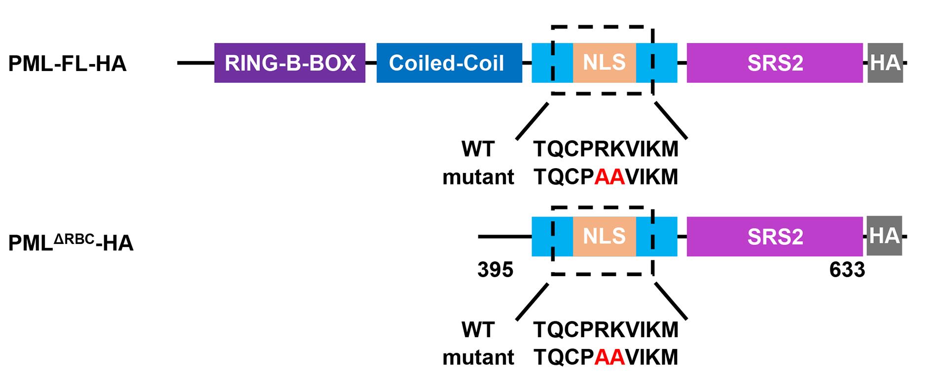
Figure 1. Illustration of promyelocytic leukemia protein (PML) variants. Schematic representation of full-length human PML isoform IV and the truncated PMLΔRBC construct. The N-terminal RING finger, B-box, and coiled-coil regions, the nuclear localization sequence (NLS), the isoform-specific C-terminal region, and the HA tag are indicated. The R486A and K487A substitutions used to generate the cytoplasmic mPML and mPMLΔRBC variants are also shown.
Materials and reagents
Biological materials
1. HEK293T cells (Shanghai Cell Bank, Type Culture Collection Committee; GNHu17)
2. Mouse primary cortical neurons
Plasmids
Note: All plasmids described in this protocol are available from the corresponding authors upon reasonable request.
1. Cytoplasmic PML variant–related plasmids:
a. p23 mPML-HA (you must clone): Expresses cytoplasmically localized PML fused with an HA tag. You can clone the coding sequence of PML into the p23-HA backbone by Gibson assembly. To promote the cytoplasmic location of PML, you can mutate the arginine 486 residue and lysine 487 residue into alanine by site-directed mutagenesis.
Note: Digesting the PCR products with DpnI to eliminate the template plasmid can improve the efficiency of plasmid construction.
b. p23 mPMLΔRBC-HA (you must clone): This construct expresses a cytoplasmically localized truncated PML fragment fused with an HA tag. To obtain the truncated PML, you can amplify the coding sequence corresponding to residues 395–633 of mPML by PCR and insert it into the p23-HA backbone by Gibson assembly.
c. pAAV-mPML-HA (you must clone): An adeno-associated virus (AAV) vector can be used to express cytoplasm-localized PML. You can clone the coding sequence of mPML and insert it into the pAAV-HA backbone by Gibson assembly.
2. Cytoplasmic protein aggregate–related plasmids:
a. pcDNA3.1-Flag-TDP-43-CTF-mNeon-NES (you must clone): This construct expresses the C-terminal fragment of TDP-43 (208-414 residues), fused with N-terminal Flag tag and mNeonGreen (mNeo) fluorescent reporter. To ensure the formation of cytoplasmic protein aggregates, the sequence is C-terminal-fused with a nuclear export signal (NES). You can amplify the coding sequences corresponding to residues 208–414 of TDP-43 with an N-terminal Flag tag by PCR. In parallel, the coding sequence of mNeonGreen is amplified by PCR with a C-terminal NES sequence. The Flag-TDP-43 (208–414 residues) fragment and the mNeonGreen-NES fragment are then inserted into the pcDNA3.1 backbone by Gibson assembly.
b. pcDNA3.1-Flag-SOD1-G93A-mNeon (you must clone): This construct expresses the SOD1-G93A mutant, which is fused with an N-terminal Flag tag and an mNeonGreen reporter. You can amplify the coding sequence of SOD1 with an N-terminal Flag tag by PCR. Simultaneously, you can amplify the coding sequence of mNeonGreen by PCR and then insert these two sequences into the pcDNA3.1 backbone by Gibson assembly. Eventually, you can mutate the glycine 93 residue into alanine by site-directed mutagenesis.
c. pcDNA3.1-VN-SOD1-G93A and pcDNA3.1-VC-SOD1-G93A (you must clone): A bimolecular fluorescence complementation (BiFC) system can be used to visualize SOD1-G93A aggregation by fluorescence imaging. To generate these constructs, amplify the coding sequence of SOD1-G93A and fuse it separately to the N-terminal fragment of Venus (VN) and the C-terminal fragment of Venus (VC). Insert the resulting VN-SOD1-G93A and VC-SOD1-G93A fragments into the pcDNA3.1 backbone by Gibson assembly. The VN and VC fragments can be amplified from the pT2-Venus plasmid (Miaoling Biology, catalog number: P0189).
d. pAAV- Flag-TDP-43-CTF-mNeon-NES (you must clone): An adeno-associated virus (AAV) vector can be used to express the Flag-tagged TDP-43-CTF. You can clone the coding sequence of Flag-TDP-43-CTF-mNeon-NES and insert it into the pAAV backbone by Gibson assembly.
Reagents
1. Dulbecco's modified Eagle medium (DMEM), high glucose (BasalMedia, catalog number: L110KJ)
2. NeurobasalTM medium (Thermo Fisher, catalog number: 21103049)
3. Fetal bovine serum (FBS), heat-inactivated (Lonsera, catalog number: S711-001S)
4. Penicillin-streptomycin (Beyotime, catalog number: C0222)
5. Dulbecco’s phosphate-buffered saline (D-PBS), Ca2+/Mg2+-free (Procell, catalog number: PB180329)
6. Trypsin-EDTA (Thermo Fisher, catalog number: 25200072)
7. Opti-MEM (BasalMedia, catalog number: L530KJ)
8. Polyethylenimine (PEI) (YEASEN, catalog number: 40816ES02)
9. B-27 supplement (Thermo Fisher, catalog number: A3582801)
10. L-glutamine (Thermo Fisher, catalog number: 25030081)
11. Poly-D-lysine (Beyotime, catalog number: C0312)
12. HBSS (Procell, catalog number: PB180323)
13. DNase I (Beyotime, catalog number: D7076)
14. Protease inhibitor cocktail (YEASEN, catalog number: 20124ES10)
15. D-PBS (Sangon Biotech, catalog number: E607009)
16. 5× SDS (Beyotime, catalog number: P0015)
17. Immunofluorescence related reagents:
a. Paraformaldehyde (PFA), 4% in D-PBS (Sigma-Aldrich, catalog number: 16005)
b. Triton X-100 (Sigma-Aldrich, catalog number: T8787)
c. Bovine serum albumin (BSA) (Sangon, catalog number: A500023)
d. Rabbit anti-HA tag antibody (Proteintech, catalog number: 51064-2-AP)
e. CoraLite488-conjugated goat anti-rabbit IgG(H+L) (Proteintech, catalog number: SA00013-2)
f. DAPI (Beyotime, catalog number: C1002)
g. Antifade mounting medium (Vectorlabs, catalog number: H-1900-10)
Solutions
1. RIPA I lysis buffer (see Recipes)
2. 2% BSA (see Recipes)
3. 0.5% PBST (see Recipes)
4. Neurobasal medium (see Recipes)
Recipes
1. RIPA I lysis buffer
| Reagent | Final concentration | Quantity or volume (for 50 mL) |
|---|---|---|
| NP40 | 1% | 0.5 mL |
| CHAPS | 0.5% | 0.25 g |
| SDS (10%) | 0.1% | 0.5 mL |
| NaCl | 150 mM | 0.435 g |
| Tris-HCl (pH 7.4, 1 M) | 50 mM | 2.5 mL |
| Protease inhibitor cocktail (100×) | 1× (add before use) | 500 μL |
Note: Add protease inhibitor cocktail immediately before use and keep the buffer on ice during cell lysis.
2. 2% BSA
| Reagent | Final concentration | Quantity or volume (for 50 mL) |
|---|---|---|
| BSA | 2% | 1 g |
| D-PBS | 1× | 50 mL |
3. 0.5% PBST
| Reagent | Final concentration | Quantity or volume (for 50 mL) |
|---|---|---|
| Triton X-100 | 0.5% | 250 μL |
| D-PBS | 1× | 50 mL |
4. Neurobasal medium
| Reagent | Final concentration | Quantity or volume (for 40 mL) |
|---|---|---|
| NeurobasalTM medium | 96.75% | 38.7 mL |
| Glutamine (200 mM) | 500 μM | 100 μL |
| B-27 | 2% | 800 μL |
| Penicillin-streptomycin | 1% | 400 μL |
Laboratory supplies
1. Cell culture multi-well plates (e.g., 12-well plate) (BIOFIL, catalog number: TCP011012)
2. 1.5 mL Eppendorf tubes (Sangon, catalog number: F607620-9001)
3. 50 mL centrifuge tube (BIOFIL, catalog number: CFT011500)
4. Cell coverslips for 12-well plate (WHB, catalog number: WHB-12-CS)
5. Cell counting chamber slides (Marienfeld, catalog number: 0650010)
6. 10 μL pipette tips (LAIBOER, catalog number: 1100103)
7. 200 μL pipette tips (LAIBOER, catalog number: 1102002)
8. 1 mL pipette tips (LAIBOER, catalog number: 1110004)
Equipment
1. Biological safety cabinet (The Baker Company, model: SG604-INT)
2. Cell incubator (Thermo, model: 371)
3. Inverted microscope (objectives: 4×, 10×, 20×) (yuehe, model: YHF40)
4. Laboratory centrifuge with rotors for 15 and 50 mL conical tubes (Eppendorf, model: 5702)
5. FinnpipetteTM F2 GLP Pipetting kit 2
6. Pipette (Fisher Scientific, catalog number: NC0085685)
7. Cell counter (Counter star, model: Mira BF)
8. Liquid carbon dioxide (CO2) tank
9. Vacuum aspirator (Yuwell, model: 7A-23D)
10. Refrigerated microcentrifuge (Eppendorf, model: 5424R)
11. Fluorescence stereomicroscope (Olympus, model: SZX16)
12. Confocal microscope (Andor, model: Dragonfly 200)
Procedure
登录/注册后免费查看全文
文章信息
稿件历史记录
提交日期: Jun 9, 2026
接收日期: Aug 12, 2026
在线发布日期: Aug 25, 2026
出版日期: Sep 20, 2026
版权信息
© 2026 The Author(s); This is an open access article under the CC BY-NC license (https://creativecommons.org/licenses/by-nc/4.0/).
如何引用
Zhu, J. X., Xia, Y., Liu, J., Cao, L., Hou, S. X. and Wang, Y. (2026). Protecting Against Cytoplasmic Protein Aggregates With Cytoplasmic PML Variants. Bio-protocol 16(18): e5815. DOI: 10.21769/BioProtoc.5815.

 Download Citation in RIS Format
分类
神经科学 > 神经系统疾病 > 神经退行性病变
分子生物学 > 蛋白质 > 靶向降解
细胞生物学 > 基于细胞的分析方法 > 蛋白互作
您对这篇实验方案有问题吗?
在此处发布您的问题,我们将邀请本文作者来回答。同时,我们会将您的问题发布到Bio-protocol Exchange,以便寻求社区成员的帮助。
 发布问题
发布问题


