


Homogeneous Time-Resolved Fluorescence-Based Assay to Screen ADP-Ribosyl Hydrolase Inhibitors
基于均相时间分辨荧光的 ADP-核糖水解酶抑制剂筛选方法

发布: 2026年09月20日第16卷第18期 DOI: 10.21769/BioProtoc.5814 浏览次数: 658
评审: Catherine HurdJagrity ChoudhuryAnonymous reviewer(s)
 PDF
PDF  Q&A
Q&A





Abstract
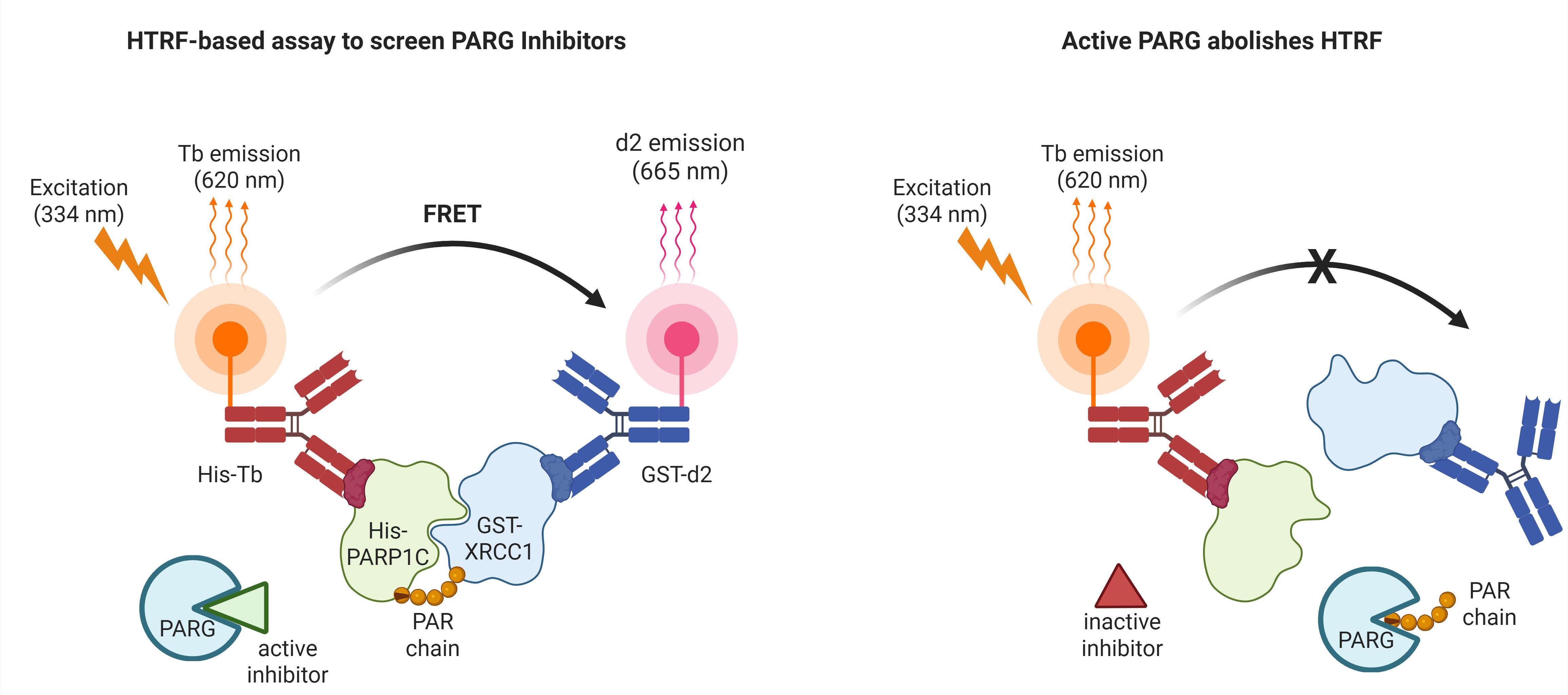
ADP ribosylation (ADPr) is a crucial post-translational modification that plays a vital role in DNA damage repair. Catalyzed by ADP ribose polymerases using NAD+ as a substrate, ADPr activates DNA repair pathways rapidly, thereby maintaining genomic integrity. The involvement of ADP ribose hydrolases in this process is significant, as they hydrolyze PAR chains, facilitating the release of ADPr-modified proteins from DNA or other proteins, which is essential for subsequent DNA repair steps. This protocol outlines a high-throughput screening method for identifying inhibitors of ADP ribose hydrolases, utilizing His-Tb-conjugated and ADPr-modified His-ADP ribose polymerase as the signal donor, and GST-d2-conjugated GST-XRCC1 as the signal receptor. The detection of time-resolved fluorescence signals enables efficient evaluation of compounds with potential therapeutic activity against cancer.
Key features
• A high-throughput screening method is provided for efficiently identifying inhibitors of ADP ribose hydrolases.
• The method employs homogeneous time-resolved fluorescence (HTRF) for sensitive detection of inhibitory effects on ADP ribose hydrolases, enabling high-throughput screening without complex separation steps.
• The research aims to accelerate the development of novel cancer therapies targeting ADP ribose hydrolases to improve treatment outcomes.
Keywords: ADP ribosylation (ADP-核糖基化) ADP ribose hydrolase (ADP-核糖水解酶)
PARG (PARG)
Homogeneous time-resolved fluorescence (均相时间分辨荧光)
ADP-ribosyl hydrolase inhibitors (ADP-核糖水解酶抑制剂)
ADP ribose hydrolase (ADP-核糖水解酶)
PARG (PARG)
Homogeneous time-resolved fluorescence (均相时间分辨荧光)
ADP-ribosyl hydrolase inhibitors (ADP-核糖水解酶抑制剂)
Graphical overview
Background
ADP ribosylation (ADPr) is an important post-translational modification that occurs in response to DNA damage, contributing to the activation and coordination of DNA repair pathways [1]. This modification is catalyzed by poly(ADP-ribose) polymerases, such as PARP1, which use NAD+ as a substrate to synthesize poly(ADP-ribose) (PAR) chains on target proteins, including PARP1 itself and nucleosomal histones [2]. PAR is subsequently recognized by DNA repair factors containing PAR-binding domains, including XRCC1, thereby facilitating their recruitment to sites of DNA damage [3]. However, once PAR-binding DNA repair factors have been recruited to DNA lesions, ADP-ribose hydrolases are required to hydrolyze PAR chains and promote the timely release and redistribution of these factors, allowing subsequent DNA repair steps to proceed [4]. If PAR chains are not efficiently hydrolyzed, PAR-binding repair factors may remain sequestered at PARylation sites and fail to execute their downstream repair functions. Therefore, the hydrolysis of PAR chains orchestrates the cellular response to DNA damage and contributes to the maintenance of genomic stability [5].
Given the pivotal role of PAR chain hydrolysis in DNA repair, it has emerged as a promising therapeutic target in cancer [5]. Poly(ADP-ribose) glycohydrolase (PARG), in particular, is the major enzyme to hydrolyze PAR chains [4]. PARG inhibition leads to PAR accumulation and impaired resolution of DNA damage signaling, which may increase the sensitivity of cancer cells to DNA-damaging agents, particularly in tumors with defective DNA repair mechanisms [6].
Targeting DNA damage repair has already provided substantial clinical benefits in ovarian and breast cancers. PARP inhibitors have been approved for selected patients with these cancers, particularly those harboring BRCA1/2 mutations or other homologous recombination repair deficiencies [1]. In ovarian cancer, PARP inhibitors have become important maintenance treatments, particularly for patients with platinum-sensitive disease and BRCA mutations or homologous recombination deficiency. In HER2-negative breast cancer, germline BRCA1/2 mutations have been established as predictive biomarkers for selecting patients for PARP inhibitor therapy. These advances demonstrate how research into ADP-ribosylation and DNA repair vulnerabilities has facilitated targeted drug discovery and biomarker-guided patient selection [7]. PARG inhibition represents a complementary approach that may be effective in homologous recombination-deficient ovarian and breast cancers and in tumors that have developed resistance to PARP inhibitors. However, the clinical efficacy of PARG inhibitors and the patient populations most likely to benefit remain to be established [5].
Several biochemical and cell-based methods have been developed to measure PARG activity and screen for PARG inhibitors. Early assays commonly monitored the degradation of radiolabeled PAR substrates using chromatographic or electrophoretic separation. Although sensitive, these methods require radioactive materials and multiple separation steps and are therefore not readily adaptable to large-scale compound screening. Nonradiometric assays were subsequently developed to quantify ADP-ribose released following PAR hydrolysis, enabling PARG inhibitor screening in multi-well formats [8]. Previous HTRF-based assays have measured PARG activity using labeled PAR substrates [9,10]. In contrast, our protocol uses ADP-ribosylated His-PARP1C as an artificial PAR substrate and monitors its interaction with GST-XRCC1, providing a distinct and reproducible approach for screening PARG inhibitors in a low-volume 384-well format. In addition, cell-based immunofluorescence assays measure the persistence or accumulation of cellular PAR following DNA damage and can identify cell-permeable PARG inhibitors [11]. However, the results of cell-based assays may be influenced by compound permeability, cellular metabolism, and other regulators of PAR homeostasis.
The introduction of high-throughput screening techniques significantly enhances the ability to quickly assess large libraries of compounds for their inhibitory effects on ADP ribose hydrolases, such as PARG. Identifying potent and selective PARG inhibitors may provide new therapeutic opportunities for patients with homologous recombination-deficient tumors, tumors characterized by high replication stress, and tumors that have developed resistance to PARP inhibitors. Such inhibitors may be used as monotherapies or in combination with DNA-damaging agents and other DNA damage response-targeted therapies, thereby potentially expanding the population of patients who may benefit from therapies targeting DNA repair vulnerabilities [7,11]. The present protocol directly monitors the PARG-dependent disruption of the interaction between ADP-ribosylated His-PARP1C and the PAR-binding protein GST-XRCC1. Its homogeneous, low-volume, 384-well format avoids radioactive substrates and post-reaction separation steps and allows up to 16 inhibitors to be evaluated simultaneously per plate. This protocol presents a novel approach to facilitate the discovery of potent inhibitors, ultimately contributing to the advancement of therapeutic strategies for cancer treatment and enhancing our understanding of the functional mechanisms of ADP ribose hydrolases in DNA damage repair.
Materials and reagents
Reagents
Note: Unless specified, all reagents are stored at room temperature (RT) (20–25 °C).
1. Super optimal broth with catabolite repression (SOC) (Sigma-Aldrich, catalog number: S1797) (-80 °C)
2. Tryptone (Oxoid, catalog number: LP0042B)
3. Yeast extract (Oxoid, catalog number: LP0021)
4. Kanamycin (Rhawn, catalog number: 25389-94-0) (2–8 °C)
5. Ampicillin (Rhawn, catalog number: 69-52-3) (2–8 °C)
6. Isopropyl β-D-1-thiogalactopyranoside) (IPTG) (Rhawn, catalog number: 367-93-1) (2–8 °C)
7. 10× phosphate-buffered saline (PBS) (Fisher Scientific, catalog number: MT-46013CM)
8. Protease inhibitor cocktail (Sigma-Aldrich, catalog number: P8849) (-20 °C)
9. Tris(hydroxymethyl)aminomethane (Tris base) (MCE, catalog number: HY-15917)
10. Bovine serum albumin (BSA) (Sigma-Aldrich, catalog number: A7906) (2–8 °C)
11. NaCl (Sigma-Aldrich, catalog number: S9888)
12. MgCl2 (Sigma-Aldrich, catalog number: M8266)
13. KCl (Sigma-Aldrich, catalog number: S7653)
14. Tween-20 (Sigma-Aldrich, catalog number: SLCL7671)
15. Dithiothreitol (DTT) (Sigma-Aldrich, catalog number: D0632) (-20 °C)
16. Dimethyl sulfoxide (DMSO) (MCE, catalog number: HY-Y0320C)
17. Imidazole (MCE, catalog number: HY-D0837)
18. Phenylmethylsulfonyl fluoride (PMSF) (MCE, catalog number: HY-B0496)
19. Ethylenedinitrilotetraacetic acid (EDTA) (MCE, catalog number: HY-Y0682A)
20. L-Glutathione reduced (Sigma-Aldrich, catalog number: G4251) (2–8 °C)
21. Bio-Safe Coomassie Stain (Bio-Rad, catalog number: 1610787)
22. Quick StartTM Bradford Protein Assay kit 2 (Bio-Rad, Catalog number: 5000202)
23. β-nicotinamide adenine dinucleotide (NAD+) (Sellect, catalog number: S2518) (-20 °C)
24. Custom DNA oligos (Millipore Sigma) (-20 °C)
25. HTRF mAb Anti-6His Gold Tb-conjugate (His-Tb) (Revvity Health Sciences Inc, catalog number: 50-103-1167) (2–8 °C)
26. mAb Anti-GST d2-conjugate (GST-d2) (Revvity Health Sciences Inc, catalog number: 50-211-7729) (2–8 °C)
27. PDD 00017273 (MCE, catalog number: HY-108360)
Solutions
1. Lysogeny broth (LB) medium (see Recipes)
2. LB agar plates (see Recipes)
3. Lysis buffer (His-tag) (see Recipes)
4. Lysis buffer (GST-tag) (see Recipes)
5. Wash buffer (His-tag) (see Recipes)
6. Elution buffer (His-tag) (see Recipes)
7. Equilibration buffer (GST-Tag) (see Recipes)
8. Elution buffer (GST-tag) (see Recipes)
9. Low salt buffer (see Recipes)
10. Size-exclusion chromatography buffer (see Recipes)
11. PARylation reaction buffer (see Recipes)
12. Assay buffer (see Recipes)
13. 10 μM custom DNA oligo (see Recipes)
Recipes
1. LB medium (liquid)
| Reagent | Final concentration | Quantity for 1 L |
|---|---|---|
| Tryptone | 10 g/L | 10 g |
| Yeast extract | 5 g/L | 5 g |
| NaCl | 10 g/L | 10 g |
| dH2O | - | To 1 L |
Note: Sterilize by autoclaving at 121 °C for 20 min. Allow to cool to RT or 37 °C before use. Store at RT (short term) or 4 °C (long term).
2. LB agar plates
For the solid medium, add 15 g/L agar to the LB medium (Recipe 1) before autoclaving. Autoclave at 121 °C for 20 min. Cool to approximately 50 °C, then add antibiotics (if needed, after cooling to ~50 °C to avoid thermal degradation). Pour approximately 20–25 mL per sterile Petri dish (10 cm diameter). Allow to solidify at RT. Store plates at 4 °C (inverted, in sealed bags) for up to 4 weeks.
3. Lysis buffer (His-tag)
| Reagent | Final concentration | Quantity for 100 mL |
|---|---|---|
| 1 M Tris-HCl (pH 8.0) | 50 mM | 5 mL |
| 5 M NaCl | 300 mM | 6 mL |
| 1 M Imidazole | 10 mM | 1 mL |
| 1 M DTT | 1 mM | 100 μL |
| 1 M PMSF | 1 mM | 100 μL |
| Protease inhibitor cocktail | As recommended | As per manufacturer |
| dH2O | - | To 100 mL |
Note: DTT and PMSF are labile and should be added to the buffer immediately before use. PMSF should be added from a fresh 100 mM stock (in isopropanol or DMSO) just before use. If using Sigma protease inhibitor cocktail P8849 (DMSO solution), add 10 μL per 1 mL of buffer (1:100, v/v) immediately before cell lysis. Do not store the buffers containing DTT, PMSF, or the protease inhibitor cocktail for extended periods.
4. Lysis buffer (GST-tag)
| Reagent | Final concentration | Quantity for 100 mL |
|---|---|---|
| 10× PBS (pH 7.4) | 1× | 10 mL |
| 1 M DTT* | 1 mM | 100 μL |
| 0.5 M EDTA | 1 mM | 200 μL |
| 1 M PMSF* | 1 mM | 100 μL |
| Protease inhibitor cocktail | As recommended | As per manufacturer |
| dH2O | - | To 100 mL |
*See note for Recipe 3.
5. Wash buffer (His-tag)
| Reagent | Final concentration | Quantity for 1 L |
|---|---|---|
| 1 M Tris-HCl (pH 8.0) | 50 mM | 50 mL |
| 5 M NaCl | 300 mM | 60 mL |
| 1 M Imidazole | 20 mM | 20 mL |
| 1 M DTT* | 1 mM | 1 mL |
| 1 M PMSF* | 1 mM | 1 mL |
| dH2O | - | To 1 L |
*See note for Recipe 3.
Note: Filter-sterilize (0.22 μm) and degas (e.g., by vacuum filtration or sonication) to remove dissolved air bubbles that could interfere with the size exclusion column.
6. Elution buffer (His-tag)
| Reagent | Final concentration | Quantity for 1 L |
|---|---|---|
| 1 M Tris-HCl (pH 8.0) | 50 mM | 50 mL |
| 5 M NaCl | 300 mM | 60 mL |
| 1 M Imidazole | 250 mM | 250 mL |
| 1 M DTT* | 1 mM | 1 mL |
| 1 M PMSF* | 1 mM | 1 mL |
| dH2O | - | To 1 L |
*See note for Recipe 3.
Note: Filter-sterilize (0.22 μm) and degas (e.g., by vacuum filtration or sonication) to remove dissolved air bubbles that could interfere with the size exclusion column.
7. Equilibration buffer (GST-Tag)
| Reagent | Final concentration | Quantity for 1 L |
|---|---|---|
| 10× PBS (pH 7.4) | 1× | 100 mL |
| 1 M DTT* | 1 mM | 1 mL |
| dH2O | - | To 1 L |
*See note for Recipe 3.
Note: Filter-sterilize (0.22 μm) and degas (e.g., by vacuum filtration or sonication) to remove dissolved air bubbles that could interfere with the size exclusion column.
8. Elution buffer (GST-tag)
| Reagent | Final concentration | Quantity for 1 L |
|---|---|---|
| 1 M Tris-HCl (pH 8.0) | 50 mM | 50 mL |
| Reduced glutathione | 20 mM | 6.15 g |
| dH2O | - | To 1 L |
Note: Prepare elution buffer immediately before use. Do not store elution buffer at 4 °C for more than a few days. Filter-sterilize (0.22 μm) and degas (e.g., by vacuum filtration or sonication) to remove dissolved air bubbles that could interfere with the size exclusion column.
9. Low salt buffer
| Reagent | Final concentration | Quantity for 1 L |
|---|---|---|
| 1 M Tris-HCl (pH 8.0) | 50 mM | 50 mL |
| 5 M NaCl | 50 mM | 10 mL |
| 1 M DTT* | 1 mM | 1 mL |
| dH2O | - | To 1 L |
*See note for Recipe 3.
Note: Filter-sterilize (0.22 μm) and degas (e.g., by vacuum filtration or sonication) to remove dissolved air bubbles that could interfere with the size exclusion column.
10. Size-exclusion chromatography buffer
| Reagent | Final concentration | Quantity for 1 L |
|---|---|---|
| 1 M Tris-HCl (pH 8.0) | 50 mM | 50 mL |
| 5 M NaCl | 150 mM | 30 mL |
| 1 M DTT* | 1 mM | 1 mL |
| dH2O | - | To 1 L |
*See note for Recipe 3.
Note: Filter-sterilize (0.22 μm) and degas (e.g., by vacuum filtration or sonication) to remove dissolved air bubbles that could interfere with the size exclusion column.
11. 2× PARylation reaction buffer
| Reagent | Final concentration | Quantity for 1 mL |
|---|---|---|
| 1 M Tris-HCl (pH 8.0) | 200 mM | 100 μL |
| 5 M NaCl | 200 mM | 20 μL |
| 0.5 M MgCl2 | 20 mM | 20 μL |
| 1 M DTT* | 20 mM | 10 μL |
| dH2O | - | to 1 mL |
*See note for Recipe 3.
Note: Store at -20 °C for up to 6 months. Thaw and keep on ice before use.
12. Assay buffer
| Reagent | Final concentration | Quantity for 10 mL |
|---|---|---|
| 1 M Tris-HCl (pH 7.5) | 100 mM | 1 mL |
| 5 M KCl | 50 mM | 100 μL |
| 10% (w/v) BSA | 0.25% | 250 μL |
| 100 mM DTT* | 1 mM | 100 μL |
| 10% (v/v) Tween-20 | 0.01% | 10 μL |
| dH2O | - | To 10 mL |
*See note for Recipe 3.
Note: BSA should be of high purity (e.g., fatty acid-free, protease-free).
13. 10 μM custom DNA oligo
| Reagent | Final concentration | Quantity for 10 mL |
|---|---|---|
| Custom DNA oligo | 10 μM custom DNA oligo | 1 mL |
| dH2O | - | To 1 mL |
Laboratory supplies
1. E. coli Rosetta (DE3) competent cells (TIANGEN, catalog number: CB108-02)
2. Petri dish (NEST, catalog number: 752101)
3. Millex 33 mm polyethersulfone (PES) 0.22 μm (Merck Millipore, catalog number: SLGPR33RB)
4. Millex 33 mm PES 0.45 μm (Merck Millipore, catalog number: SLHPR33RS)
5. White, low-volume 384-well plates (Greiner, catalog number: 784075)
6. HisTrapTM HP His tag protein purification columns (Ni-NTA) (Cytiva, catalog number: 17524801)
7. High-resolution GST-tagged protein purification with GSTrapTM HP (glutathione Sepharose) columns (Cytiva, catalog number: 17528201)
8. HiTrapTM Q HP anion exchange chromatography column (Q Sepharose) (Cytiva, catalog number: 17115401)
9. SuperdexTM 200 Increase 10/300 GL small-scale SEC column (Superdex 200 Increase) (Cytiva, catalog number: 28990944)
10. PierceTM Protein Concentrators PES, 3K or 5K MWCO, 0.5–100 mL (Thermo Fisher, catalog number: 88515)
Equipment
1. Autoclave (ZEALWAY, model: GR-85DA)
2. Incubator shaker (Shanghai Minquan, model: MQD-B3R)
3. UV-visible spectrophotometer (Shanghai Metash Instruments, model: UV-6000)
4. Sonicator (Shanghai Jingxin, model: XM-650T)
5. HTRF-compatible plate reader (Thermo Varioskan LUX with TRF module, or equivalent)
6. Nano Drop (Thermo, model: NanoDropTM One/One C)
7. Plate shaker (Cence, model: TDZ5-WS)
8. Multidrop Pico8 liquid handler (Thermo Multidrop Pico8 or equivalent)
9. AKTA (GE, model: AKTA Pure 25M)
Software and datasets
1. GraphPad Prism 8.4.3 (or equivalent)
Procedure
登录/注册后免费查看全文
文章信息
稿件历史记录
提交日期: Jun 25, 2026
接收日期: Aug 10, 2026
在线发布日期: Aug 25, 2026
出版日期: Sep 20, 2026
版权信息
© 2026 The Author(s); This is an open access article under the CC BY-NC license (https://creativecommons.org/licenses/by-nc/4.0/).
如何引用
Yu, A. and Wu, D. (2026). Homogeneous Time-Resolved Fluorescence-Based Assay to Screen ADP-Ribosyl Hydrolase Inhibitors. Bio-protocol 16(18): e5814. DOI: 10.21769/BioProtoc.5814.

 Download Citation in RIS Format
分类
癌症生物学 > 基因组不稳定性及突变 > 药物发现和分析
生物化学 > 蛋白质 > 翻译后修饰
分子生物学 > DNA > DNA 损伤和修复
您对这篇实验方案有问题吗?
在此处发布您的问题,我们将邀请本文作者来回答。同时,我们会将您的问题发布到Bio-protocol Exchange,以便寻求社区成员的帮助。
 发布问题
发布问题


