


Determining the Age of Every Cell Within Each Budding Yeast Microcolony Combining Single-Cell Microencapsulation With Confocal Microscopy
结合单细胞微囊化与共聚焦显微镜测定出芽酵母微菌落中每个细胞的年龄

发布: 2026年09月05日第16卷第17期 DOI: 10.21769/BioProtoc.5813 浏览次数: 303
评审: Joyce ChiuAnonymous reviewer(s)
 PDF
PDF  Q&A
Q&A
参见作者原研究论文
The authors used this protocol in:

Jan 2026





Abstract
Isogenic populations of Saccharomyces cerevisiae exhibit significant proliferative heterogeneity, with individual cells within a clonal culture displaying divergent growth rates and metabolic states. Investigating the origins of this variation requires a method to reconstruct the individual histories of cells within the population. This protocol describes a method for single-cell microencapsulation in alginate microspheres to create a physically stable, traceable, three-dimensional genealogical environment. By utilizing the alginate matrix to prevent daughter cell migration, the replicative history of a founder cell can be mathematically reconstructed. This is achieved by correlating the total cell count (N) within a developed microcolony with the total number of accumulated bud scars (n) visualized via confocal microscopy.
Key features
• Enables non-destructive 3D reconstruction of yeast genealogies, preserving spatial architecture and mapping all division events from a single founder cell.
• Implements a robust mathematical formula to determine the founder's replicative age, linking past mitotic history to current microcolony growth dynamics.
• Combines Flow Focusing® microencapsulation with precise confocal Z-stack analysis to review lineage-specific phenotypic heterogeneity in isogenic populations.
• Ideal for studying transgenerational inheritance and growth-rate diversification, providing a traceable, three-dimensional genealogical ecosystem for single-cell research.
Keywords: Saccharomyces cerevisiae (酿酒酵母) Microencapsulation (微囊化)
Bud scars (芽痕)
Replicative age (复制年龄)
Confocal microscopy (共聚焦显微镜)
Microencapsulation (微囊化)
Bud scars (芽痕)
Replicative age (复制年龄)
Confocal microscopy (共聚焦显微镜)
Graphical overview
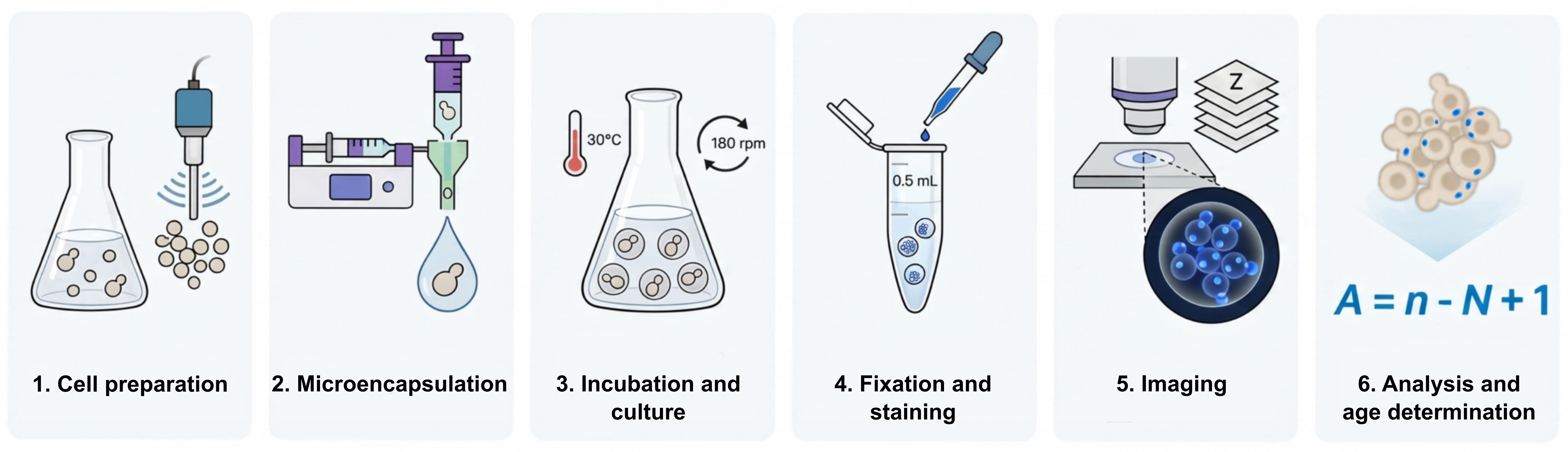
3D reconstructive genealogy and replicative age determination process
Background
Phenotypic and proliferative heterogeneity in yeast manifests as variations in cell size, cycle duration, and fitness, even in the absence of genetic or environmental flux. To study these phenomena, it is essential to determine the replicative age (A), the number of divisions a cell has undergone, of the ancestor, or founder cell, that initiated a specific lineage.
Single-cell microencapsulation in alginate provides a critical advantage over traditional liquid or agar cultures by transforming a microcolony into a traceable 3D ecosystem. The alginate scaffold ensures the physical stability of the lineage, preventing the drift of daughter cells and ensuring that every cell counted within the volume is a direct descendant of the original encapsulated founder. Because bud and birth scars are permanent chitinous markers left on the cell surface after every division, they provide a physical record of reproductive history. By calculating the total number of scars in a 3D volume relative to the total number of cells, the initial age of the founder cell can be accurately back-calculated.
Materials and reagents
Biological materials
1. Yeast strain BY4741 (EUROSCARF, Y00000) [1]
Reagents
1. D(+)-glucose anhydrous (VWR BDH Chemicals, catalog number: 24379.363)
2. Yeast extract (Condalab, catalog number: 1702.00)
3. Bacteriological peptone (Condalab, catalog number: 1616.05)
4. Adenine (adenine hemisulfate salt) (Sigma, catalog number: A9126-100G)
5. Alginate, alginic acid sodium salt from brown algae (Sigma, catalog number: 71238-250G)
6. Tri-sodium citrate dihydrate (Na3C6H5O7·2H2O) (VWR BDH Chemicals, catalog number: 27833.294)
7. CaCl2·2H2O (Sigma, catalog number: 223506-2.5KG)
8. Calcofluor White M2R (Fluorescent Brightener 28) (Sigma, catalog number: F3543)
9. Ethanol 96% (VWR BDH Chemicals, catalog number: 20823.327_T)
10. Tris base (Sigma, catalog number: T1503)
11. HCl 37% (PanReac AppliChem, catalog number: 131020.1211)
Solutions
1. Glucose 20% (w/v) (see Recipes)
2. Adenine 0.2% (w/v) (see Recipes)
3. YPAD (see Recipes)
4. Alginate 1.66% (w/v) (see Recipes)
5. CaCl2 3% (w/v) (see Recipes)
6. Citrate 10% (w/v) (see Recipes)
7. Calcofluor stock solution 1 μg/μL (see Recipes)
8. Ethanol 70% (see Recipes)
9. 1 M Tris-HCl pH 7.5. (see Recipes)
10. Tris-HCl-CaCl2 buffer solution (see Recipes)
11. Calcofluor working solution 0.1 μg/μL (see Recipes)
Recipes
1. Glucose 20% (w/v)
| Reagent | Final concentration | Quantity or volume |
|---|---|---|
| Glucose | 200 g/L | 20 g |
| Distilled water | - | Up to 100 mL |
| Total | - | 100 mL |
2. Adenine 0.2% (w/v)
| Reagent | Final concentration | Quantity or volume |
|---|---|---|
| Adenine | 2 g/L | 0.2 g |
| Distilled water | - | 100 mL |
| Total | - | 100 mL |
3. YPAD
| Reagent | Final concentration | Quantity or volume |
|---|---|---|
| Yeast extract | 10 g/L | 1 g |
| Bacterial peptone | 20 g/L | 2 g |
| Adenine (0.2% w/v) | 0.002 g/L | 8 mL |
| Glucose (20% w/v) | 20 g/L | 10 mL |
| Distilled water | - | 82 mL |
| Total | - | 100 mL |
4. Alginate 1.66% (w/v)
| Reagent | Final concentration | Quantity or volume |
|---|---|---|
| Alginate | 16.6 g/L | 0.166 g |
| Distilled water | - | 10 mL |
| Total | - | 10 mL |
5. CaCl2 3% (w/v)
| Reagent | Final concentration | Quantity or volume |
|---|---|---|
| CaCl2·2H2O | 30 g/L (anhydrous CaCl2) | 7.95 g |
| Distilled water | - | Up to 200 mL |
| Total | - | 200 mL |
6. Citrate 10% (w/v)
| Reagent | Final concentration | Quantity or volume |
|---|---|---|
| Na3C6H5O7·2H2O | 100 g/L (anhydrous Na3 citrate) | 56.98 g |
| Distilled water | - | Up to 500 mL |
| Total | - | 500 mL |
7. Calcofluor stock solution
| Reagent | Final concentration | Quantity or volume |
|---|---|---|
| Fluorescent Brightener 28 | 1 mg/mL | 1 mg |
| Distilled water | - | 1 mL |
| Total | - | 1 mL |
8. Ethanol 70% (v/v)
| Reagent | Final concentration | Quantity or volume |
|---|---|---|
| Ethanol 96% | 70% (v/v) | 364.6 mL |
| Distilled water | - | 135.4 mL |
| Total | - | 500 mL |
9. 1 M Tris-HCl pH7.5
| Reagent | Final concentration | Quantity or volume |
|---|---|---|
| Tris base | 1 M | 12.11 g |
| Distilled water | - | 70 mL |
| HCl 37% | - | Variable |
| Total | - | 100 mL (complete after pH adjustment with HCl) |
Sterilize by autoclaving.
10. Tris-HCl-CaCl2 buffer solution (2:1)
| Reagent | Final concentration | Quantity or volume |
|---|---|---|
| 1 M Tris-HCl pH 7.5 | 0.67 M | 60 mL |
| CaCl2 3% | 1% | 30 mL |
| Total | - | 90 mL |
11. Calcofluor working solution
| Reagent | Final concentration | Quantity or volume |
|---|---|---|
| Calcofluor stock solution (1 mg/mL) | 0.1 mg/mL | 50 μL |
| Tris-HCl-CaCl2 buffer solution (2:1) | - | 450 μL |
| Total | - | 500 μL |
Laboratory supplies
1. Microcentrifuge tubes 1.5 mL
2. Conical tubes 50 mL
3. Conical tubes 15 mL
4. Glass bottles (different sizes)
5. Nebulizers (Ingeniatrics®, catalog number: CLNEB002000)
6. Sterilization filters 0.2 μm (Corning, catalog number: 431219) for connection to the nebulizer inlet and sterilization of small-volume solutions
7. Sterilization filters 0.22 μm (Millipore® Express PLUS, Steritop® 45 mm Neck Size, 0.22 μm PES, 500 mL, catalog number: S2GPT05RE) for filter-sterilization of large-volume solutions
8. Cell strainer 40 μm (Corning, Falcon® 40 μm Cell Strainer, catalog number: CLS352340)
9. Syringe 5 mL (BD, EmeraldTM, catalog number: 307731)
10. Syringe 50 mL (BD, PlastipakTM, catalog number: 300866)
11. Micropipette tips 5–200 μL (DeltaLab, Daslab, catalog number: 162001)
12. Micropipette tips 100–1,000 μL (DeltaLab, Daslab, catalog number: 162222)
13. Glass beaker 50 mL (Schott Duran)
14. Microscope slides
15. Coverslips
16. Fluorescence immersion oil
Equipment
1. Bioencapsulation device (Ingeniatrics®, model: Cellena® Flow Focusing®)
2. Shaker (Eppendorf, model: New BrunswickTM Innova® 2300 Open Air Shaker)
3. Spectrophotometer (Eppendorf® 6135, Model Basic)
4. Sonicator (Diagenode, model: Bioruptor UCD-200TM-EX)
5. Centrifuge (Eppendorf, model: 5424)
6. Optical microscope (Leica, model: ICC50 HD)
7. Confocal microscope (Nikon, model: A1R+)
8. Bunsen burner
9. Micropipettes 0.2–2 μL, 100–1,000 μL, 2–20 μL, 20–200 μL (Gilson, PIPETMAN G, catalog number: 15664077)
10. Spoon
11. Autoclave
12. pH meter
13. Portable UV lamp (see General notes)
Software and datasets
1. Software Leica LAS Application Suite Version 3.0.0
2. Software Nikon NIS Elements Version 4.15
3. Microsoft® Excel Version 16.109.3
4. Fiji-ImageJ Version 2.16.0
5. All confocal microscopy images used to validate this protocol and perform the bud scar and microcolony genealogy analyses were deposited in the BioStudies database (http://www.ebi.ac.uk/biostudies/) and are available under the accession number S-BSST1071.
Procedure
登录/注册后免费查看全文
文章信息
稿件历史记录
提交日期: Jun 25, 2026
接收日期: Jul 27, 2026
在线发布日期: Aug 24, 2026
出版日期: Sep 5, 2026
版权信息
© 2026 The Author(s); This is an open access article under the CC BY-NC license (https://creativecommons.org/licenses/by-nc/4.0/).
如何引用
Ruger-Herreros, C., Delgado-Román, I., García-Marcelo, M. J., Chávez, S. and Muñoz-Centeno, M. C. (2026). Determining the Age of Every Cell Within Each Budding Yeast Microcolony Combining Single-Cell Microencapsulation With Confocal Microscopy. Bio-protocol 16(17): e5813. DOI: 10.21769/BioProtoc.5813.

 Download Citation in RIS Format
分类
微生物学 > 微生物细胞生物学 > 细胞染色
细胞生物学 > 细胞成像 > 共聚焦显微镜
细胞生物学 > 单细胞分析 > 细胞载体
您对这篇实验方案有问题吗?
在此处发布您的问题,我们将邀请本文作者来回答。同时,我们会将您的问题发布到Bio-protocol Exchange,以便寻求社区成员的帮助。
 发布问题
发布问题


