


From Bacterial Cellulose Production by Komagataeibacter xylinus to Bacterial Cellulose Nanoparticles: A Standardized Enzymatic Approach
从木糖驹形氏杆菌生产细菌纤维素到制备细菌纤维素纳米颗粒:一种标准化酶法


发布: 2026年09月05日第16卷第17期 DOI: 10.21769/BioProtoc.5812 浏览次数: 327
评审: DIPANWITA BHATTACHARYAPritish MitraJessica Davis
 PDF
PDF  Q&A
Q&A
参见作者原研究论文
The authors used this protocol in:
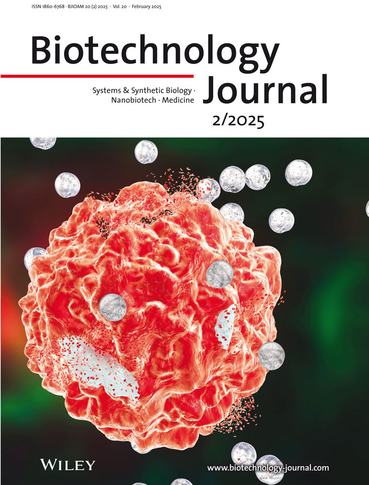
Feb 2025





Abstract
Bacterial cellulose (BC) is a renewable biopolymer valued for its exceptional purity, biocompatibility, and mechanical strength, with broad applications in biomedicine and sustainable materials. However, achieving reproducible BC production and downstream processing remains a major challenge. Inoculum preparation is particularly difficult to standardize because cellulose-producing strains form pellicles that sequester cells, making optical density measurements unreliable. In addition, recovery and drying procedures can alter fiber accessibility, and enzymatic hydrolysis conditions are often inconsistently defined and lack proper enzyme activity assessment. These issues contribute to substantial variability in BC-derived nanoparticle yields. This protocol describes the production of BC from Komagataeibacter xylinus DSMZ 6513, including culture medium preparation, inoculum generation, and scaling up under static cultivation conditions. It further details BC pellicle purification using NaOH, followed by pulping, freeze-drying, and milling to ensure material stability during storage and use. BC hydrolysis is performed with commercially available cellulase from Trichoderma reesei, with enzyme activity quantified prior to each reaction to ensure reproducibility. This standardized approach enables the reproducible production of bacterial cellulose nanoparticles (BCNPs). The protocol also includes minimal morphological characterization methods. By standardizing culture, recovery, and hydrolysis steps, the workflow reduces experimental variability and improves comparability across laboratories. Overall, it provides an accessible and reproducible method for generating BC and BCNPs of consistent quality without the need for specialized instrumentation.
Key features
• Standardized inoculum preparation that does not rely on optical density measurements, ensuring reproducible starting conditions for cellulose-producing strains.
• Controlled recovery and freeze-drying procedures that preserve fiber accessibility and promote consistent enzymatic hydrolysis outcomes.
• Use of commercially available Trichoderma reesei cellulase (≥700 units/g), with enzyme dosage defined by measured activity according to a publicly available Megazyme protocol.
• Minimal but robust morphological characterization methods to assess material quality.
Keywords: Bacterial cellulose (细菌纤维素) Komagataeibacter xylinus (木糖驹形氏杆菌)
Bacterial cellulose nanoparticles (细菌纤维素纳米颗粒)
Enzymatic hydrolysis (酶水解)
Cellulase activity (纤维素酶活性)
Nanomaterial characterization (纳米材料表征)
Sustainable biomaterials (可持续生物材料)
Enzyme-assisted nanoparticle synthesis (酶辅助纳米颗粒合成)
Antimicrobial material platforms (抗菌材料平台)
Dynamic light scattering (DLS) (动态光散射(DLS))
Scanning electron microscopy (SEM) (扫描电子显微镜(SEM))
Transmission electron microscopy (TEM) (透射电子显微镜(TEM))
Komagataeibacter xylinus (木糖驹形氏杆菌)
Bacterial cellulose nanoparticles (细菌纤维素纳米颗粒)
Enzymatic hydrolysis (酶水解)
Cellulase activity (纤维素酶活性)
Nanomaterial characterization (纳米材料表征)
Sustainable biomaterials (可持续生物材料)
Enzyme-assisted nanoparticle synthesis (酶辅助纳米颗粒合成)
Antimicrobial material platforms (抗菌材料平台)
Dynamic light scattering (DLS) (动态光散射(DLS))
Scanning electron microscopy (SEM) (扫描电子显微镜(SEM))
Transmission electron microscopy (TEM) (透射电子显微镜(TEM))
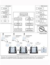
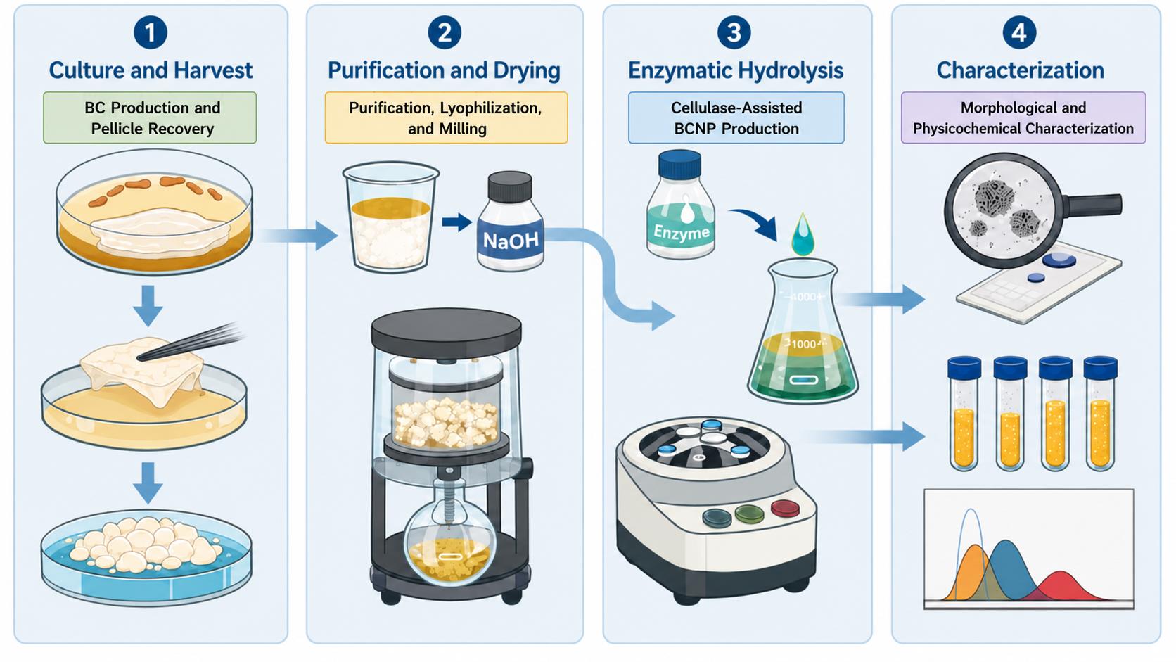
Graphical overview
Background
Nanocellulose has emerged as a versatile class of biomaterials with growing relevance in biotechnology, nanomedicine, and sustainable materials science [1]. Among its various forms, bacterial cellulose (BC) has attracted considerable attention due to its high purity, crystallinity, mechanical strength, and excellent biocompatibility [2,3]. Unlike plant-derived cellulose, BC is synthesized extracellularly by bacteria such as Komagataeibacter xylinus, producing a highly ordered three-dimensional nanofiber network free of lignin and hemicellulose. This intrinsic purity simplifies downstream processing and makes BC particularly suitable for biomedical applications [4].
Both BC and its nanoscale derivatives have been widely explored for applications including wound dressings, tissue engineering scaffolds, drug delivery systems, and antimicrobial materials, owing to their large surface area, mechanical stability, and capacity to interact with biological molecules [1,5]. Bacterial cellulose nanoparticles (BCNPs), in particular, represent promising nanocarriers for therapeutic agents. Their nanoscale dimensions and physicochemical properties enable efficient loading of bioactive molecules while maintaining high biocompatibility and environmental sustainability. Multiple strategies have been developed to obtain cellulose nanoparticles from bulk cellulose fibers. Traditional methods often rely on strong acid hydrolysis or other aggressive chemical treatments, which can produce hazardous waste, require extensive purification, and alter the resulting nanoparticles’ physicochemical properties [6]. Enzymatic hydrolysis using cellulases has recently emerged as a more sustainable alternative, enabling controlled fragmentation of cellulose macrofibers under milder conditions and reducing environmental impact [5]. Indeed, the successful exploitation of BCNPs in biomedical and biotechnological applications requires reliable control over critical parameters, including particle size, size distribution, morphology, and surface properties, which directly influence their performance as functional nanomaterials. Therefore, standardized and reproducible production strategies are essential to ensure consistent BCNP characteristics and facilitate their comparison across different studies and their translation toward practical applications.
Recent studies have shown that BCNPs generated through cellulase-mediated hydrolysis can serve as efficient platforms for the delivery of antimicrobial peptides (AMPs), improving peptide stability and enabling the development of sustainable antimicrobial formulations. For example, BCNPs produced from K. xylinus were functionalized with a human AMP via non-covalent interactions, yielding nanomaterials that inhibited bacterial growth while maintaining good biocompatibility with human cells [7]. Despite these advances, reproducibility in BC production and processing remains a major challenge. Sources of variability include differences in microbial growth kinetics, nutrient composition, oxygen availability, and cultivation scale, which can affect BC yield, fiber organization, and the final properties of the material. Furthermore, variations in purification strategies and drying procedures may influence cellulose crystallinity, aggregation state, and subsequent enzymatic accessibility. Therefore, the development of standardized protocols that define critical parameters throughout BC production and nanoparticle generation is essential to ensure consistent material quality and facilitate the translation of BC-based nanomaterials into research and industrial applications [3,6]. In particular, inoculum standardization, pellicle recovery, and control of enzymatic hydrolysis conditions can strongly influence nanoparticle yield and morphology. Many published procedures lack standardized approaches for culture preparation, enzyme activity assessment, and downstream handling of BC macrofibers, limiting inter-laboratory reproducibility [3,6].
The protocol presented here provides a standardized workflow for producing BC and generating BCNPs through controlled enzymatic hydrolysis. It includes reproducible procedures for culture preparation, pellicle purification, freeze-drying, and milling, as well as quantification of cellulase activity prior to hydrolysis. Compared with previously described approaches, this protocol emphasizes reproducibility, accessibility, and minimal instrumentation, while remaining compatible with common nanoparticle characterization techniques.
Beyond BCNPs production, this workflow supports a wide range of downstream applications, including antimicrobial formulations, nanocarriers for bioactive molecules, and functional biomaterials for biomedical and biotechnological use.
Materials and reagents
Biological materials
1. Bacterial strain: Komagataeibacter xylinus DSMZ 6513, gram-negative, aerobic, non-motile acetic acid bacterium characterized by the ability to produce extracellular bacterial cellulose as a pellicle; German Collection of Microorganisms and Cell Cultures GmbH (Leibniz Institute DSMZ, Germany)
2. Cellulase enzyme: Cellulase from Trichoderma reesei, aqueous solution ≥700 units/g (Merck Life Science S.r.l., catalog number: C2730)
Reagents
Note: All chemicals used were standard laboratory-grade reagents suitable for microbiological and biochemical applications.
1. Acetic acid (Merck, catalog number: A6283)
2. Sodium hydroxide (NaOH), pellets, anhydrous (Merck, catalog number: 221465)
3. Peptone (Merck, catalog number: 70028)
4. Yeast extract (Merck, catalog number: Y1625)
5. Disodium phosphate (Na2HPO4) (Merck, catalog number: 71699)
6. Citric acid monohydrate (Merck, catalog number: C1909)
7. D-(+)-Glucose (Merck, catalog number: G8270)
8. Agar (for solid media) (Merck, catalog number: A1296)
9. Sodium acetate trihydrate (Merck, catalog number: 29152900)
10. Azo-CM-Cellulose powder (Megazyme, catalog number S-ACMC)
11. Milli-Q water (or equivalent ultrapure water system)
Solutions
1. Hestrin–Schramm (HS) liquid medium (see Recipes)
2. HS agar (see Recipes)
3. 2 M sodium acetate buffer, pH 5.0 (see Recipes)
4. 1 M NaOH (see Recipes)
Note: Solutions for the Azo-CM-Cellulose assay were prepared according to the publicly available Megazyme protocol (Megazyme Ltd., Ireland; catalog number S-ACMC), including all buffers and dilutions required for enzyme activity quantification.
Recipes
1. HS liquid medium, pH 4.5
| Reagent | Final concentration | Quantity or volume |
|---|---|---|
| Peptone | 5 g/L | 5 g |
| Yeast extract | 5 g/L | 5 g |
| Na2HPO4 | 2.7 g/L | 2.7 g |
| Citric acid monohydrate | 1.15 g/L | 1.15 g |
| D-(+)-Glucose (separately autoclaved) | 20 g/L | 40 mL concentrated solution* |
| Milli-Q water | n/a | 960 mL |
| Total | n/a | 1,000 mL |
*Glucose is prepared as a concentrated solution and autoclaved separately to prevent Maillard reactions.
1. For 1 L of liquid culture medium, in a graduated cylinder, dissolve 5 g of peptone, 5 g of yeast extract, 2.7 g of disodium phosphate, and 1.15 g of citric acid in 960 mL of Milli-Q water. Transfer the solution into a heat-resistant bottle (Sol. A).
2. In a separate clean cylinder, dissolve glucose in Milli-Q water at a concentration of 500 mg/mL and transfer the solution into a heat-resistant bottle (Sol. B). The solution is viscous compared to water but remains fluid and easy to handle.
3. Autoclave Sol. A and Sol. B separately. After sterilization by autoclaving at 121 °C for 20 min, aseptically add 40 mL of sterilized Sol. B to Sol. A and mix thoroughly. This yields 1 L of culture medium with a final glucose concentration of 20 g/L.
4. Use immediately for inoculation or store under sterile conditions at room temperature.
2. HS agar
| Reagent | Final concentration | Quantity or volume |
|---|---|---|
| Peptone | 5 g/L | 5 g |
| Yeast extract | 5 g/L | 5 g |
| Na2HPO4 | 2.7 g/L | 2.7 g |
| Citric acid monohydrate | 1.15 g/L | 1.15 g |
| Agar | 20 g/L | 20 g |
| D-(+)-Glucose (separately autoclaved) | 20 g/L | 40 mL concentrated solution* |
| Milli-Q water | n/a | 960 mL |
| Total | n/a | 1,000 mL |
*Glucose is prepared as a concentrated solution and autoclaved separately to prevent Maillard reactions.
1. For 1 L of solid culture medium, add 20 g of agar to Sol. A (see Recipe 1) before autoclaving.
2. Proceed as for liquid culture medium.
3. After autoclaving, and while still hot, aseptically combine Sol. A and Sol. B. Immediately pour 10 mL into each Petri dish and allow to solidify at room temperature. A volume of 10 mL is intentionally selected because it is sufficient to uniformly cover the bottom of a 90 mm Petri dish while producing a relatively thin agar layer. This facilitates the excision of the agar disc used for inoculum preparation compared with the thicker agar layer obtained using the more conventional pouring volume of 20 mL.
4. It is recommended to prepare multiple Petri dishes at once to avoid re-melting or reheating the solidified medium.
5. After solidification, store Petri dishes at 4 °C under sterile conditions until use.
3. 2 M sodium acetate buffer, pH 5.0
| Reagent | Final concentration | Quantity or volume |
|---|---|---|
| Sodium acetate trihydrate | 2 M | 272.2 g |
| Acetic acid (glacial) | adjust pH | as needed |
| Milli-Q water | n/a | to 1 L |
Sterilize all stock solutions by filtering them through a 0.22 μm membrane under a laminar flow hood. Store all stock solutions at 4 °C. Dilute sodium acetate buffer to a 0.1 M working concentration immediately before use in enzymatic hydrolysis. Check the pH after dilution, as minor variations may affect enzyme activity.
4. 1 M NaOH, pH ~14.0
| Reagent | Final concentration | Quantity or volume |
|---|---|---|
| Sodium hydroxide pellets (anhydrous) | 1 M | 40 g/L |
| Milli-Q water | n/a | to 1 L |
Store the 1 M NaOH stock solution at room temperature. Dilute 1:10 to obtain a 0.1 M NaOH solution immediately before use for BC purification.
Laboratory supplies
1. 50 mL Falcon tubes (Merck, catalog number: CLS430829)
2. 100 mL Erlenmeyer flasks (Merck, catalog number: Z744843)
3. Sterile 1 L Erlenmeyer flasks (Merck, catalog number: SLW1135/26D)
4. Autoclavable bottles, 500 mL (Merck, catalog number: DWK61110P-500)
5. Autoclavable bottles, 1 L (Merck, catalog number: DWK61110T1000)
6. Micropipettes (Gilson), P20, P200, P1000 (Colaver, catalog number: 43GI3600)
7. Sterile pipette tips for Gilson P20, P200, P1000 (Merck, catalog numbers: F196087, F196088, F196089)
8. Sterile plastic disposable pipettes, 5 and 10 mL (Merck, catalog numbers: CLS70775N, CLS707710N)
9. Sterile L-shaped plastic loop, disposable (Merck, catalog number: HS8171A)
10. Sterile 90 mm Petri dishes (Merck, catalog number: Z717223)
11. Heat-resistant glass beakers, 250 and 400 mL (Merck, catalog numbers: BR91236, BR91241)
12. 40 mL glass flat-bottom tubes (Merck, catalog number: 27184)
13. Sterile 1.5 mL polypropylene centrifuge tubes (Merck, catalog number: HS4323)
14. 1 L graduated cylinders (Merck, catalog number: CLS3022P1L)
15. Magnetic stir bars (Merck, catalog number: Z745058)
16. Sterile 0.22 μm filters (Merck, catalog number: WHA10463607)
17. Stainless steel forceps (Merck, catalog number: Z168777)
18. Aluminum foil (Merck, catalog number: Z691577)
19. Parafilm M laboratory film (Merck, catalog number: P7793)
20. Disposable microcuvettes for dynamic light scattering (DLS) measurements (Malvern Panalytical, catalog number: ZEN0040)
21. Folded capillary zeta cell (Malvern Panalytical, catalog number: DTS1070)
22. Aluminum SEM pin stub mounts (Merck, catalog number: 933155)
23. Black conductive adhesive tabs for SEM (Ted Pella, catalog number: 16084-3)
24. Round glass microscope slides (Merck, catalog number: 63413)
25. 200-Mesh carbon-coated copper TEM grids (Merck, catalog number: 930369)
Equipment
1. Laminar flow hood (NuAire, Inc. Biological Safety Cabinet, ID 81718, model: NU-440-600E)
2. Chemical fume hood (Dynamika 150, ID 007)
3. Magnetic stirrer with heating plate and temperature probe (Four E’S Scientific Co., Ltd., ID LS52P051052)
4. Freeze-dryer (Lyoques, Hosmotic, ID C61644)
5. Milli-Q water purification system (Merck KGaA, Millipore Sigma, model: Q-Gard 1)
6. Analytical balance (0.1 mg precision) (Mettler-Toledo International Inc., model: XPR204S; ID XPR204S)
7. pH meter (Jenway, Cole-Parmer Instrument Company Ltd., ID 3510)
8. Hot plate with thermocouple (Kartell S.p.A., model: TechnoKartell TK22)
9. Countertop jar blender (Munro Instruments Ltd., catalog number: 800 G)
10. IKA® M 20 universal mill (IKA-Werke GmbH & Co. KG, catalog number: Z645141)
11. Water bath (GFL Gesellschaft für Labortechnik mbH, ID GFL_20001)
12. Orbital incubator (Stuart Equipment, Cole-Parmer Ltd., model: SI600)
13. Vortex mixer (Kartell S.p.A., model: TechnoKartell TK3S; ID 1892)
14. UV lamp (integrated UV sterilization lamp of biological safety cabinet) (NuAire Inc., model: NU-440-600E; ID 81718)
15. Refrigerated microcentrifuge (Thermo Fisher Scientific Inc., ID Fresco 21)
16. FisherbrandTM Elmasonic Select 30 ultrasonic bath (Thermo Fisher Scientific Inc., FEI Company, Waltham, Massachusetts, USA)
17. Zetasizer Nano ZSP system for DLS and electrophoretic light scattering (ELS) measurements (Malvern Panalytical Ltd., Worcestershire, UK)
18. Sputter coater for SEM sample preparation (Denton Vacuum LLC Desk V, Moorestown, NJ, USA)
19. Scanning electron microscope (FEI Nova NanoSEM 450; Thermo Fisher Scientific Inc., FEI Company, Waltham, Massachusetts, USA)
20. Transmission electron microscope (FEI Tecnai G2 200 kV; Thermo Fisher Scientific Inc., FEI Company, Waltham, Massachusetts, USA)
Software and datasets
1. Zetasizer Software 8.02 (Malvern Panalytical Ltd., Malvern, Worcestershire, UK), www.malvernpanalytical.com
2. GraphPad Prism version 8.4.3 for Windows (GraphPad Software, LLC, San Diego, California, USA), www.graphpad.com
3. Microsoft Excel version 365 (Microsoft Corporation, Redmond, Washington, USA), www.microsoft.com
Procedure
登录/注册后免费查看全文
文章信息
稿件历史记录
提交日期: Jun 4, 2026
接收日期: Jul 27, 2026
在线发布日期: Aug 24, 2026
出版日期: Sep 5, 2026
版权信息
© 2026 The Author(s); This is an open access article under the CC BY-NC license (https://creativecommons.org/licenses/by-nc/4.0/).
如何引用
Schibeci, M., Gaglione, R., Piccolo, E., Della Ventura, B. and Arciello, A. (2026). From Bacterial Cellulose Production by Komagataeibacter xylinus to Bacterial Cellulose Nanoparticles: A Standardized Enzymatic Approach. Bio-protocol 16(17): e5812. DOI: 10.21769/BioProtoc.5812.

 Download Citation in RIS Format
分类
生物工程 > 合成生物学
生物物理学 > 生物工程 > 纳米材料
微生物学 > 微生物生物化学 > 糖类
您对这篇实验方案有问题吗?
在此处发布您的问题,我们将邀请本文作者来回答。同时,我们会将您的问题发布到Bio-protocol Exchange,以便寻求社区成员的帮助。
 发布问题
发布问题


