


A Modified Slide-Embedded Scanning Electron Microscopy Preparation Method to Visualize Antagonistic Interactions Between Trichoderma viride and Fusarium sp.
用于观察绿色木霉与镰刀菌拮抗互作的改良载玻片包埋扫描电镜制样方法

(§Technical contact: sandra.tomichen2023@vitstudent.ac.in) 发布: 2026年09月05日第16卷第17期 DOI: 10.21769/BioProtoc.5805 浏览次数: 376
评审: Jessica DavisVandana MishraNidia Maldonado Carmona
 PDF
PDF  Q&A
Q&A





Abstract
Mycoparasitism is an important mechanism of fungal antagonism in which one fungus parasitizes another. This type of interaction plays a major role in the biocontrol activity of Trichoderma spp. against phytopathogenic fungi. Detailed visualization of these interactions is essential for understanding the structural mechanisms involved in fungal antagonism, including hyphal attachment, coiling, penetration, and cellular distortion. Scanning electron microscopy (SEM) is widely used for structural examination of fungal interactions; however, conventional preparation methods such as filter paper systems, membrane overlays, and agar block techniques often result in structural distortion, fragile sample handling, and difficulty in locating defined interaction zones. Here, we describe a modified slide-embedded technique for SEM visualization of mycoparasitic interactions between filamentous fungi. The protocol is adapted from previously reported slide culture approaches and involves embedding pre-cut sterile glass slide fragments directly into potato dextrose agar (PDA), followed by sequential inoculation of Fusarium sp. and Trichoderma viride. Fungal interactions occurring directly on the glass surface are subsequently subjected to fixation with 2.5% glutaraldehyde, graded ethanol dehydration, sputter coating, and SEM observation. Compared with conventional methods, the present approach provides improved handling stability, better preservation of native hyphal architecture, reduced deformation during processing, and easier localization of interaction zones during microscopy. The protocol also enables clear visualization of early antagonistic events such as hyphal coiling, penetration, and surface colonization. Due to its simplicity, reproducibility, and minimal technical complexity, this method serves as a practical and efficient approach for SEM-based investigation of fungal–fungal interactions and can be readily adapted for studying diverse mycoparasitic systems.
Key features
• A simple, cost-effective method to visualize filamentous fungus–fungus interactions, particularly mycoparasitism, using minimal reagents and basic laboratory equipment.
• A standardized inoculation strategy with pathogen inoculation 48 h before the mycoparasite ensures reproducible fungal interaction and mycoparasitic development.
• Blunt-cut square glass slide pieces embedded in PDA enable easy sample handling, storage, transport, and direct preparation for scanning electron microscopy.
• SEM-compatible fixation and ethanol dehydration preserve fungal ultrastructure, enabling high-resolution visualization of hyphal coiling, distortion, and other mycoparasitic interactions.
Keywords: Trichoderma viride (绿色木霉) Fusarium sp. (镰刀菌)
Mycoparasitism (重寄生)
Antagonism (拮抗作用)
Scanning Electron Microscopy (扫描电子显微镜)
Fusarium sp. (镰刀菌)
Mycoparasitism (重寄生)
Antagonism (拮抗作用)
Scanning Electron Microscopy (扫描电子显微镜)
Graphical overview
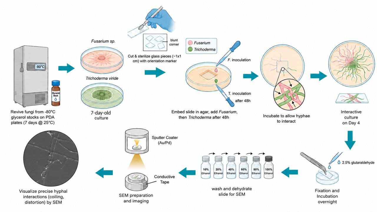
Graphical overview of the slide-embedded technique for scanning electron microscopy (SEM) visualization of mycoparasitic interactions between Trichoderma viride and Fusarium sp. Fungal cultures are revived from glycerol stocks, and a sterile square glass slide piece with a single blunt-cut edge is embedded in potato dextrose agar (PDA). Fusarium sp. is inoculated first and allowed to grow for 48 h, followed by inoculation of T. viride at the advancing hyphal margin. After interaction, the embedded slide is fixed, dehydrated through a graded ethanol series, sputter-coated with Au/Pd, and examined by SEM to visualize mycoparasitic structures such as hyphal coiling and distortion.
Background
Mycoparasitism is one of the major mechanisms underlying fungus–fungus antagonism, in which one living fungus derives nutrients from another living fungal host. Species of Trichoderma are among the most extensively studied mycoparasitic fungi because of their broad host range and strong antagonistic activity against several phytopathogenic fungi [1]. Fungicolous fungi are commonly categorized as symbionts, saprophytes, neutral associates, or mycoparasites depending on the nature of their interaction with other fungi [2,3]. Mycoparasitism is considered an ancestral trait of Trichoderma spp., contributing significantly to their effectiveness as biological control agents against plant pathogens.
In addition to mycoparasitism, Trichoderma spp. exhibit multiple biocontrol mechanisms, including antibiosis, induced systemic resistance (ISR), and competitive exclusion. These fungi behave predominantly as necrotrophs toward their hosts through the secretion of cell wall–degrading enzymes and secondary metabolites that suppress pathogenic fungal growth. Important enzymes produced by Trichoderma spp. include β-glucanases, proteases, cellulases, hemicellulases, and small secreted proteins, whereas secondary metabolites such as gliotoxin contribute to antagonistic activity [4,5].
Visualization of filamentous fungal interactions using scanning electron microscopy (SEM) plays an important role in understanding the structural and mechanistic aspects of mycoparasitism, including hyphal coiling, penetration, attachment, and distortion of host hyphae. Conventionally, SEM studies of fungal interactions have employed filter paper–based systems in which fungi grow over removable substrates prior to fixation and imaging [6]. However, these methods often present several limitations, including fragility of the substrate, difficulty during handling, and distortion of delicate hyphal structures during fixation and dehydration procedures.
To overcome these challenges, alternative techniques such as slide culture methods, cellophane membrane overlays, and coverslip-based systems have been developed to improve structural preservation and facilitate clearer visualization of fungal interaction zones [7,8]. Among these, the slide culture technique is considered one of the most reliable and feasible approaches for studying localized fungal interactions because of its simple setup and improved preservation of fungal morphology [8].
The present protocol describes a modified slide-embedded technique adapted from the novel slide culture approach reported by Bhat 2017 for studying mycoparasitic interactions between filamentous fungi [9]. In this method, pre-cut sterile glass slide fragments are embedded directly into PDA medium, allowing fungal growth and interaction to occur on a stable glass surface suitable for SEM analysis. Compared with conventional methods, this approach offers several advantages: (i) simplified experimental setup without the need for elaborate slide culture assemblies; (ii) improved sample handling and reduced structural deformation during SEM preparation; (iii) enhanced preservation of hyphal interactions; (iv) better visualization of early antagonistic events such as hyphal coiling and penetration; and (v) consistent localization of interaction zones using orientation-marked slide fragments. Consequently, this modified slide-embedded method provides a practical, reproducible, and user-friendly approach for SEM-based visualization of fungal mycoparasitic interactions. In this study, we investigate and visualize the mycoparasitic interaction between Fusarium sp. and the biocontrol fungus Trichoderma viride using SEM.
Materials and reagents
Biological materials
1. Trichoderma viride TVI (Elamala Biotech Lab, Chellarcovil, Kerala)
2. Fusarium sp. isolate STSP (Elamala Biotech Lab, Chellarcovil, Kerala)
Reagents
1. Potato dextrose agar (PDA) (HiMedia, catalog number: MH096)
2. Acetone 99% (Hyma, catalog number: ASA2019)
3. Absolute ethanol 99.9% (MSB Chemical Limited, catalog number: 5268-39); ethanol solutions (10%, 20%, 40%, 60%, and 80%) were prepared in Milli-Q water and syringe-filtered (0.22 μm)
3. Glutaraldehyde 25% aq. solution (SRL, catalog number: 92577)
4. Sodium phosphate dibasic dihydrate (SRL, catalog number: 87258)
6. Sodium phosphate monobasic dihydrate (SRL, catalog number: 40597)
7. Milli-Q water
Solutions
1. 0.1 M phosphate buffer (pH 7.4) (see Recipes)
2. Glutaraldehyde fixative (see Recipes)
Recipes
1. 0.1 M phosphate buffer (pH 7.4)
| Reagent | Final concentration | Quantity or volume |
|---|---|---|
| Sodium phosphate dibasic dihydrate | 75.407 mM | 1.342 g |
| Sodium phosphate monobasic dihydrate | 24.593 mM | 0.383 g |
| Milli-Q water | n/a | Make up to 100 mL |
| Total | n/a | 100 mL |
Dissolve both components in 80 mL of Milli-Q water. Adjust the pH to 7.4 using 1 N HCl or 1 N NaOH and make up the final volume to 100 mL with Milli-Q water. Store at room temperature and chill the required volume to 4 °C before use. This solution can be stored at room temperature for approximately 1–2 weeks if prepared and handled aseptically. Without sterilization (e.g., filtration or autoclaving), it should be used within 24–48 h. When properly sterilized and stored in tightly sealed containers, it remains stable for up to 2 months. Because phosphate buffers are prone to microbial contamination, discard the buffer if cloudiness, particulates, or visible microbial growth is observed. For maximum stability and extended shelf life, store the buffer at 4 °C.
Note: Avoid using KOH to adjust the pH, as excess potassium may alter the buffer composition, disturb the Na+/K+ balance, and affect cellular integrity.
2. Glutaraldehyde fixative
| Reagent | Final concentration | Quantity or volume |
|---|---|---|
| Glutaraldehyde 25% aq. solution | 2.5% | 1 mL |
| 0.1 M phosphate buffer (pH 7.4) | 10 mM | 1 mL |
| Milli-Q water | n/a | 8 mL |
| Total | n/a | 10 mL |
Freshly prepare this solution and then syringe-filter it through a 0.22 μm filter before use. Chill all components to 4 °C prior to preparation (see Troubleshooting 1).
Caution: Toxic if inhaled, ingested, or in contact with skin.
Laboratory supplies
1. Petri dish 100 mm (Borosil Scientific, catalog number: 3160077)
2. Conical flask 500 mL (Borosil Scientific, catalog number: 4980024)
3. Kimtech Science® KimwipesTM (Kimberly-Clark Professional, catalog number: 34155)
4. Inoculation loop (HiMedia, catalog number: LA019)
5. Syringe filter 0.22 μm (Sartorius, catalog number: 16532)
6. Disposable Pasteur pipettes (Genaxy, catalog number: GEN-153)
7. Parafilm M (Amcor, catalog number: PM992)
8. Microtips, 200–1,000 μL (Tarsons, catalog number: 521020)
9. Sterile tissue culture dish 60 mm (Tarsons, catalog number: 960020)
10. Sterile single-use syringe 10 mL
11. Forceps
12. Microscopic glass slide
13. Double-sided adhesive tape
14. Permanent marker
15. Rubber band
16. Newspaper
17. Non-absorbent cotton
Equipment
1. Scanning electron microscope (Carl Zeiss, model: EVO 18 Research)
2. Glass cutter (generic)
3. pH meter (HANNA instruments, model: HI98107)
4. Autoclave (EPS, model: EPS/VA-22)
5. Test tube rack (generic)
6. Laminar air flow (Clean Air Products, model: CAP412)
7. Microbiological incubator (Orbitek, model: LE)
8. Brightfield microscope (Carl Zeiss, model: Axioscope 5)
9. Bunsen burner (generic)
10. Refrigerator (2–8 °C) (generic)
11. Micropipettes 100–1,000 μL (Eppendorf, model: Research Plus Manual Single-channel pipette)
Procedure
登录/注册后免费查看全文
文章信息
稿件历史记录
提交日期: May 14, 2026
接收日期: Jul 16, 2026
在线发布日期: Aug 14, 2026
出版日期: Sep 5, 2026
版权信息
© 2026 The Author(s); This is an open access article under the CC BY-NC license (https://creativecommons.org/licenses/by-nc/4.0/).
如何引用
Tomichen, S. and Panchal, S. (2026). A Modified Slide-Embedded Scanning Electron Microscopy Preparation Method to Visualize Antagonistic Interactions Between Trichoderma viride and Fusarium sp.. Bio-protocol 16(17): e5805. DOI: 10.21769/BioProtoc.5805.

 Download Citation in RIS Format
分类
微生物学 > 微生物细胞生物学 > 细胞成像
微生物学 > 微生物-宿主相互作用 > 真菌
环境生物学 > 真菌
您对这篇实验方案有问题吗?
在此处发布您的问题,我们将邀请本文作者来回答。同时,我们会将您的问题发布到Bio-protocol Exchange,以便寻求社区成员的帮助。
 发布问题
发布问题


