


Semi-Automated Multiplex Workflow for Functional In Vitro Testing of Chemotherapeutic Treatments in Primary, Patient-Derived Cancer Organoids

发布: 2026年12月20日第16卷第24期 DOI: 10.21769/BioProtoc.5803 浏览次数: 238
评审: Philipp WörsdörferNarendra Verma
 PDF
PDF  Q&A
Q&A
参见作者原研究论文
The authors used this protocol in:

Sep 2024





Abstract
Most existing preclinical models have been limited in their predictive value to mimic patients’ responses, which is a major drawback in drug development and the identification of predictive biomarkers. To overcome these limitations, patient-derived three-dimensional in vitro models have been proposed. One of them is the organoid model, which preserves the original cellular heterogeneity and recapitulates epithelial architecture and functionality. Recently, studies using patient-derived organoids for drug screening applications have increased in quantity, and organoids have already been applied to pancreatic, colon, and lung cancers and female gynecological malignancies. Here, we established a multiplex workflow to analyze longitudinal therapeutic effects of anti-cancer therapeutics on organoid growth, viability, and cytotoxicity by combining state-of-the-art viability measurement with automated live cell imaging. This workflow can be used for the prediction of patient-specific treatment response, high-throughput screening of potential anticancer drugs, and downstream analysis to identify novel therapeutic targets.
Key features
• Multiplex workflow combines automated live cell imaging and metabolic readouts for viability quantification.
• It uses commercial and state-of-the-art readouts that serve as the gold standard for 3D cell cultures.
• It enables the differentiation of cytotoxic and cytostatic effects of chemotherapeutics.
• It allows for functional, precision medicine and the comparison of patient responses to chemotherapy.
Keywords: Organoids Personalized medicine
Functional precision medicine
Automated imaging
Multiplexing
Cell Titer Glo 3D
Personalized medicine
Functional precision medicine
Automated imaging
Multiplexing
Cell Titer Glo 3D
Graphical overview
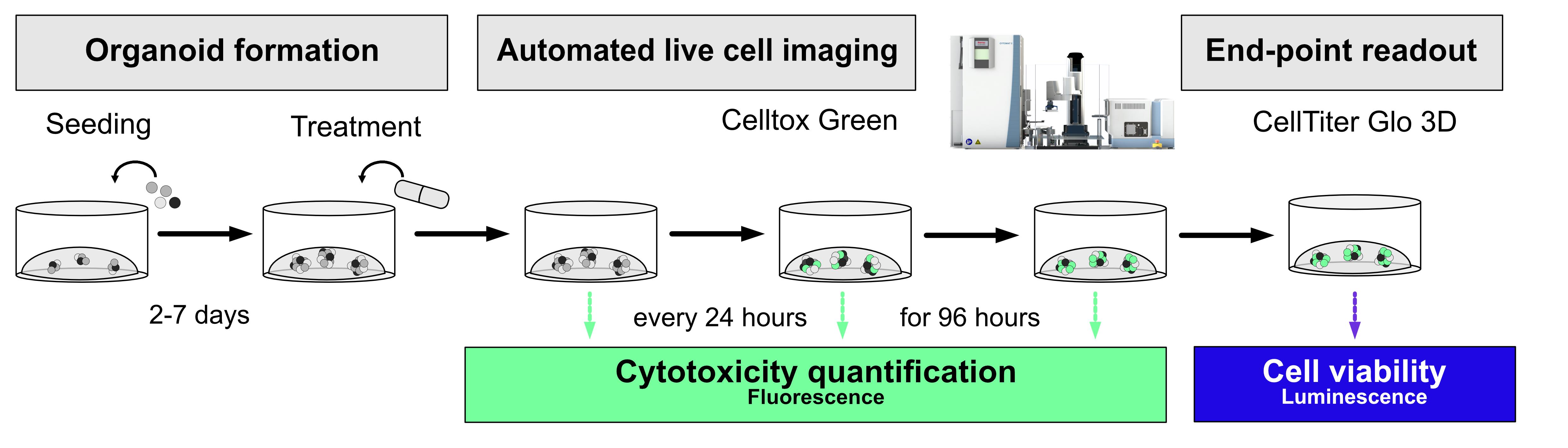
Summary of the described workflow. Organoids are seeded in 96-well plates and grown for 2–7 days, depending on organoid entity and growth characteristics. Then, cells are treated with different concentrations of chemotherapeutics, supplemented with CellTox Green cytotoxicity assay, and transferred to an automated imaging system (i.a. by SYNENTEC). Plates are imaged at the t0 timepoint and every 24 h to compare chemotherapeutic influence on cell growth and cytotoxicity. After 96 h, the CellTiter-Glo 3D cell viability assay is performed for end-point viability readouts.
Background
Despite advantages in prevention, detection, and treatment, cancer remains a major cause of death in Western countries, accounting for millions of new diagnoses and deaths each year. Lung cancer remains the leading cause of cancer-related deaths, followed by colorectal, breast, prostate, and pancreatic cancer [1]. Personalized medicine has the potential to improve treatment efficacy and overcome therapy resistance by characterizing the molecular profile of the patient’s tumor and identifying targetable pathways [2,3]. As conventional 2D in vitro models are limited in their ability to mimic patient-specific tumor biology and therefore often fail to adequately predict patient-specific therapy responses, multiple 3D models have been proposed to overcome these limitations [4]. One of them is the organoid model. Organoids are three-dimensional in vitro models that mimic the structural, genetic, and functional characteristics of their original tissue and preserve cellular heterogeneity. Patient-derived organoids can be generated from resected tumor tissue or fine-needle biopsies [2]. Expansion through serial passaging enables the generation of sufficient biological material for large-scale drug screening and downstream analysis, even from small resected specimens. Since their first description by Sato et al. [5], organoids have revolutionized the field of stem cell research, and their application has also expanded to cancer research. To date, organoids have been successfully established for various tumor entities, including pancreatic ductal adenocarcinoma (PDAC), ovarian cancer, head and neck squamous cell carcinoma, and lung cancer [6–13].
Here, we establish a medium-throughput, multiplex workflow including automated imaging monitoring longitudinal (chemo-) therapeutic effects on growth and cytotoxicity that is paired with gold-standard, state-of-the-art viability measurement using CellTiter-Glo 3D®. Using this workflow, drug response profiles can be easily detected, and morphological changes, i.e., loss of organoid integrity and cytotoxicity, are monitored in parallel. The CellTiter-Glo® 3D cell viability assay (Promega) confirms and complements results generated by imaging approaches and serves as the end-point readout. The protocol described here, therefore, enables the parallel, complementary measurement of various parameters that all share significance for functional precision medicine of primary cancers. Longitudinal imaging enables differentiation of cytostatic and cytotoxic features of used (chemo-) therapeutics, while end-point measurements using gold-standard assays enable comparison with other published datasets. Application of multimodal and longitudinal imaging of patient avatar models like organoids will enable high-throughput screens for alternative anticancer therapeutics, thereby facilitating an essential step toward patient-specific therapy.
Materials and reagents
Reagents
1. Extracellular matrix (ECM), i.a., Matrigel (Corning, catalog number: 354230); aliquot and store at -80 °C
Note: The protocols listed here were established using Matrigel. The use of other substitutes may affect organoid growth.
2. Advanced DMEM/F12 (Gibco, catalog number: 12634010); store at 4 °C
3. DPBS (PanBioTech, catalog number: P04-36500); store at 4 °C
4. TrypLE Express (Gibco, catalog number: 12605010); store at 4 °C
5. 1 M HEPES buffer (PanBioTech, catalog number: P05-01100); store at 4 °C
6. GlutaMax (Gibco, catalog number: 35050038); store at 4 °C
7. Penicillin/Streptomycin (Gibco, catalog number: 15140122); aliquot and store at -20 °C
8. Bovine serum albumin (BSA) (Carl Roth, catalog number: 3854); store at 4 °C
9. CellTox Green cytotoxicity assay (Promega, catalog number: G8731); store at -20 °C
10. CellTiter-Glo 3D cell viability assay (Promega, catalog number: G9682); store at -20 °C
11. Puromycin (InvivoGen, catalog number: ant-pr-1); store at -20 °C
12. Dimethyl sulfoxide (DMSO) (Sigma-Aldrich, catalog number: D2650); store at room temperature (RT)
Solutions
1. Advanced DMEM/F12+++ (ADF+++) (see Recipes)
2. Coating solution (see Recipes)
Recipes
1. ADF+++
| Reagent | Final concentration | Quantity or volume |
|---|---|---|
| Advanced DMEM/F12 | - | 500 mL |
| HEPES | 10 mM | 5 mL |
| GlutaMax | 1× | 5 mL |
| Penicillin/Streptomycin | 1% (v/v) | 5 mL |
Note: Keep at 4 °C for a maximum of four weeks. Invert the bottle to mix.
2. Coating solution
| Reagent | Final concentration | Quantity or volume |
|---|---|---|
| BSA | 0.1% | 2 mL of a 10% BSA stock |
| DPBS | Fill up to 200 mL |
Note: Filter the freshly made solution through a 0.22-μm filter to avoid contamination and keep at 4 °C for a maximum of three months. Invert the bottle to mix.
Laboratory supplies
1. 24-well tissue culture plates (TPP/Faust, catalog number: 92024; or comparable product)
2. 15, 50 mL conical centrifuge tubes (TPP/Faust, catalog numbers: 91015, 91050; or comparable product)
3. 1.5, 2 mL Eppendorf tubes (Sarstedt, catalog numbers: 72.706, 72.695.500; or comparable product)
4. 10, 200, 1,000 μL ART barrier pipette tips (Thermo Scientific, catalog numbers: 10098960, 10029040, 10313272; or comparable product)
5. Vacuum filtration system rapid Filtermax (0.22 μm) (TPP, catalog number: 99505; or comparable product) for filtration of coating solution
6. Duran glass bottles (500, 1,000 mL) (Schott, catalog numbers: 4459, 5455; or comparable product) for coating solution stock
7. Sterile 96-well flat clear bottom white polystyrene TC-treated microplates (Corning, catalog number: 3903; or comparable product)
8. B Braun solo cone Luer syringes (1/20 mL) (Braun, catalog number: 12752637; or comparable product)
9. Single-use 18 G syringe needles (blunt, 40 mm) (VWR, BD Medical, catalog number: BDAM303129)
Equipment
1. Safety cabinet for BSL-2 work, Safe 2020 Class II Biological Safety Cabinet (Thermo Scientific, catalog number: 51026640; or comparable device)
2. Shaking water bath (Lauda, model: Hydro H 20 S; or comparable device)
3. Centrifuge for 50/15 mL tubes (Hettich, model: Rotina 420R; or comparable device)
4. Incubator with temperature, CO2, and humidity control (Binder, model: CB-UL; or comparable device)
5. Standard benchtop brightfield microscope for cell monitoring (Zeiss, model: Axiolab 5; or comparable device)
6. Automation Suite composed of CYTOMAT 2 C-LiN, SYBOT-1000, and CELLAVISTA 4K (SYNENTEC)
Note: Alternatively, automated imagers can also be used as standalones like the CELLAVISTA, NYONE (both SYNENTEC), or the Incucyte systems (Sartorius).
7. Standard laboratory refrigerator
8. Standard laboratory -20 °C freezer
9. Standard laboratory -80 °C freezer
10. Box, bucket, or crate for ice
11. Standard laboratory pipettes (P1000, P100, and P10)
Software and datasets
1. YT-SOFTWARE (SYNENTEC GmbH, v25.9.1; license required) for image analysis
2. Scheduler (SYNENTEC GmbH, v26.0.0; license required) for automated image scheduling
3. Excel (Microsoft, 2021; license required) or LibreOffice packages (v26.2; free to use)
4. Prism (GraphPad, v11.0.0; license required) or R (v4.5.2; free to use) [14] for downstream analysis
Procedure
登录/注册后免费查看全文
文章信息
稿件历史记录
提交日期: Jun 23, 2026
接收日期: Aug 2, 2026
在线发布日期: Aug 13, 2026
出版日期: Dec 20, 2026
版权信息
© 2026 The Author(s); This is an open access article under the CC BY license (https://creativecommons.org/licenses/by/4.0/).
如何引用
Brauer, A., Grundt, F., Grohmann, F., Verkooyen, C., Weimer, J. P., Maass, N., Van Mackelenbergh, M. T., Sebens, S., Bauerschlag, D. O., Hedemann, N. and Holthaus, D. (2026). Semi-Automated Multiplex Workflow for Functional In Vitro Testing of Chemotherapeutic Treatments in Primary, Patient-Derived Cancer Organoids. Bio-protocol 16(24): e5803. DOI: 10.21769/BioProtoc.5803.

 Download Citation in RIS Format
分类
癌症生物学
细胞生物学 > 细胞活力 > 细胞存活
您对这篇实验方案有问题吗?
在此处发布您的问题,我们将邀请本文作者来回答。同时,我们会将您的问题发布到Bio-protocol Exchange,以便寻求社区成员的帮助。
 发布问题
发布问题


