


Stepwise Generation of Vascularized Multilayered 3D Organotypic Skin Models
逐步构建血管化多层三维组织型皮肤模型

(§Technical contact: hzhai3@huskers.unl.edu) 发布: 2026年08月20日第16卷第16期 DOI: 10.21769/BioProtoc.5802 浏览次数: 584
评审: Jessica DavisShanmugaPriyaa MadhukaranPankaj Mogha
 PDF
PDF  Q&A
Q&A
参见作者原研究论文
The authors used this protocol in:

Jan 2026





Abstract
Skin models play critical roles in understanding disease mechanisms and advancing therapeutic development. However, conventional systems based on 2D cell cultures, in vivo animal models, and ex vivo tissue explants are limited by insufficient physiological complexity, interspecies differences, and restricted accessibility, respectively. Advances in biofabrication technologies have enabled the engineering of 3D skin equivalents that better balance biological complexity and experimental scalability. Here, we present a biofabrication protocol inspired by the regenerative processes of wound healing to construct vascularized 3D organotypic skin models in a stepwise manner. The approach integrates bioprinting for precise spatial organization of cellular compartments with guided cell self-organization to achieve native-like tissue complexity and heterogeneity. Through a programmable culture strategy, tissue maturation proceeds sequentially through keratinocyte proliferation and collective migration, microchannel endothelialization, basal-to-suprabasal differentiation, and progressive extracellular matrix remodeling within a fibrin-based scaffold. The resulting tissue constructs comprise stratified epidermal layers positioned atop a vascularized, fibroblast-remodeled dermal matrix. Beyond reproducing key structural features of human skin, this protocol recapitulates cellular processes associated with tissue regeneration, providing a dynamic platform for investigating disease pathogenesis, progression, and therapeutic responses.
Key features
• This protocol provides detailed procedures for preparing cells and biomaterials used in bioink formulation.
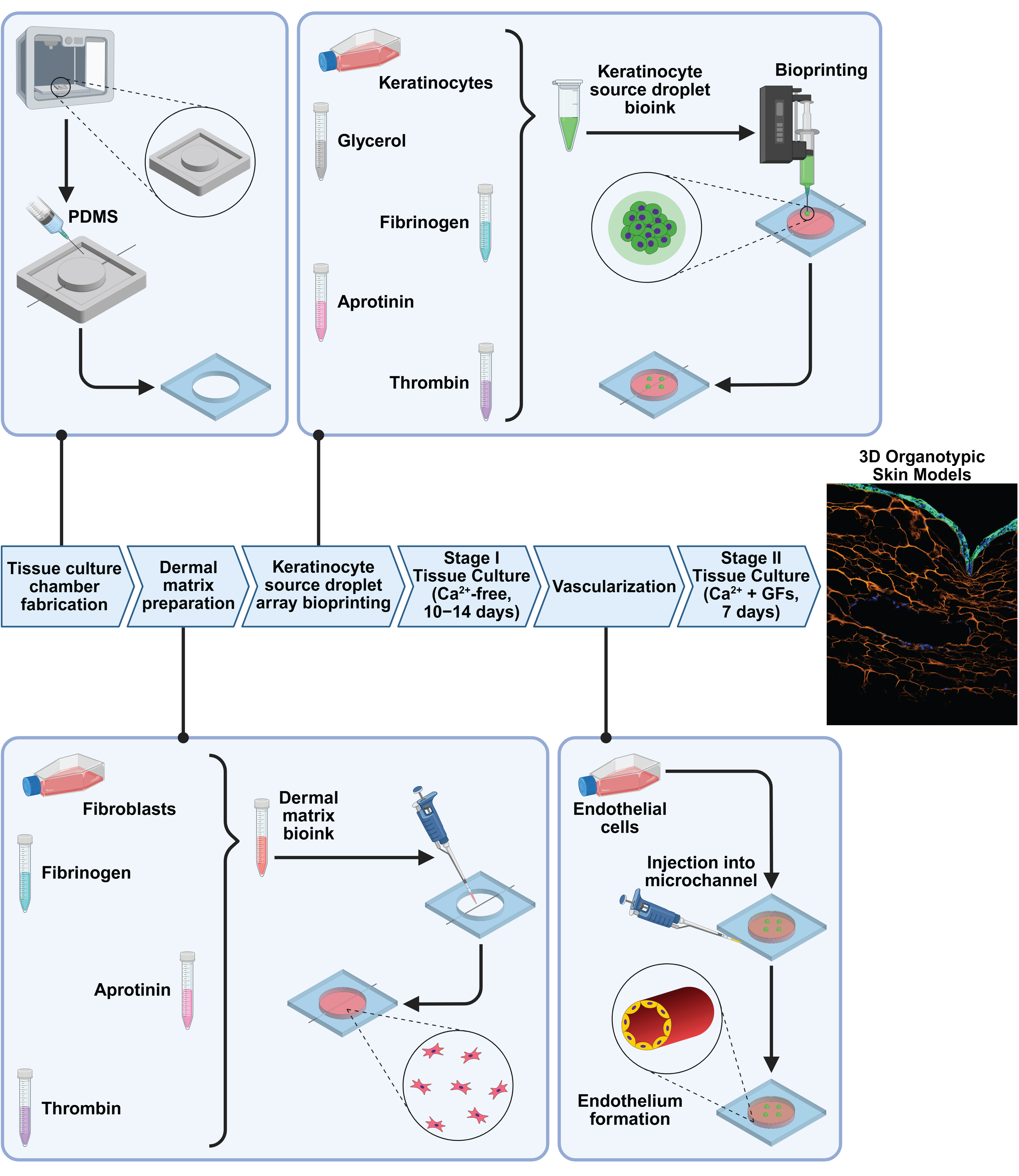
• This protocol outlines a stepwise biofabrication process for integrating keratinocytes, fibroblasts, and endothelial cells into a multicellular skin model.
• This protocol employs PolyJet 3D printing for tissue culture chamber fabrication and extrusion-based 3D bioprinting for spatial placement of cell-laden compartments.
• This protocol leverages a dynamic, wound healing–inspired cell self-organization process under a programmable culture strategy to promote tissue maturation and architectural development.
Keywords: Organotypic skin models (组织型皮肤模型) Bioprinting (生物打印)
Stratified epidermis (分层表皮)
ECM remodeling (细胞外基质重塑)
Vascularization (血管化)
Programmable culture (程序化培养)
Bioprinting (生物打印)
Stratified epidermis (分层表皮)
ECM remodeling (细胞外基质重塑)
Vascularization (血管化)
Programmable culture (程序化培养)
Graphical overview
Background
Skin serves as the body’s primary protective barrier and interface with the external environment [1]. Dysfunction of this barrier is associated with numerous diseases, including cancer, autoimmune disorders, and chronic wounds, and increases susceptibility to both acute and chronic microbial infections [2]. Experimental models that faithfully recapitulate the architecture and function of human skin are therefore essential for understanding disease mechanisms and for developing safe and effective therapeutics [3–5]. However, currently available model systems each present significant limitations. 2D monolayer cultures fail to capture the structural complexity, multicellular interactions, and microenvironmental cues present in native tissues [6–8]. Although animal models provide the physiological context, ethical concerns and interspecies differences limit their translational relevance to human disease [9–11]. Human ex vivo skin explants retain native tissue architecture and function, but their use is constrained by limited tissue availability and the lack of robust methods to maintain long-term viability and functionality. Furthermore, repeated biopsy collection can negatively affect patients' quality of life and well-being [12–15].
Advances in tissue engineering have expanded the ability to reconstruct human skin equivalents in vitro using human-derived cells [16,17]. Conventional skin equivalents commonly employ Transwell-based coculture systems, in which keratinocytes and fibroblasts are cultured to mimic epidermal and dermal compartments. However, these models generally lack perfusable vasculature and provide limited spatial control over cellular organization [18,19]. Microfluidic skin-on-a-chip platforms have enabled the integration of epidermal, stromal, and vascular compartments in more physiologically relevant configurations, yet many rely on porous membranes to separate cell populations and create artificial tissue interfaces [20,21]. More recently, 3D bioprinting has emerged as a powerful tool for spatially organizing multiple cell types within continuous hydrogel matrices without introducing synthetic barriers. By depositing cell-laden bioinks with high spatial precision, bioprinting can generate vascularized, fibroblast-populated dermal compartments together with overlying keratinocyte layers [22–25]. Nevertheless, the limited resolution of current bioprinting technologies makes it challenging to reproduce the highly organized, multilayered epidermis characterized by progressive basal-to-suprabasal differentiation. In parallel, skin organoids derived through stem cell self-organization can recapitulate aspects of epidermal stratification and cellular heterogeneity [26–28]. However, organoid formation often depends on poorly defined animal-derived matrices such as Matrigel and provides limited control over the biochemical and biomechanical properties of the microenvironment, particularly the fibroblast-regulated extracellular matrix (ECM) within the dermis. Consequently, faithfully reproducing both the structural and functional complexity of stratified epidermis and dynamically remodeled dermis remains a major challenge.
Physiological wound healing provides a blueprint for skin regeneration, encompassing coordinated processes of re-epithelialization, ECM remodeling, and angiogenesis. This protocol describes a dynamic biofabrication strategy that integrates 3D bioprinting with guided cell self-organization to recapitulate these regenerative processes and stepwise reconstruct vascularized, multilayered human skin tissues in vitro. From an engineering perspective, 3D printing is used to fabricate customized culture chambers that support integration of multiple cellular compartments, while bioprinting precisely positions cell populations according to native skin anatomy within a fibrin-based scaffold. From a biological perspective, a stagewise culture strategy with media optimized for distinct phases of tissue maturation promotes sequential development of tissue barriers through keratinocyte proliferation and collective migration, microchannel endothelialization, epidermal stratification, and fibroblast-driven ECM remodeling. This coordinated approach supports both long-term tissue viability and physiological function. It is worth noting that this protocol focuses on the epithelialization phases of basal layer formation and basal-to-suprabasal transition under submerged culture. However, its modular architecture and programmable culture workflow can be readily extended to generate full-thickness skin models by introducing an air–liquid interface (ALI) to promote terminal cornification and epidermal maturation.
As proof of concept, the resulting organotypic skin models have been applied to recapture skin disease pathogenesis and evaluate therapeutic candidates in our recent studies [29,30]. More broadly, this protocol illustrates an alternative paradigm for tissue biofabrication. Rather than relying solely on direct physical placement of cells and biomaterials to recreate tissue architecture, it combines engineering-guided assembly with physiological self-organization to restore structural and functional complexity beyond the capabilities of current fabrication technologies alone.
Materials and reagents
Biological materials
1. HaCaT cells, a widely used human keratinocyte line (kindly provided by Prof. Animesh A. Sinha); HaCaT cells tagged with green fluorescent protein on E-cadherin (GFP–E-cad–HaCaT cells) (generated in-house with the help of Prof. James K. Wahl III [29])
2. Normal adult human dermal fibroblasts (NHDF-Ad) (Lonza, catalog number: CC-2511) (passages ≤12)
3. Human umbilical vein endothelial cells (HUVEC) from pooled donors (Lonza, catalog number: C2519A) (passages ≤9)
Reagents
1. DMEM, high glucose, no glutamine, no calcium (Ca2+-free DMEM) (Thermo Fisher Scientific, Gibco, catalog number: 21068028)
2. Fetal bovine serum (FBS) (Thermo Fisher Scientific, Gibco, catalog number: A3160501)
3. GlutaMAX supplement (100×) (Thermo Fisher Scientific, Gibco, catalog number: 35050061)
4. Penicillin-streptomycin (Pen-Strep), 10,000 U/mL (Thermo Fisher Scientific, Gibco, catalog number: 15140122)
5. EGM-2 Endothelial Cell Growth Medium-2 BulletKit (Lonza, catalog number: CC3162)
6. FGM-2 Fibroblast Growth Medium-2 BulletKit (Lonza, catalog number: CC-3132)
7. Fibrinogen, human plasma (MilliporeSigma, catalog number: 341576-M)
8. Aprotinin (MilliporeSigma, catalog number: A4529)
9. Thrombin, human plasma (≥1,000 NIH units/mg protein) (MilliporeSigma, catalog number: T7009)
10. Glycerol, ≥99% (GC) (MilliporeSigma, catalog number: G2025)
11. Trypsin neutralizing solution (ATCC, catalog number: PCS-999-004)
12. Trypsin-EDTA for primary cells (ATCC, catalog number: PCS-999-003)
13. DPBS, no calcium, no magnesium (Thermo Fisher Scientific, Gibco, catalog number: 14190250)
14. Isopropanol (MilliporeSigma, catalog number: I9516)
15. Distilled water (sterilized)
Solutions
1. Ca2+-free DMEM medium (see Recipes)
2. FGM-2 medium (see Recipes)
3. EGM-2 medium (see Recipes)
4. Fibrinogen solution (see Recipes)
5. Thrombin stock solution (100×) (see Recipes)
6. Glycerol solution (50% v/v solution) (see Recipes)
7. Aprotinin stock solution (40×) (see Recipes)
8. DMEM-A medium (see Recipes)
Recipes
1. Ca2+-free DMEM medium
| Reagent | Final concentration | Quantity or volume |
|---|---|---|
| Ca2+-free DMEM | 88% | 44 mL |
| FBS | 10% | 5 mL |
| GlutaMax supplement (100×) | 1% | 0.5 mL |
| Pen-Strep (10,000 U/mL) | 1% | 0.5 mL |
| Total | 100% | 50 mL |
Thaw the pre-aliquoted frozen FBS and Pen-Strep stock solutions at room temperature for approximately 1 h or at 2–8 °C overnight. Supplement Ca2+-free DMEM with GlutaMax, FBS, and Pen-Strep. Store the complete medium at 2–8 °C for up to 1 week.
2. FGM-2 medium
| Reagent | Final concentration | Quantity or volume |
|---|---|---|
| FBM basal medium | 98% | 500 mL |
| FGM-2 SingleQuots supplement pack | 2% | 11.5 mL |
Thaw the frozen FGM-2 SingleQuots supplement pack, containing FBS (10 mL), insulin (0.5 mL), hFGF-B (0.50 mL), and GA-1000 (0.50 mL), at room temperature for approximately 1 h or at 2–8 °C overnight. Add the thawed supplements to FBM basal medium and gently swirl to mix. Store the complete medium at 2–8 °C for up to 1 week or at -20 °C for up to 1 month.
3. EGM-2 medium
| Reagent | Final concentration | Quantity or volume |
|---|---|---|
| EBM-2 basal medium | 97% | 500 mL |
| EGM-2 SingleQuots supplement pack | 3% | 15.2 mL |
Thaw the frozen EGM-2 SingleQuots supplement pack (10 mL of FBS, 0.20 mL of hydrocortisone, 2 mL of hFGF-B, 0.50 mL of VEGF, 0.50 mL of R3-IGF-1, 0.50 mL of ascorbic acid, 0.50 mL of hEGF, 0.50 mL of GA-1000, and 0.50 mL of heparin) at room temperature for ~1 h or at 2–8 °C overnight. Transfer the thawed supplements to EBM-2 basal medium and gently swirl to mix. Store the complete medium at 2–8 °C for up to 1 week or at -20 °C for up to 1 month.
4. Fibrinogen solution
| Reagent | Final concentration | Quantity or volume |
|---|---|---|
| Ca2+-free DMEM medium (Recipe 1) | 100% | 2 mL |
| Fibrinogen | 25 mg/mL | 50 mg |
Dissolve 50 mg of fibrinogen powder in 2 mL of Ca2+-free DMEM medium. Swirl to dissolve completely and place on ice. Freshly prepare the fibrinogen solution immediately before use to minimize enzyme-independent aggregation, non-fibrous gelation, and precipitation. Because fibrinogen powder can be difficult to weigh accurately, adjust the volume of Ca2+-free DMEM as needed to achieve a final fibrinogen concentration of 25 mg/mL.
5. Thrombin stock solution (100×)
| Reagent | Final concentration | Quantity or volume |
|---|---|---|
| DPBS | 100% | 2.5 mL |
| Thrombin | 100 U/mL | 250 U |
Dilute 250 U of thrombin powder in 2.5 mL of DPBS to prepare a stock solution. Mix thoroughly until the powder is completely dissolved. Dispense the solution into 100 μL aliquots and store at -80 °C. Thaw aliquots before use and avoid repeated freeze–thaw cycles to preserve enzyme activity.
6. Glycerol solution (50% v/v)
| Reagent | Final concentration | Quantity or volume |
|---|---|---|
| Ca2+-free DMEM medium (Recipe 1) | 50% | 1 mL |
| Glycerol | 50% | 1 mL |
Mix glycerol and Ca2+-free DMEM medium at a 1:1 (v/v) ratio. Filter-sterilize the solution and store at 2–8 °C.
7. Aprotinin stock solution (40×)
| Reagent | Final concentration | Quantity or volume |
|---|---|---|
| DPBS | 100% | 5 mL |
| Aprotinin | 1 mg/mL | 5 mg |
Dissolve 5 mg of aprotinin powder in 5 mL of DPBS and mix until completely dissolved. Dispense the solution into 250 μL aliquots and store at -80 °C. Thaw aliquots before use and avoid repeated freeze–thaw cycles to preserve enzyme activity.
8. DMEM-A medium
| Reagent | Final concentration | Quantity or volume |
|---|---|---|
| Ca2+-free DMEM medium (Recipe 1) | 97.5% | 9.75 mL |
| Aprotinin (40× solution) | 0.025 mg/mL | 0.25 mL |
| Total | 100% | 10 mL |
Thaw a frozen aliquot of 40× aprotinin solution at room temperature for ~1 h or at 2–8 °C overnight. Add the thawed solution to the complete Ca2+-free DMEM medium and gently swirl to mix. Store the supplemented medium at 2–8 °C for up to 1 week.
Laboratory supplies
1. Tissue culture flasks (25 and 75 cm2 growth areas)
2. Tissue culture dishes (35 and 100 mm diameters)
3. Centrifuge tubes (0.2, 1.5, 15, and 50 mL)
4. Micropipettes (10, 20, 100, 200, and 1,000 μL) and compatible tips
5. Syringe filters (28 mm diameter, 0.2 μm pore size)
6. Vacuum tube-top filter (50 mL capacity, 0.2 μm pore size)
7. SylgardTM 184 silicone elastomer kit (Krayden, catalog number: DC4019862)
8. RTV silicone sealant (Henkel, model: Loctite® SI 595)
9. Needles, 30 gauge (1.5 in, 1 in, and 0.5 in lengths) (Fisnar, catalog numbers: 8001114, 8001104, and 8001094)
10. Coverslips (170 μm thickness, 22 × 22 mm)
11. Clear silicone sheet (250 μm thickness)
12. Dispensing tips, 32 gauge (Nordson EFD, catalog number: 7018462)
13. Dispensing syringe barrels, 3 cc (Fisnar, catalog number: 8001001)
14. Hemocytometer (Bulldog Bio, catalog number: DHC-N420)
15. Other general lab supplies: single-use spatula, disposable glass Pasteur pipets, syringes (3 and 10 mL), razor blades, and double-sided tape
Equipment
1. Cell culture and maintenance: Class II, Type A2 biosafety cabinet, CO2 incubator, autoclave, liquid nitrogen storage system, and inverted microscope
2. General lab equipment: water bath, centrifuge, refrigerator (2–8 °C), freezer (-20 and -80 °C), vacuum desiccator, oven, and vortex mixer
3. 3-axis inline gantry robot (Fisnar, model: F5200N.2)
4. Fluid dispenser (Nordson EFD, model: UltimusPlus II)
5. PolyJet 3D printer (Stratasys, model: Objet500 Connex 3)
6. Thinky mixer (Thinky, model: AR-100)
7. Plasma cleaner (Harrick Plasma, model: PDC-001)
8. Inverted fluorescence microscope for 3D cell culture assays (Leica, model: THUNDER Imager)
9. TEER (WPI, model: EVOM Manual)
10. Nanoindentor (KLA, model: iNano)
11. Peristaltic pump (Ismatec, model: Reglo ICC 4-Channel)
Software and datasets
1. GrabCAD Print associated with the PolyJet printer (Stratasys)
2. Robot Edit associated with the dispensing robot (Fisnar)
Procedure
登录/注册后免费查看全文
文章信息
稿件历史记录
提交日期: Jun 23, 2026
接收日期: Jul 27, 2026
在线发布日期: Aug 7, 2026
出版日期: Aug 20, 2026
版权信息
© 2026 The Author(s); This is an open access article under the CC BY-NC license (https://creativecommons.org/licenses/by-nc/4.0/).
如何引用
Readers should cite both the Bio-protocol article and the original research article where this protocol was used:
- Zhai, H., Jin, X., Biegert, M., Zahs, B., Yang, R. and Meng, F. (2026). Stepwise Generation of Vascularized Multilayered 3D Organotypic Skin Models. Bio-protocol 16(16): e5802. DOI: 10.21769/BioProtoc.5802.
- Zhai, H., Jin, X., Moghaddam, A. O., Spieker, M. F., Seiffert-Sinha, K., Sinha, A. A., Yang, R. and Meng, F. (2026). 3D organotypic skin models recapitulate autoantibody-driven pemphigus pathomechanisms and targeted therapeutic response. Sci Adv. 12(2): eadz5003. https://doi.org/10.1126/sciadv.adz5003

 Download Citation in RIS Format
分类
细胞生物学 > 细胞工程 > 组织工程
细胞生物学 > 细胞分离和培养 > 3D细胞培养
生物工程 > 生物打印
您对这篇实验方案有问题吗?
在此处发布您的问题,我们将邀请本文作者来回答。同时,我们会将您的问题发布到Bio-protocol Exchange,以便寻求社区成员的帮助。
 发布问题
发布问题


