


Quantitative Analysis of Axonal Degeneration and TDP-43 Aggregation in Compartmentalized Human iPSC-Derived Motor Neuron–Myotube Co-cultures
人 iPSC 来源运动神经元—肌管分区共培养体系中轴突变性与 TDP-43 聚集的定量分析

(§Technical contact: gsanand09@gmail.com; talpery@tauex.tau.ac.il) 发布: 2026年09月05日第16卷第17期 DOI: 10.21769/BioProtoc.5801 浏览次数: 457
评审: Marion HoggThirupugal Govindarajan
 PDF
PDF  Q&A
Q&A
参见作者原研究论文
The authors used this protocol in:

Nov 2025





Abstract
Amyotrophic lateral sclerosis (ALS) is characterized by early and spatially restricted pathology in motor axons, including distal degeneration and accumulation of aggregation-prone proteins such as TDP-43. However, a major limitation in the field has been the lack of approaches that enable robust, quantitative, and compartment-specific analysis of these early axonal events, particularly in human-relevant systems. Here, we describe an integrated experimental and analytical framework that enables quantitative dissection of axonal degeneration and protein aggregation, specifically within distal motor axons. By combining compartmentalized human co-cultures with a dedicated image analysis strategy, this approach enables selective and quantitative analysis of pathological processes specifically within axons, independent of surrounding tissues such as muscle and other cellular compartments. This framework captures both structural degeneration and protein aggregation dynamics at subcellular resolution, enabling spatially resolved quantitative analysis of disease-relevant changes along axons. Importantly, the analytical framework is not limited to TDP-43 but is broadly applicable to diverse aggregation-prone proteins, thereby providing a generalizable platform to study axonal pathology across neurodegenerative diseases. Together, this work provides a scalable approach for investigating axonal pathology as an early and measurable feature of neurodegeneration, with potential applications in mechanistic studies and therapeutic targeting in ALS and related disorders.
Key features
• Compartmentalized human induced pluripotent stem cell (iPSC)-derived motor neuron–myotube co-cultures for modeling distal axonal pathology.
• Microfluidic separation of somatic and distal axonal compartments enabling spatial perturbation and analysis.
• Quantitative imaging of neurofilament heavy chain (NFH)-associated axonal degeneration and pTDP-43 accumulation.
• Semi-automated workflow for a reproducible, scalable, and modular pipeline for image quantification.
Keywords: Human iPSC-derived motor neuron–myotube co-culture (人 iPSC 来源运动神经元—肌管共培养) Axonal degeneration (轴突变性)
TDP-43 aggregation (TDP-43 聚集)
Microfluidic co-culture (微流控共培养)
Automated image analysis (自动化图像分析)
Amyotrophic lateral sclerosis (人 iPSC 来源运动神经元—肌管共培养)
Axonal degeneration (轴突变性)
TDP-43 aggregation (TDP-43 聚集)
Microfluidic co-culture (微流控共培养)
Automated image analysis (自动化图像分析)
Amyotrophic lateral sclerosis (人 iPSC 来源运动神经元—肌管共培养)
Graphical overview
Background
Distal axonal degeneration is among the earliest pathological features of amyotrophic lateral sclerosis (ALS). Evidence from animal models and induced pluripotent stem cell (iPSC)-derived neurons indicates that motor axons undergo progressive distal retraction and cytoskeletal disruption, consistent with a “dying-back” mechanism of neurodegeneration [1–6]. At the molecular level, cytoplasmic mislocalization and aggregation of TDP-43 within motor axons has emerged as a pathological hallmark of ALS, with phosphorylated TDP-43 (pTDP-43) accumulating in distal axonal compartments [7,8] and intramuscular nerve bundles during early disease stages [9,10]. We have previously shown that axonal TDP-43 condensates impair local protein synthesis and contribute to neurodegeneration, underscoring the relevance of quantifying axonal TDP-43 pathology in disease models [7,11]. Notably, axon-localized protein aggregation extends beyond TDP-43 and ALS and is also observed in other neurodegenerative disorders, including tau aggregation in tauopathies [12,13], α-synuclein accumulation in Parkinson’s disease [14,15], and SOD1 aggregates [1,6] or neurofilament accumulation in familial forms of ALS [6,16–19]. Together, these findings highlight the necessity of not only detecting but rigorously quantifying axonal phenotypes, including protein aggregation and structural degeneration within human model systems, to facilitate both mechanistic studies and therapeutic development.
Human iPSC-derived neuromuscular models, including self-organizing 2D systems, rapid differentiation-based co-cultures, and 3D organoids, have improved scalability and provided valuable insight into motor neuron–muscle connectivity [20–23]. However, many of these models lack physical compartmentalization, limiting quantitative analysis of distal axonal pathology. In non-compartmentalized co-cultures, neuronal somata, proximal neurites, distal axons, and myotubes are intermingled within contractile muscle cultures, making it difficult to assign pathology specifically to distal axons. This heterogeneity also renders morphology-based features, such as fragmentation or punctate protein accumulation, difficult to segment reproducibly against the underlying muscle background [24–26]. Consequently, most existing models are better suited for endpoint functional readouts than for spatially resolved tracking of progressive distal axonal pathology.
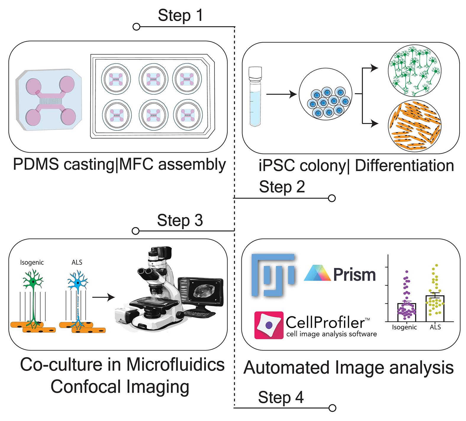
To address these limitations, we developed a compartmentalized human iPSC-derived motor neuron–myotube co-culture in microfluidic chambers, integrated with a semi-automated, compartment-resolved image analysis workflow. Similar human co-culture systems in microfluidic devices have been used to model ALS-relevant phenotypes and therapeutic responses, supporting the broader utility of this platform design [27]. In this system, motor neuron somata are confined to a proximal compartment, while chemotactic and volumetric gradients direct axons through microgrooves into a distal chamber containing myotubes [28,29]. Both motor neurons and myotubes were differentiated from the corresponding isogenic or ALS genotypes and cultured in a compartment-specific manner. Cultures are maintained for approximately 4–6 weeks, sufficient for co-culture maturation and emergence of ALS-relevant phenotypes, including neurofilament heavy chain (NFH)-associated axonal degeneration and pTDP-43 accumulation. A practical challenge in image-based analysis of neuromuscular co-cultures is that axons extend across or over myotubes, creating a heterogeneous background that interferes with signal detection and feature segmentation. Our workflow addresses this by employing channel-specific segmentation and spatial masking to restrict quantification to NFH-positive axonal regions. By integrating Fiji/ImageJ preprocessing with CellProfiler-based object identification, the pipeline effectively isolates axonal signal from underlying muscle background, enabling reproducible quantification of axonal phenotypes even in dense co-culture regions. This semi-automated approach reduces manual bias and supports blinded analysis across large imaging datasets.
We previously applied this microfluidic neuromuscular system and image analysis workflow to demonstrate that muscle-derived miR-126 regulates axonal TDP-43 condensates in ALS models [30]. As a proof-of-principle, modulation of miR-126 levels in ALS and isogenic co-cultures showed that the semi-automated pipeline can sensitively quantify resulting distal axonal degeneration and axonal pTDP-43 pathology. Furthermore, the workflow is highly modular. The segmentation approach uses thresholding, object identification, and spatial masking rather than a single predefined marker. This allows the pipeline to be adapted to quantify other aggregation-prone proteins, including SOD1, tau, and α-synuclein, by adjusting segmentation parameters. Overall, this platform provides a practical and versatile framework for investigating compartment-specific axonal pathology across a broad spectrum of neurodegenerative conditions.
Materials and reagents
Biological materials
Human iPSC lines used in this study included the KOLF2.1J parental/isogenic control line and a CRISPR/Cas9-engineered KOLF2.1J-derived TARDBP/TDP-43M337V homozygous mutant line (The Jackson Laboratory, catalog numbers: JIPSC001000 for KOLF2.1J and JIPSC001106 for TARDBP/TDP-43M337V SNV/SNV).
1. For the motor neuron differentiation system, stable iPSC lines (KOLF2.1J and TDP-43M337V) of doxycycline-inducible expression of human NGN2, ISL1, and LHX3 (hNIL) were generated using the PiggyBac Tet-On PB-TO-hNIL plasmid.
2. For myogenic differentiation, stable lines (KOLF2.1J and TDP-43M337V) of doxycycline-inducible expression of human MyoD1, together with Oct3/4 silencing, were generated using the PiggyBac Tet-On PB-TO-MYOD1-shOct4 plasmid.
3. PB-TO-hNIL (Addgene plasmid #172113) and PB-TO-MYOD1-shOct4 (Addgene plasmid #182309) were gifts from the iPSC Neurodegenerative Disease Initiative (iNDI) and Michael Ward.
Reagents
1. Accutase (Merck, catalog number: SCR005); store aliquots (5 mL) at -20 °C
2. Agrin (R&D, catalog number: 6624-AG-050); prepare as a 100 μg/mL solution by reconstituting 50 μg in 500 μL of filtered 1× PBS; store aliquots (10, 20, and 50 μL) at -80 °C
3. AR-grade Ethanol 70% (Biolab-chemicals, catalog number: 000522030500); store at room temperature
4. AR-grade 2-propanol (Biolab-chemicals, catalog number: 001626052100); store at room temperature
5. B27 (50×) (Gibco, catalog number: 17504044); store aliquots at -20 °C
6. B27 plus supplement (50×) (Gibco, catalog number: A3582801); store aliquots at -20 °C
7. BDNF (Alomone Labs, catalog number: B-250); prepare as a 10 μg/mL stock solution by dissolving 10 μg in 1 mL of nuclease-free water containing 2.5 μL of 4% BSA (final concentration: 0.01% w/v) dissolved in 1× PBS; store aliquots at -80 °C
8. Bovine serum albumin (BSA) (Sigma, catalog number: A3311); for immunofluorescence (IF) staining procedures, prepare as 50 mg/mL in filtered 1× PBS and store aliquots (500 μL) at -20 °C; use a final concentration of 1 mg/mL
9. Beta-mercaptoethanol, 55 mM (Gibco, catalog number: 21985023); store at 4 °C; Caution: Light-sensitive and irritant.
10. BrdU (5-Bromo-2′-deoxyuridine) (Sigma, catalog number: B9285); prepare as a 40 mM stock solution by dissolving 50 mg in 4.07 mL of nuclease-free water; store aliquots at -20 °C
11. CHIR99021 (Cayman, catalog number:13122-5); prepare as 2.5 mg/mL in 100% DMSO; store aliquots at -20 °C
12. Compound E (Stem Cell Technologies, catalog number: 73952); prepare as a 1 mM solution by dissolving 1 mg in 2.03 mL of DMSO; light-sensitive; store aliquots at -20 °C
13. Cryostor10 (Biolife, catalog number: 210102); store at 2–8 °C, protected from light; once opened, aliquot aseptically into 10 mL volumes in sterile 15 mL tubes and use before the product expiry date, provided sterility is maintained
14. CultureOne Supplement (Gibco, catalog number: A3320201); store aliquots at -20 °C
15. Dulbecco's phosphate-buffered saline (DPBS), 10×; no calcium, no magnesium; (Gibco, catalog number: 1420067); store at room temperature; Dilute with ultrapure water to prepare a 1× working solution
16. DMEM/F12, HEPES (Gibco, catalog number: 31330038); store at 4 °C
17. Dimethyl Sulfoxide (DMSO) (Sigma-Aldrich, catalog number: D5879); store at room temperature
18. Doxycycline hyclate (Sigma, catalog number: D9891); prepare as a 2 mg/mL solution by dissolving powder in nuclease-free water; light-sensitive; store aliquots at -20 °C
19. EDTA, 0.5 M, pH 8.0 (Gibco, catalog number:15575020); store at room temperature
20. ECM gel (Sigma, catalog number: E6909-5 ml); store aliquots at -20 °C; for coating, thaw one aliquot on ice or at 4 °C; dilute the 100 μL ECM by adding 900 μL of DMEM, achieving to a final volume of 1 mL; transfer this solution to a 15 mL conical tube and add an additional 9 mL of DMEM to obtain a final volume of 10 mL (1:100 dilution); ECM-coated culture surface is used for myogenic differentiation
21. GDNF (Alomone Labs, catalog number: G-240); prepare as a 10 μg/mL stock solution by dissolving 10 μg in 1 mL of nuclease-free water containing 2.5 μL of 4% BSA (final concentration: 0.01% w/v) dissolved in 1× PBS; store aliquots at -80 °C
22. Goat serum (Jackson ImmunoResearch, catalog number: 005-000-121); rehydration using 10 mL of double-distilled water yields 100% serum (60 mg/mL); for IF, use 5% (v/v) solution (1:20 dilution from rehydrated volume); store aliquots at -20 °C
23. GlutaMAX (Gibo, catalog number: 35050038); store at 4 °C
24. IGF-1 (Peprotech, catalog number: 100-11); prepare as 100 μg/mL by reconstituting 100 μg in 1 mL of nuclease-free water (+2.5 μL of 4% BSA); store aliquots at -80 °C
25. Insulin, human recombinant (Sigma, catalog number: 11376497001); prepare as 50 mg/mL by reconstituting in nuclease-free water; store aliquots at -20 °C
26. Laminin (Gibco, catalog number: 23017015); store aliquots at -80 °C; prepare a 15 μg/mL solution from 1 mg/mL stock solution for coating solution
27. L-ascorbic acid (Sigma, catalog number: A4403); prepare as 500 μg/mL in nuclease-free water; store aliquots at -20 °C
28. Matrigel (Corning, catalog number: 356234); store aliquots (100 μL) at -20 °C; for coating, thaw one aliquot on ice or at 4 °C; dilute the 100 μL Matrigel in cold DMEM (900 μL) to a final volume of 1 mL; transfer this solution to a 15 mL conical tube and add an additional 9 mL of DMEM to obtain a final volume of 10 mL (1:100 dilution); Matrigel-coated culture surfaces are used for routine iPSC colony culture and maintenance
29. Complete mTeSR1TM 1 medium (Stem cell technologies, catalog number: 85850); contains basal medium and 5× supplement; follow manufacturer’s instruction for preparing complete medium; store aliquots (40 mL) at -20 °C; before use, thaw overnight at 4 °C
30. N2 supplement (Gibco, catalog number: 17502048); store aliquots at -20 °C
31. NEAA (Biological Industries, catalog number: 01-340-1B); store aliquots at 4 °C
32. Neurobasal A (Gibco, catalog number:10888022); store at 4 °C
33. Neurobasal (Gibco, catalog number: 21103049)
34. NT-3, human recombinant (Alomone, catalog number: N-260); prepare as 5 μg/mL stock solution by dissolving 5 μg in 1 mL of nuclease-free water containing 2.5 μL of 4% BSA (0.1 mg/mL) dissolved in filtered 1× PBS; store aliquots at -20 °C
35. Paraformaldehyde aqueous (PFA) solution, EM grade (Electron Microscopy Sciences, catalog number: BN15714); inside fume hood, open a 32% ampule (10 mL) of PFA, divide into 0.5 mL aliquots, and store at -20 °C; to prepare the required volume of 4% PFA, thaw an aliquot at room temperature inside the fume hood and dilute 32% with 1× PBS
36. Penicillin/streptomycin (Pen-Strep) (10,000 U/mL) (Gibco, catalog number: 15140-122); store aliquots (5 mL) at -20 °C; thaw aliquots at 4 °C overnight, store at 4 °C, and use for media preparation within 1–2 weeks; 1% (v/v) is generally recommended
37. Poly-DL-ornithine hydrobromide (Sigma, catalog number: P8638); prepare as 1.5 mg/mL stock solution by dissolving 25 mg of powder in 16.67 mL of 1× PBS; store aliquots at -20 °C
38. Puromycin dihydrochloride (Sigma, catalog number: P8833); prepare as 10 mg/mL in nuclease-free water; store aliquots at -80 °C for up to 1 year
39. ProLong Gold Antifade Mountant ± DAPI [Molecular Probes, catalog numbers: P36934 (-DAPI), P36935 (+DAPI)]
40. Sodium pyruvate (Gibco, catalog number:11360070); store at 4 °C
41. Sonic Hedgehog (Peprotech, catalog number:100-45); prepare as 50 μg/mL in nuclease-free water; store aliquots at -80 °C
42. TritonTM X-100 (Sigma-Aldrich, catalog number: T8787-250)
43. Sylgard 184 Silicone Elastomer kit (Dow, catalog number: 4019862)
44. Y-27632 dihydrochloride (ROCK1/2 inhibitor) (Cayman, catalog number: 10005583); prepare as a 10 mM stock solution by dissolving 5 mg in 1.561 mL of nuclease-free water; store aliquots at -80 °C; ROCK1/2 inhibitor is added only to the plating medium on the day of cell seeding; it is omitted from all subsequent medium changes unless otherwise stated
45. Mouse anti-Titin (DSHB, catalog number: 9d10); store aliquots at -20 °C
46. Chicken anti-neurofilament heavy chain NFH (Abcam, catalog number: ab72996); store aliquots at -20 °C
47. Rabbit anti-pTDP43 (Proteintech, catalog number: 22309-1-AP); store aliquots at -20 °C
48. DAPI (4′,6-Diamidino-2-phenylindole dihydrochloride) (Sigma, catalog number: D8417); prepare as 1 mg/mL stock solution with nuclease-free water; store aliquots at -20 °C
49. Goat anti-rabbit 405 (Abcam, catalog number: ab175654); store aliquots at -20 °C
50. Goat anti-chicken 488 (Abcam, catalog number: ab150173); store aliquots at -20 °C
51. Goat anti-rabbit 647 (Abcam, catalog number: ab150083); store aliquots at -20 °C
52. Goat anti-rabbit 594 (Jackson ImmunoResearch, catalog number: 111-585-144); store aliquots at -20 °C
Solutions
1. Blocking solution (see Recipes)
2. DMEM/F12 + P/S (see Recipes)
3. mTeSR1, iPSC culture medium (see Recipes)
4. Myotube induction medium (IM-Myo) (see Recipes)
5. Myotube maturation medium (MM-Myo) (see Recipes)
6. Neuronal induction medium (IM-MN) (see Recipes)
7. Neuronal maturation medium (MM-MN) (see Recipes)
8. Polydimethylsiloxane (PDMS) mixture (see Recipes)
9. Triton permeabilization solution (see Recipes)
Recipes
1. Blocking solution
| Reagent | Final concentration | Quantity or volume |
|---|---|---|
| BSA (50 mg/mL) | 1 mg/mL | 20 μL |
| Normal goat serum, 6% (w/v), stock | 5% (v/v) | 50 μL |
| 1× PBS | 1× | 930 μL |
| Total | 1 mL |
For IF staining procedures.
2. DMEM/F12+ P/S 1%
| Reagent | Final concentration | Quantity or volume |
|---|---|---|
| DMEM/F12 | 1× | 49.5 mL |
| Pen-Strep | 1% (v/v) | 500 μL |
| Total | 50 mL |
Prepare this exclusively to flush PDMS debris in microfluidic chamber (MFC) assembly and store at 4 °C. Do not use this for cell culture purposes.
3. mTeSR1TM 1 + P/S 1%
| Reagent | Final concentration | Quantity or volume |
|---|---|---|
| mTeSRTM 1 | 1× | 40 mL |
| Pen-Strep | 1% (v/v) | 500 μL |
| Total | ~40 mL |
4. Induction medium for myotube (IM-Myo), days 1–6, distal compartment of co-culture
| Reagent | Final concentration | Quantity or volume | |
| Basal | DMEM/F12 with HEPES | 1× | 49 mL |
| Sodium pyruvate, 100× | 1× | 0.5 mL | |
| NEAA, 100× | 1× | 0.5 mL | |
1. Prepare basal medium, filter, and store at 4 °C for a maximum of 1 week. | |||
| Factor cocktail | aβ-mercaptoethanol, 55 mM | 110 μM | 100 μL |
| Insulin, 10 mg/mL | 10 μg/mL | 50 μL | |
| Doxycycline, 1,000× | 1× | 50 μL | |
| bROCK inhibitor (RI), Y-27632, 1,000× | 1× | 50 μL | |
| Total | 50 mL | ||
aAdd freshly before use.
bAdd Rock inhibitor for plating on day 0 and day 3.
5. Induction medium for myotube (IM-Myo), days 7–9, distal compartment of co-culture
Add 10 μL of CHIR99021 (15 mM stock) to 50 mL of IM-Myo medium (without ROCK inhibitor) to achieve a final concentration of 3 μM.
6. Maturation medium for myotubes (MM-Myo), distal compartment of co-culture
| Reagent | Final concentration | Quantity or volume | |
| Basal | Neurobasal A | 1× | 48 mL |
| B27 plus supplement, 100× | 1× | 1 mL | |
| NEAA, 100× | 1× | 0.5 mL | |
| Gluta-MAX, 100× | 1× | 0.5 mL | |
1. Prepare basal medium, filter, and store at 4 °C for a maximum of 1 week. | |||
| Factor cocktail | aCultureOne supplement, 100× | 1× | 500 μL |
| NT-3 (5 μg/mL) | 20 ng/mL | 200 μL | |
| Doxycycline, 1,000× | 1× | 50 μL | |
| aSonic Hedgehog, Shh (50 μg/mL) | 50 ng/mL | 50 μL | |
| Agrin (100 μg/mL) | 100 ng/mL | 50 μL | |
| L-ascorbic acid, 200 mM | 200 μM | 50 μL | |
| IGF-1 (100 μg/mL) | 10 ng/mL | 5 μL | |
| Laminin (50 μg/mL) | 50 ng/mL | 50 μL | |
| Total | 50 mL | ||
aRemove CultureOne and Shh from basal medium after 2 weeks.
7. Induction medium for motor neuron (IM-MN); proximal compartment of co-culture
| Reagent | Final concentration | Quantity or volume | |
| Basal | DMEM/F12 with HEPES | 1× | 48.5 mL |
| N2 supplement, 100× | 1× | 0.5 mL | |
| NEAA, 100× | 1× | 0.5 mL | |
| Gluta-MAX, 100× | 1× | 0.5 mL | |
1. Prepare basal medium, filter, and store at 4 °C for a maximum of 1 week. | |||
| Factor cocktail | aROCK inhibitor, Y-27632, 1,000× | 1× | 50 μL |
| Doxycycline, 1,000× | 1× | 50 μL | |
| Compound E, 1mM | 0.2 μM | 10 μL | |
| bBrdU, 40 mM | 40 μM | 50 μL | |
| Total | 50 mL | ||
aAdd Rock inhibitor for plating on DIV 0–3.
bAdditional components only for plating on DIV 3.
8. Maturation medium for motor neuron (MM-MN); proximal compartment of co-culture
| Reagent | Final concentration | Quantity or volume | |
| Basal | Neurobasal | 1× | 47.5 mL |
| B27 supplement, 50× | 1× | 1 mL | |
| N2 supplement, 100× | 1× | 0.5 mL | |
| NEAA, 100× | 1× | 0.5 mL | |
| Gluta-MAX, 100× | 1× | 0.5 mL | |
1. Prepare basal medium, filter, and store at 4 °C for a maximum of 1 week. | |||
| Factor cocktail | Laminin (1mg/mL) | 1 μg/mL | 50 μL |
| CultureOne supplement, 100× | 1× | 500 μL | |
| BDNF (10 μg/mL) | 10 ng/mL | 50 μL | |
| GDNF (10 μg/mL) | 10 ng/mL | 50 μL | |
| NT-3 (5 μg/mL) | 10 ng/mL | 100 μL | |
| Total | 50 mL | ||
9. PDMS mixture (9:1 mix)
| Reagent | Final concentration | Quantity or volume |
|---|---|---|
| PDMS base (part A) | n/a | 45 g |
| PDMS curing reagent (part B) | n/a | 5 g |
| Total | 50 g |
Note: SylgardTM 184 Silicone Elastomer kit contains two highly viscous PDMS components. When dispensing the PDMS into a 50 mL polypropylene tube for weighing, the material may flow slowly; proceed patiently. Creating a small hole in the bottle cap can help improve control and facilitate dispensing.
10. Triton permeabilization solution (0.1%)
| Reagent | Final concentration | Quantity or volume |
|---|---|---|
| BSA, (50 mg/mL) | 1 mg/mL | 20 μL |
| Normal goat serum, 6% (w/v), stock | 5% (v/v) | 50 μL |
| 1× PBS | 1× | 930 μL |
| Triton (20%) | 0.1% | 5 μL |
| Total | 1 mL |
Laboratory supplies
1. P2, P20, P200, and P1000 micropipettes (Gilson)
2. Pipette-holder (O-pette, ORNAT)
3. Stripette 5, 10, and 25 mL serological pipettes, polystyrene, individually wrapped (Corning, catalog numbers: 4487, 4101, and 4489, respectively)
4. Sterile polystyrene 6-well, 24-well plates (Corning, catalog numbers: 3516, 3526)
5. Sterile 1.5-mL polypropylene microcentrifuge tubes (Axygen, catalog number: MCT-175-C)
6. Sterile glass coverslips, thickness 1.0 (0.13–0.16), 22 × 22 mm (Marienfeld, catalog number: 0101050)
7. Sterile, FluoroDish, glass bottom, clear wall, individually wrapped (WPI, catalog number: FD35-100)
8. Sterile surgical forceps
9. Pasteur pipette 15 cm glass (Beith Dekel, catalog number: 9411015)
10. 100 μL to 1,250 mL filter pipette tips (Axygen, catalog number: TF-1000-R-S)
11. 20–200 μL filter pipette tips (SRS-17370-X)
12. 2–20 μL filter pipette tips (SRS-30370T)
13. 1–10 μL filter pipette tips (SRS-30340)
14. Syringe driven filters, disposable, 0.22 μm, cellulose acetate (Cytiva, catalog number:10462200)
15. Vacuum bottle filter, 250 mL, 0.22 μm, PES (Corning, catalog number: 431097)
16. Vacuum bottle filter, 500 mL, 0.22 μm, PES (SORFA, catalog number: SPE-22-500)
17. Centrifuge for 15-mL and 50-mL conical tubes (Corning, catalog numbers: 430052 and 430290)
18. Cryotube vials (Thermo Scientific, catalog number: NU-368632)
19. Mr. FrostyTM freezing container (Thermo Scientific, catalog number: 5100-0001)
20. ibidi Immersion Oil 2 (ibidi, catalog number: 50102)
21. Lint-free tissue for optical lenses
Equipment
Cell culture
1. -20 °C and -80 °C freezers
2. 5% CO2, 95% humidity cell culture incubator (Thermo Scientific, model: Heracell 150i)
3. Hemocytometer Neubauer Improved, depth 0.1 mm 0.0025 mm2, glass (Bar Naor, catalog number: BN442-2)
4. Class II, Type A2 biological safety cabinet (Thermo Scientific, model: 1300 Series)
5. Vacuum aspirator and aspirating pipettes
6. Water bath set at 37 °C (Lab Companion, model: BW-10H)
7. High-speed centrifuge (iCen-24)
MFC preparation
8. Vertical rotating mixer
9. HB-500 Minidizer Oven (Biolab, catalog number: 85996695033002)
10. Vacuum desiccator (Nalgene, catalog number: D2797-1EA)
11. Biopsy punch 6-mm (WPI, catalog number: 504533)
12. Intelli-mixer rotator (ELMI, catalog number: RM-2L)
13. Plasmatic Systems, Inc. Plasma Preen II #973
Imaging systems
14. EVOS FL imaging system (Invitrogen, catalog number: AMF300)
15. FLoid Cell Imaging Station (Invitrogen, catalog number: 4471136)
16. Olympus Benchtop CKX31 inverted microscope equipped with phase-contrast 10× objective (Olympus)
17. Andor BC-43 benchtop spinning disk confocal system controlled by Andor Fusion software version 2.3a; equipped with on-stage humidified incubation chamber maintaining 37 °C, 5% CO2
Software and datasets
1. Fiji (ImageJ v1.54p), released February 2025; used for image visualization and preprocessing [31]
2. CellProfiler (v4.2.8); used for segmentation and quantitative image analysis. Both previous versions (v4.2.6 or v4.2.8) were tested and found to be functionally equivalent and fully compatible with the analysis pipeline. The most recent release (v4.2.8) was issued in September 2024
3. CellProfiler [32] is a free, open-source software for cell image analysis and supports the development of custom image-processing modules.
4. All analysis code has been deposited on GitHub: https://github.com/PerlsonLab/Bioprotocol
Procedure
登录/注册后免费查看全文
文章信息
稿件历史记录
提交日期: Mar 22, 2026
接收日期: Jul 22, 2026
在线发布日期: Aug 6, 2026
出版日期: Sep 5, 2026
版权信息
© 2026 The Author(s); This is an open access article under the CC BY-NC license (https://creativecommons.org/licenses/by-nc/4.0/).
如何引用
Subramaniam, A. G., de Andrade Gensas, L. K., Gradus-Pery, T. and Perlson, E. (2026). Quantitative Analysis of Axonal Degeneration and TDP-43 Aggregation in Compartmentalized Human iPSC-Derived Motor Neuron–Myotube Co-cultures. Bio-protocol 16(17): e5801. DOI: 10.21769/BioProtoc.5801.

 Download Citation in RIS Format
分类
神经科学 > 神经系统疾病 > 神经退行性病变
细胞生物学 > 细胞分离和培养 > 微流体细胞培养
干细胞 > 多能干细胞 > 基于细胞的分析方法
您对这篇实验方案有问题吗?
在此处发布您的问题,我们将邀请本文作者来回答。同时,我们会将您的问题发布到Bio-protocol Exchange,以便寻求社区成员的帮助。
 发布问题
发布问题


