


An Automated, Ventana Discovery Platform-based Imaging Workflow for Simultaneous Quantification of B Cells, Plasma Cells, and Plasmablasts in FFPE Human Tissues
基于 Ventana Discovery 平台的自动化成像流程:同步定量 FFPE 人组织中的 B 细胞、浆细胞和浆母细胞

发布: 2026年09月05日第16卷第17期 DOI: 10.21769/BioProtoc.5797 浏览次数: 310
评审: Alessandro DidonnaSunjay J FernandesAnonymous reviewer(s)
 PDF
PDF  Q&A
Q&A
参见作者原研究论文
The authors used this protocol in:

Mar 2026





Abstract
Accurate, sensitive quantification of B-lineage cells is critical for pharmacodynamic evaluation of B cell–targeted therapies in lupus nephritis (LN) clinical trials. While high-dimensional discovery platforms offer broad profiling, they often lack the sensitivity, quantitative rigor, and throughput needed for precise cell enumeration in renal trial needle biopsies. Traditional immunostaining is hampered by CD20-directed therapeutic masking or downregulation, inadequate sensitivity of CD19 in FFPE tissue, and confounding renal tubular CD138 expression. This protocol details an automated, fit-for-purpose, 5-plex sequential tyramide signal amplification (TSA)-based immunofluorescence assay (CD38, CD79a, CD19, Ki-67, CD138) developed on the Ventana Discovery Ultra platform for deployment on single tissue sections. The workflow anchors B-cell detection on CD79a to ensure sensitivity and utilizes CD38 as an obligate co-marker for CD138+ antibody-secreting cells (ASCs) to definitively exclude the CD138+ epithelial background. Following acquisition via fluorescence whole-slide imaging, a digital analysis pipeline utilizing InstanSeg-based automated segmentation rigorously classifies cell phenotypes to generate precise spatial densities (cells/mm2). This validated protocol maximizes data yield from scarce clinical biopsies while providing high-precision quantitative monitoring of longitudinal therapeutic depletion in the renal microenvironment.
Key features
• Automated trial scalability: Details an automated, TSA-based 5-plex assay optimized for the Ventana Discovery Ultra platform, ensuring high-throughput, reproducible B-lineage quantification in multicenter cohorts.
• Robust lineage identification: Establishes CD79a as an anchor for sensitive B-cell detection in FFPE tissue, avoiding CD20 therapeutic masking/downregulation and CD19 epitope instability.
• High-specificity ASC enumeration: Resolves confounding CD138+ renal tubular background using an obligate CD38/CD138 co-labeling strategy to definitively identify ASCs within the renal microenvironment.
• Single-section phenotypic resolution: Combines multiplexing and InstanSeg segmentation on one 4-μm section to simultaneously quantify B-lineage subsets in tissue-limited renal needle biopsies.
Keywords: Lupus nephritis (狼疮性肾炎) B cells (B 细胞)
Plasma cells (浆细胞)
Plasmablasts (浆母细胞)
Antibody-secreting cells (ASCs) (抗体分泌细胞(ASC))
Quantitative-multiplex immunofluorescence (定量多重免疫荧光)
FFPE tissue (FFPE 组织)
Spatial density (空间密度)
B cells (B 细胞)
Plasma cells (浆细胞)
Plasmablasts (浆母细胞)
Antibody-secreting cells (ASCs) (抗体分泌细胞(ASC))
Quantitative-multiplex immunofluorescence (定量多重免疫荧光)
FFPE tissue (FFPE 组织)
Spatial density (空间密度)
Graphical overview
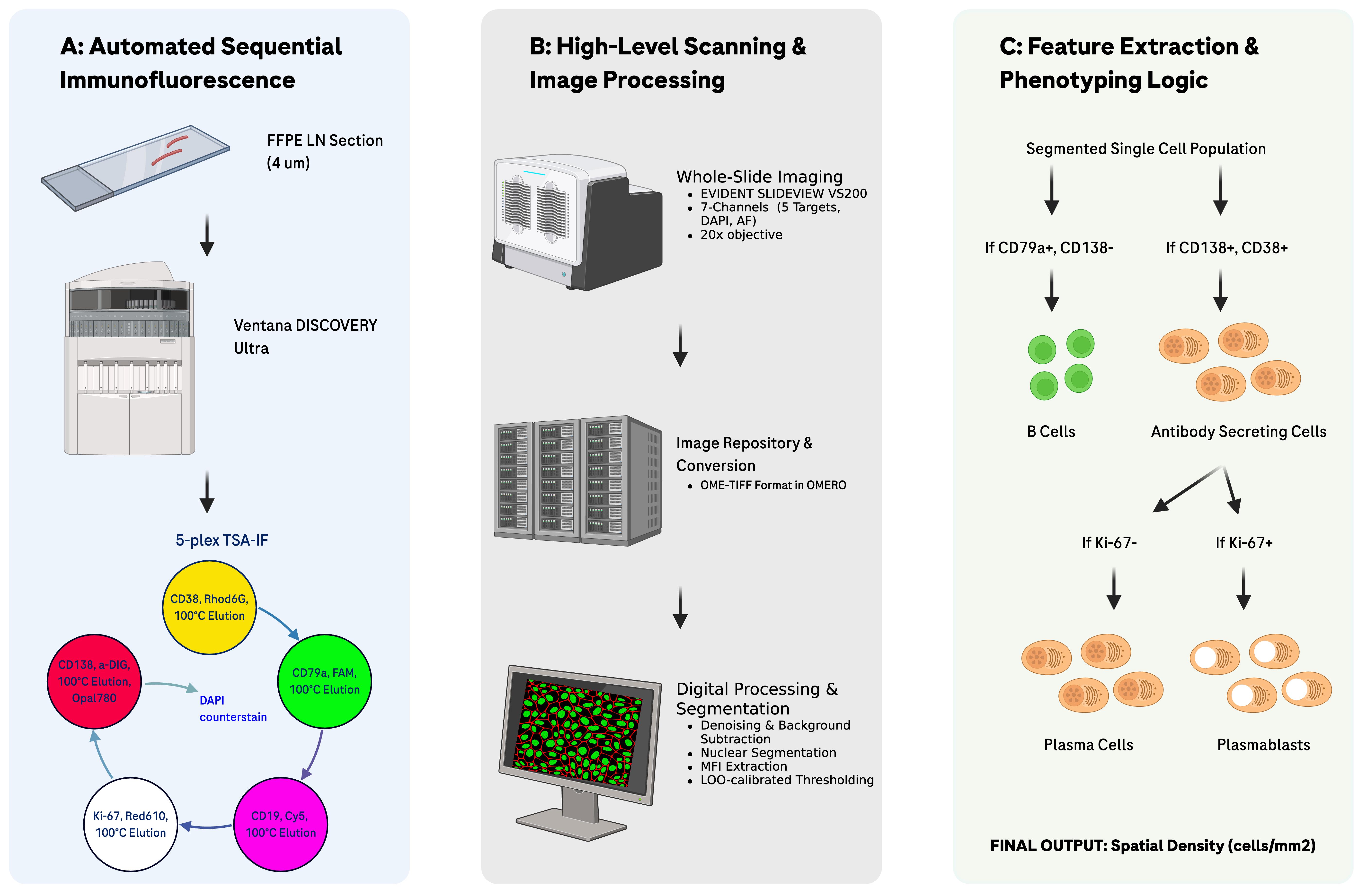
Automated multiplex immunofluorescence and digital phenotyping workflow. (A) Formalin-fixed, paraffin-embedded (FFPE) tissue sections (4 μm) undergo automated 5-plex tyramide signal amplification immunofluorescence (CD38, CD79a, CD19, Ki-67, CD138, plus DAPI) on the Ventana DISCOVERY Ultra. (B) Slides are digitized via 7-channel whole-slide imaging (20×) on the EVIDENT SLIDEVIEW VS200 and imported into OMERO. Digital processing includes median blur filtering, background subtraction, InstanSeg cell segmentation, and single-cell MFI calculation. (C) A decision logic tree classifies segmented single cells into B cells (CD79a+/CD138-) and antibody-secreting cells (ASCs; CD138+/CD38+). ASCs are further stratified into plasma cells (Ki-67-) and plasmablasts (Ki-67+) to yield final spatial densities (cells/mm2).
Background
Accurate spatial enumeration of B-lineage cells within the renal microenvironment is essential for understanding the pathogenesis of lupus nephritis (LN), where B cells and antibody-secreting cells (ASCs), which include plasma cells (PCs) and plasmablasts (PBs), mediate kidney injury [1]. Monitoring these populations in formalin-fixed, paraffin-embedded (FFPE) kidney tissue is critical for evaluating the pharmacodynamic efficacy of B cell–targeted therapies in clinical trials. Traditional immunohistochemical (IHC) approaches often rely on single markers such as CD20 for B cells and CD138 for plasma cells. However, in the context of CD20-directed depletion therapies, CD20 is unreliable for quantifying total B-lineage burden because it can be masked or internalized following therapeutic depletion [2,3]. Additionally, while CD19 is a canonical B-cell marker often used in flow cytometry studies [4], its detection in FFPE tissue lacks the sensitivity required for rigorous quantitative analysis due to poor epitope preservation. CD138 (Syndecan-1), often used as a standalone marker for ASC identification, is unreliable in renal tissue due to substantial endogenous CD138 expression on renal tubular epithelium, creating a significant risk of false-positive ASC identification [5,6]. While high-dimensional spatial platforms offer broad inflammatory cell profiling, they often lack the throughput and quantitative rigor required for precise cell enumeration in large multicenter clinical cohorts. Furthermore, reliance on serial sections for multiple markers is often unfeasible for scarce LN needle biopsy material.
This multiplex protocol (see Graphical overview) provides an automated, sequential tyramide signal amplification (TSA) workflow [7] (CD38, CD79a, CD19, Ki-67, CD138) developed on the Ventana Discovery Ultra platform. Key advantages include establishing CD79a as a requisite anchor, which remains detectable, even when CD20 is lost or masked [8,9], providing far greater sensitivity than CD19 in FFPE sections [10,11]. To ensure specific renal ASC identification, the protocol employs an obligate CD38/CD138 co-labeling strategy to definitively distinguish them from the confounding renal tubular epithelium. Finally, the workflow allows for the efficient use of tissue by combining all markers and InstanSeg-based digital segmentation [12] on a single 4-μm section, maximizing data yield from material-constrained clinical trial samples.
A limitation of this methodology is the requirement for specific automated staining hardware and digital analysis expertise. While validated specifically for LN, this robust, fit-for-purpose workflow is adaptable for any FFPE tissue where high-precision B-lineage enumeration is required. Furthermore, the method is agile and amenable to swapping in or out these markers with other markers of interest.
Materials and reagents
Biological materials
1. FFPE human tissue (validation samples were de-identified clinical remnants acquired from Avaden Biosciences; however, the protocol is broadly applicable to clinical trials, clinical remnants, or other commercial specimens)
a. Target tissue: Designed for lupus nephritis (LN) needle biopsies, but adaptable to other tissue types; if feasible, standardization of fixation (e.g., 16–24 h in 10% neutral buffered formalin at ambient temperature), automated processing, and embedding in histology-grade paraffin is recommended
b. Control tissue: Secondary lymphoid tissue with germinal centers (e.g., palatine tonsil) to validate B-cell maturation spectrum, including B cell (CD19, CD79a), ASC (CD38, CD138), and proliferation Ki-67 markers; tonsillar epithelium provides a reference for the epithelial CD138 expression
Reagents
Note: All reagents should be stored at 4 °C unless otherwise noted.
1. Discovery CC1 (predilute) (Roche Diagnostics, catalog number: 950-500); store at room temperature (RT)
2. DISCOVERY inhibitor (Roche Diagnostics, catalog number: 760-4840)
3. CD38 (clone: SPC32) antibody (Leica Biosystems, catalog number: NCL-L-CD38-290)
4. Bovine serum albumin (BSA) (Fisher Scientific, SKU #501657297)
5. Phosphate-buffered saline (PBS) 1×, sterile-filtered (Life Technologies, SKU #J61196.AP); store at RT
6. Goat Ig block (Roche Diagnostics, catalog number: 760-6008)
7. OmniMap anti-mouse HRP (Roche Diagnostics, catalog number: 760-4310)
8. Discovery Rhodamine 6G (Roche Diagnostics, catalog number: 760-244)
9. Ultra CC2 (Roche Diagnostics, catalog number: 950-223); store at RT
10. CD79a (clone: SP18) antibody (Roche Diagnostics, catalog number: 790-4432)
11. OmniMap anti-rabbit HRP (Roche Diagnostics, catalog number: 760-4311)
12. Discovery FAM (Roche Diagnostics, catalog number: 760-243)
13. CD19 (clone: LE-CD19) antibody (Agilent, catalog number: M7296)
14. Discovery Cy5 (Roche Diagnostics, catalog number: 760-238)
15. Ki-67 (clone: D3B5) antibody (Cell Signaling, catalog number: 12202S); store at -20 °C
16. Discovery Red610 (Roche Diagnostics, catalog number: 760-245)
17. CD138 (clone: B-A38) antibody (Roche Diagnostics, catalog number: 760-4248)
18. Opal 780 Reagent Pack (Akoya Biosciences, catalog number: FP1501001KT); store at -20 °C in powder form
19. DAPI nucleic acid stains (ThermoFisher, catalog number: D1306); store at -20 °C
20. 1× Plus automation amplification diluent (Perkin Elmer, catalog number: FP1609)
21. Antibody diluent/block (Perkin Elmer, catalog number: ARD1001EA)
22. Dawn ultra liquid dish detergent (Procter & Gamble, catalog number: 24386798)
23. ProLong Gold antifade reagent (Invitrogen, catalog number: P36930)
Solutions
1. 3% BSA (see Recipes)
2. CD38 antibody solution (see Recipes)
3. CD19 antibody solution (see Recipes)
4. Ki-67 antibody solution (see Recipes)
5. TSA-DIG stock solution (see Recipes)
6. TSA-DIG working solution (see Recipes)
7. Anti-DIG Opal 780 stock solution (see Recipes)
8. Anti-DIG Opal 780 working solution (see Recipes)
9. DAPI solution (see Recipes)
10. Slide cleaning solution (see Recipes)
Recipes
Notes:
1. Recipes for working antibody solutions and DAPI are calculated for a 10-slide batch. This volume specifically accounts for the 1-mL dead volume required by Ventana reagent dispensers to prevent bubble introduction.
2. All solutions should be stored at 4 °C, except the slide cleaning solution.
1. 3% BSA
| Reagent | Final concentration | Quantity or volume |
|---|---|---|
| BSA | 3% | 30 g |
| PBS | 1× | 1 L |
| Total | n/a | 1 L |
Filter-sterilize (0.2 μm). Stable at 4 °C for up to 1 year.
2. CD38 antibody solution
| Reagent | Final concentration | Quantity or volume |
|---|---|---|
| CD38 stock solution | 46 μg/mL | 20 μL |
| 3% BSA (Recipe 1) | n/a | 1,980 μL |
| Total | 0.46 μg/mL | 2 mL |
Stable at 4 °C for up to 2 weeks.
3. CD19 antibody solution
| Reagent | Final concentration | Quantity or volume |
|---|---|---|
| CD19 stock solution | 83 μg/mL | 20 μL |
| 3% BSA (Recipe 1) | n/a | 1,980 μL |
| Total | 0.83 μg/mL | 2 mL |
Stable at 4 °C for up to 2 weeks.
4. Ki-67 antibody solution
| Reagent | Final concentration | Quantity or volume |
|---|---|---|
| Ki-67 stock solution | 270 μg/mL | 2.22 μL |
| 3% BSA (Recipe 1) | n/a | 1997.78 μL |
| Total | 0.3 μg/mL | 2 mL |
Stable at 4 °C for up to 2 weeks.
5. TSA-DIG stock solution
| Reagent | Final concentration | Quantity or volume |
|---|---|---|
| Opal TSA-DIG | n/a | 1 vial |
| Dimethyl sulfoxide (DMSO) | n/a | 75 μL |
| Total | n/a | 75 μL |
Opal TSA-DIG and DMSO are part of the Opal 780 Reagent Pack. Stable at 4 °C for up to 1 month.
6. TSA-DIG working solution
| Reagent | Final concentration | Quantity or volume |
|---|---|---|
| Opal TSA-DIG stock solution (Recipe 5) | n/a | 20 μL |
| 1× Plus automation amplification diluent | n/a | 1,980 μL |
| Total | 1:100 | 2 mL |
Store at 4 °C; it should be used within 3 weeks of preparation for the best outcome.
7. Anti-DIG Opal 780 stock solution
| Reagent | Final concentration | Quantity or volume |
|---|---|---|
| Anti-DIG Opal 780 | n/a | 1 vial |
| Deionized H2O | n/a | 300 μL |
| Total | n/a | 300 μL |
Anti-DIG Opal 780 is part of the Opal 780 Reagent Pack. Stable at 4 °C for up to 1 month.
8. Anti-DIG Opal 780 working solution
| Reagent | Final concentration | Quantity or volume |
|---|---|---|
| Anti-DIG Opal 780 stock solution (Recipe 7) | n/a | 80 μL |
| Antibody Diluent/Block | n/a | 1920 μL |
| Total | 1:25 | 2 mL |
Prepare in an ASSY, LP PREP KIT dispenser. Store at 4 °C; it should be used within 3 weeks of preparation for the best outcome.
9. DAPI solution
| Reagent | Final concentration | Quantity or volume |
|---|---|---|
| DAPI stock solution | 5 mg/mL | 8 μL |
| Deionized H2O | n/a | 1 mL |
| PBS | 1× | 199 mL |
| Total | 0.2 μg/mL | 200 mL |
Prepare in an ASSY, LP PREP KIT dispenser. Stable at 4 °C for up to 1 month.
10. Slide cleaning solution
| Reagent | Final concentration | Quantity or volume |
|---|---|---|
| Dawn Ultra liquid dish soap | n/a | 50 mL |
| Deionized H2O | n/a | 150 mL |
| Total | 1:4 | 200 mL |
Store at RT.
Laboratory supplies
1. Superfrost Plus glass slides (Thermo Fisher Scientific, Waltham, MA, USA, catalog number 4951PLUS-001 or equivalent)
Critical: Use of high-adhesion slides is critical to prevent tissue detachment during sequential heat-induced epitope retrieval cycles.
2. DISCOVERY, REAGENT DISP. CARD, Detection 1 (Roche Diagnostics, catalog number: 05280192001)
3. DISCOVERY, REAGENT DISP. CARD, Detection 2 (Roche Diagnostics, catalog number: 05280206001)
4. NEXES REAGENT DISP. CARD, CNTRSTAIN 1 (Roche Diagnostics, catalog number: 05271720001)
5. ASSY, LP PREP KIT (Roche Diagnostics, catalog number: 7475144001); brown dispenser to prevent light damage to the solution
6. Nexes Primary Antibody 1 Dispenser (Roche Diagnostics, catalog number: 05269857001)
7. Nexes Primary Antibody 2 Dispenser (Roche Diagnostics, catalog number: 05269865001)
8. Nexes Primary Antibody 3 Dispenser (Roche Diagnostics, catalog number: 05269873001)
9. 24 × 50 mm No. 1 SuperSlip coverslip (VWR, catalog number: 48404-453)
10. Acrodisc PF syringe filter with 0.8/0.2 μm Supor membrane non-pyrogenic, sterile (Pall Life Sciences, catalog number: 4658)
Equipment
1. Desert Chamber Oven (Biocare Medical, model: DRY2008US)
2. Ventana DISCOVERY ULTRA (Roche Diagnostics, catalog number: 05987750001)
3. SLIDEVIEW VS200 Universal Whole Slide Imaging Scanner (EVIDENT, model: VS200), including the following filter cubes:
a. DAPI LED Filter Set 378/52X/BS409/447/60M zeropixel w/ BX3 cube (EVIDENT, model: OSF-DAPIZLEDBX3)
b. CFP Zero LED Filter (EVIDENT, model: OSF-CFPZLEDBX3)
c. FITC LED Filter Set 474/27X/BS495/525/45M zeropixel w/ BX3 cube (EVIDENT, model: OSF-FITCZLEDBX3)
d. ET-Gold/Yellow Filter Set 546/10×, BS556, 572/23M w/ BX3 cube (EVIDENT, model: OCT-49304BX3)
e. ET-Red#1 FISH Filter Set 580/25×, BS600, 625/30M w/ BX3 cube (EVIDENT, model: OCT-49306BX3)
f. ET-CY5 Filter Set 640/30×, BS660, 690/50M w/ BX3 cube (EVIDENT, model: OCT-49009BX3)
g. Cy7 LED Filter Set 735/28×/BS757/809/81M zeropixel w/ BX3 cube (EVIDENT, model: OSF-CY7ZLEDBX3)
Software and datasets
Staining and scanning
1. Ventana NexES, VSS (Roche Diagnostics, version 12.5.4; included with the instrument)
2. VS200 ASW (EVIDENT, version 4.3; included with the instrument)
Data analysis
The digital analysis pipeline described herein (including image processing, segmenting whole-slide images, and single-cell feature extraction) was successfully performed on a high-performance computing node (HPC) with 128 GB RAM, an Intel Xeon Gold 6248 CPU, and an NVIDIA V100 GPU with 32 GB VRAM to efficiently run InstanSeg and handle large OME-TIFF files locally.
1. NGFF-Converter (Glencoe Software, version 2.0.2; freely available at https://www.glencoesoftware.com/products/ngff-converter/)
2. OMERO (Glencoe Software, version OMERO.web 5.29.2; freely available at https://pypi.org/project/omero-web/)
3. InstanSeg (v0.1.0; freely available at https://github.com/instanseg/instanseg)
4. Custom Python analysis scripts (Threshold Explorer and tissue area calculations). Scripts were executed in Python 3.12. The specific repository version 0.1.0, source code, and full environment dependencies (listed in pyproject.toml) are freely available at https://github.com/Genentech/sp_pipeline_public. Please refer to the repository’s README file for the installation of dependencies.
Procedure
登录/注册后免费查看全文
文章信息
稿件历史记录
提交日期: May 29, 2026
接收日期: Jul 20, 2026
在线发布日期: Aug 12, 2026
出版日期: Sep 5, 2026
版权信息
© 2026 The Author(s); This is an open access article under the CC BY-NC license (https://creativecommons.org/licenses/by-nc/4.0/).
如何引用
Chang, P. S., Chan, C., Jesudason, R., Rost, S. and Austin, C. D. (2026). An Automated, Ventana Discovery Platform-based Imaging Workflow for Simultaneous Quantification of B Cells, Plasma Cells, and Plasmablasts in FFPE Human Tissues. Bio-protocol 16(17): e5797. DOI: 10.21769/BioProtoc.5797.

 Download Citation in RIS Format
分类
免疫学 > 免疫细胞染色 > 免疫检测
细胞生物学 > 组织分析 > 组织染色
生物信息学与计算生物学
您对这篇实验方案有问题吗?
在此处发布您的问题,我们将邀请本文作者来回答。同时,我们会将您的问题发布到Bio-protocol Exchange,以便寻求社区成员的帮助。
 发布问题
发布问题


