


Analysis of Bacterial-Mediated c-di-AMP Degradation by Thin-Layer Chromatography
利用薄层色谱分析细菌介导的 c-di-AMP 降解

发布: 2026年08月20日第16卷第16期 DOI: 10.21769/BioProtoc.5796 浏览次数: 429
评审: Qing TangAnonymous reviewer(s)
 PDF
PDF  Q&A
Q&A
参见作者原研究论文
The authors used this protocol in:
May 2026





Abstract
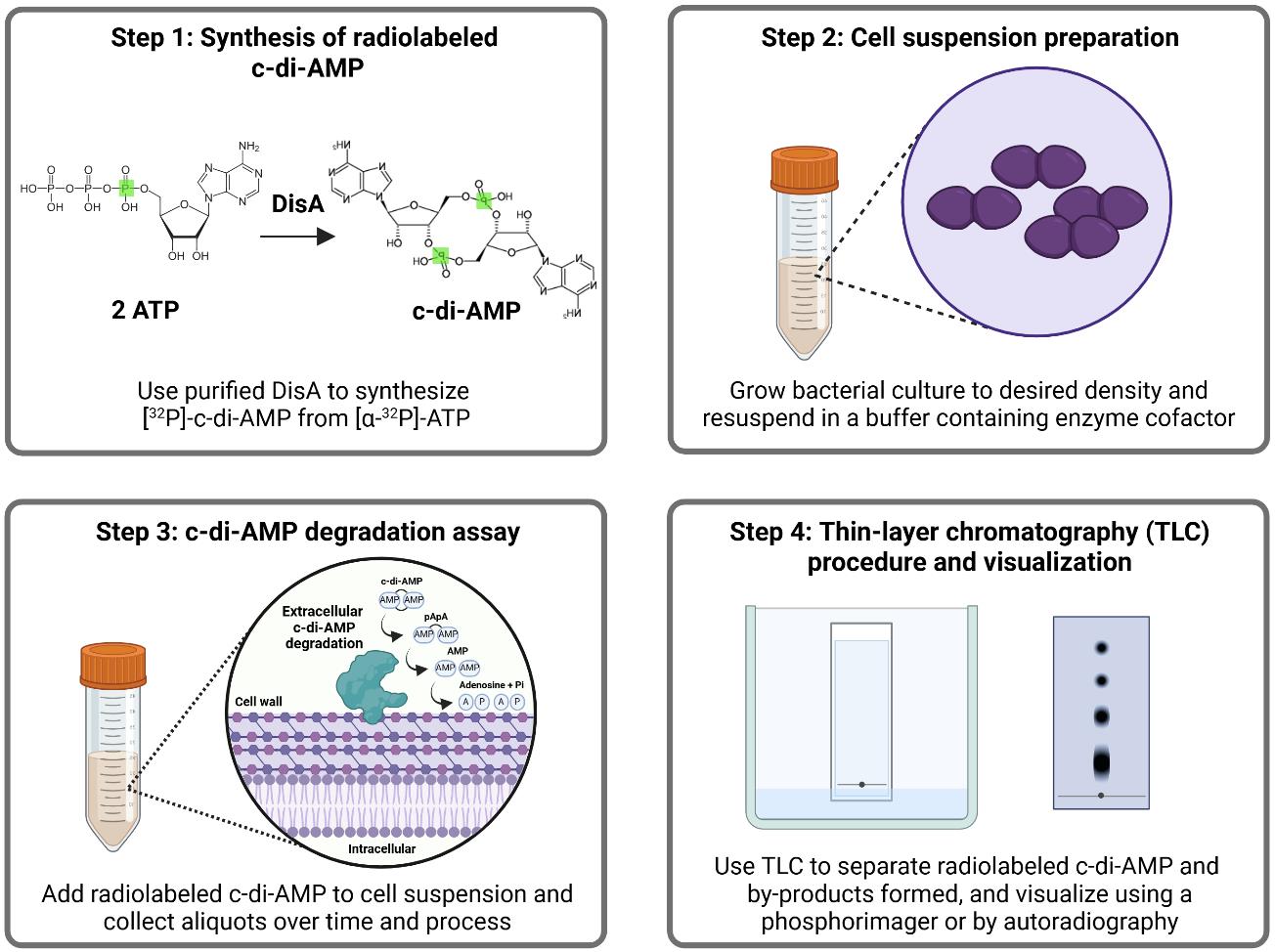
Cyclic di-AMP is a bacterial second messenger nucleotide required for the regulation of numerous cellular functions, including potassium and osmolyte homeostasis, DNA repair, cell wall integrity, central metabolism, and stress adaptation. This second messenger is synthesized from two ATP molecules by diadenylate cyclases (DAC) and degraded by cytoplasmic and surface-associated phosphodiesterases (PDE) to phosphoadenylyl adenosine (5′ pApA), adenosine monophosphate (AMP), and, in some instances, adenosine and inorganic phosphate (Pi). Levels of c-di-AMP in bacteria can be determined using different methods, including liquid chromatography–mass spectrometry (LC-MS/MS), enzyme-linked immunosorbent assay (ELISA), and luminescent and fluorescent biosensors. Thin-layer chromatography (TLC) is another method routinely used to monitor c-di-AMP synthesis and degradation by purified DAC and PDE enzymes and is particularly useful for monitoring c-di-AMP degradation products. Here, we devised a TLC-based method to monitor extracellular c-di-AMP stability and degradation by intact bacterial cells using radiolabeled c-di-AMP. We show that bacterial strains of Enterococcus faecalis and Streptococcus agalactiae that possess surface-associated PDEs can rapidly degrade extracellular c-di-AMP. In addition, we demonstrate that this method can be used to indirectly identify alternative enzyme substrates through competition assays. We propose that this TLC-based assay is an efficient method to analyze bacterial-mediated degradation of c-di-AMP and is amenable to testing other radiolabeled nucleotides.
Key features
• This method makes use of TLC, a well-established technique, to visualize c-di-AMP degradation products.
• Bypasses the need to obtain purified protein and instead uses live bacterial cells to monitor extracellular substrate degradation at the cell surface interface.
• This protocol can be adapted to other organisms and cell types and to detect other radiolabeled substrates.
Keywords: Thin-layer chromatography (薄层色谱) Bacteria (细菌)
Radiolabeled nucleotides (放射性标记核苷酸)
c-di-AMP (c-di-AMP)
Nucleotide degradation (核苷酸降解)
Bacteria (细菌)
Radiolabeled nucleotides (放射性标记核苷酸)
c-di-AMP (c-di-AMP)
Nucleotide degradation (核苷酸降解)
Graphical overview
Background
Second messengers are small regulatory molecules, many of them nucleotides, that are produced to modulate cell adaptation to specific stimuli or stressors. While second messengers are conserved across the different domains of life, they are particularly important to bacteria, with some of them deemed essential for cell viability and virulence expression. Some examples of second messenger nucleotides shown to modulate bacterial adaptation include (p)ppGpp, cyclic AMP (cAMP), cyclic di-GMP (c-di-GMP), 3′3′-cyclic GMP-AMP (3′3′-cGAMP), and cyclic di-AMP (c-di-AMP). Cyclic di-AMP was serendipitously discovered in 2008, during a crystallization study of the DNA integrity scanning protein (DisA) of Thermotoga maritima [1]. Considered an essential poison [2] due to its essentiality and toxicity at elevated levels, c-di-AMP regulates a variety of cellular processes, including osmoregulation, cell wall synthesis and homeostasis, central metabolism, DNA integrity, and stress responses [3–9]. Not surprisingly, c-di-AMP regulation has also been linked to bacterial pathogenesis through its roles in mediating antibiotic tolerance and regulating virulence factor expression [5,8,9]. As a second messenger, c-di-AMP binds to specific proteins, such as transcriptional regulators, various transporters, and riboswitches, exerting allosteric control over their activities [3–9].
Intracellular levels of c-di-AMP are maintained by diadenylate cyclases (DAC) that synthesize c-di-AMP from two molecules of ATP and by phosphodiesterases (PDE) that degrade c-di-AMP to 5′ phosphoadenylyl adenosine (pApA), adenosine monophosphate (AMP), and, in some instances, adenosine (Ado) and inorganic phosphate (Pi) [3–9]. In addition, multidrug efflux (MDF)-type transporters have been implicated in c-di-AMP export [10] and, once outside the cell, c-di-AMP is rapidly degraded by extracellular phosphodiesterases (ePDE) identified in a few bacterial species [11–14]. The ability of bacteria to modulate extracellular c-di-AMP levels is particularly relevant during infection, given that c-di-AMP is a potent pathogen-associated molecular pattern (PAMP) molecule. Despite major progress in c-di-AMP research since its discovery, our current understanding of the extent of its influence on bacterial physiology and its role as a PAMP remains limited. Thus, developing new approaches to detect and quantify c-di-AMP and its degradation products may potentially accelerate future discoveries.
Currently, c-di-AMP can be detected and quantified using several methods, including liquid chromatography–mass spectrometry (LC-MS/MS), enzyme-linked immunosorbent assay (ELISA), luminescent and fluorescent biosensors, and thin-layer chromatography (TLC). Benefits of LC-MS/MS include high sensitivity, reproducibility, and the ability to determine the presence of different types of nucleotides within one sample. However, performing this analysis requires highly specialized equipment and knowledge of analytical chemistry techniques. The ELISA-based method is also sensitive and does not require sophisticated instrumentation, yet it can only quantify c-di-AMP and does not provide information on c-di-AMP degradation products. More recently, c-di-AMP-responsive riboswitches have been used to develop sensitive biosensors that allow real-time monitoring of c-di-AMP fluctuations. Still, c-di-AMP biosensors are not easily adaptable from species to species and often require additional genetic manipulations.
Here, we describe an adaptation of the traditional TLC-based method, which normally requires purified protein, to assess extracellular degradation of c-di-AMP using live bacteria. Our goal was to investigate EecP, a unique cell wall–anchored nucleotidase in E. faecalis that degrades c-di-AMP extracellularly, which was challenging to purify despite several attempts with different expression systems [11]. Therefore, this method was developed to analyze the degradation of c-di-AMP by live bacterial cells of Enterococcus faecalis and Streptococcus agalactiae strains. Even so, this accessible and low-cost method can be easily adapted to test a variety of other organisms, including Gram-negative bacteria, archaea, yeasts, and eukaryotic cells. Furthermore, the method can also be used to monitor degradation of other nucleotides and to identify factors that interfere with extracellular nucleotide levels.
Materials and reagents
Biological materials
Bacterial strains
| Strain | Relevant characteristics | Source |
|---|---|---|
| E. faecalis | ||
| OG1RF | Laboratory/reference strain Rifr, Fusr | Lab stock |
| V583 | Laboratory/reference strain, Vanr | Lab stock |
| E. coli | ||
| Rosetta (DE3) pLysS pSpeedET:: Akr1c13 | Host for AKR1C13, protein herein referred to as RECON, protein expression, Kanr | Woodward Lab at the University of Washington [15] |
| Rosetta (DE3) pLysS pET20b:: disA | Host for DisA protein expression (disA from Bacillus subtilis), Ampr | Woodward Lab at the University of Washington [1,16] |
| S. agalactiae | ||
| COH1 | Laboratory/reference strain, serotype III | Brady Lab at the University of Florida |
| NGBS93 | WGS clinical blood isolate, serotype V, BioSample: SAMN03329865 | Fittipaldi Lab at the University of Montreal |
Reagents
1. Luria-Bertani (LB) broth, Lennox (Fisher Bioreagents, catalog number: BP1427)
2. Todd Hewitt broth (THB) (Becton Dickinson, catalog number: 249240)
3. Chemically defined medium (CDM) (see recipe in [11])
4. Kanamycin monosulfate (Fisher Scientific, CAS number: 25389-94-0)
5. Ampicillin sodium salt (Sigma-Aldrich, CAS number: 69-52-3)
6. D-(+)-Glucose (Sigma-Aldrich, CAS number: 50-99-7)
7. Isopropyl β-D-thiogalactoside (IPTG) (Fisher Scientific, CAS number: 367-93-1)
Note: Prepare 1 M stock in sterile ddH2O and freeze aliquots at -20 °C. Do not re-use thawed aliquots.
8. Halt protease inhibitor cocktail (100×) (Thermo Scientific, catalog number: 78429)
9. Ni-NTA agarose (Marvelgent Biosciences, catalog number: 11-0224-100)
10. Sodium phosphate monobasic (NaH2PO4) (ICN Biomedicals, CAS number: 10049-21-5)
11. Sodium chloride (NaCl) (Fisher Scientific, CAS number: 7647-14-5)
12. Imidazole (Acros Organics, CAS number: 288-32-4)
13. Triton X-100 (Fisher Bioreagents, CAS number: 9002-93-1)
14. Tris base (Fisher Bioreagents, CAS number: 77-86-1)
15. Manganese (II) chloride (MnCl2·4H2O) (Sigma-Aldrich, CAS number: 13446-34-9)
16. Magnesium chloride (MgCl2·6H2O) (Fisher Bioreagents, CAS number: 7791-18-6)
17. Glycerol (Fisher Bioreagents, CAS number: 56-81-5)
18. [α-32P]-ATP, 3,000 Ci/mmol, 10 mCi/mL, 250 μCi (Revvity Health Sciences, catalog number: BLU003H250UC)
19. c-di-AMP (InvivoGen, catalog number: tlrl-nacda)
20. c-di-GMP (InvivoGen, catalog number: tlrl-nacdg)
21. 2′3′-cGAMP (InvivoGen, catalog number: tlrl-nacga23-02)
22. Methanol (Fisher Chemical, CAS number: 67-56-1)
23. Ammonium sulfate [(NH4)2SO4] (Sigma-Aldrich, CAS number: 7783-20-2)
24. Monopotassium phosphate (KH2PO4) (Fisher Bioreagents, CAS number: 7778-770)
25. Phosphoric acid (H3PO4) (Sigma-Aldrich, CAS number: 7664-38-2)
26. Hydrochloric acid (HCl) (Fisher Chemical, CAS number: 7664-01-0)
27. Sodium hydroxide (NaOH) (Fisher Chemical, CAS number: 1310-73-2)
28. Pierce BCA Protein Assay kit (Thermo Scientific, catalog number: 23225)
29. Econo-Safe Biodegradable counting cocktail (Research Products International, catalog number: 111175)
30. Double-distilled water (ddH2O)
Solutions
1. Protein purification and storage buffers (see Recipes)
a. Equilibration/lysis buffer
b. Wash buffer 1
c. Wash buffer 2
d. Wash buffer 3
e. Elution buffer
f. Protein storage/binding buffer
2. Reaction buffer (see Recipes)
3. TLC mobile phase buffer (see Recipes)
Recipes
Note: All buffers can be prepared at room temperature with constant stirring unless specified. Adjust the pH of components or buffers as needed using either HCl or NaOH before adding water up to the final volume. Filter-sterilize components as needed and store at room temperature unless otherwise specified.
1. Protein purification and storage buffers
To prepare buffers for protein purification, first prepare each stock component separately as follows:
| Stock components | Molecular weight (g/mol) | Preparation |
|---|---|---|
| 1 M NaH2PO4 | 137.99 | • Dissolve 68.99 g of NaH2PO4 in 300 mL of ddH2O. • Add ddH2O to obtain a final volume of 500 mL. |
| 5 M NaCl | 58.44 | • Dissolve 146.1 g of NaCl in 300 mL of ddH2O. • Add ddH2O to obtain a final volume of 500 mL. |
| 1 M imidazole | 68.08 | • Dissolve 34.04 g of imidazole in 300 mL of ddH2O. • Add ddH2O to obtain a final volume of 500 mL. |
| 1 M MgCl2 | 203.30 | • Dissolve 101.65 g of MgCl2 in 300 mL of ddH2O. • Add ddH2O to obtain a final volume of 500 mL. |
| 1 M Tris-Cl pH 7.5 | 121.136 (Tris base) | • Dissolve 60.568 g of Tris base in 300 mL of ddH2O. • Add ddH2O to obtain a final volume of 500 mL after pH adjustment with HCl. |
| 1 M MnCl2 | 197.9 | • Dissolve 98.95 g of MnCl2 in 300 mL of ddH2O. • Add ddH2O to obtain a final volume of 500 mL. |
a. Equilibration/lysis buffer (pH 7.5)
| Stock components | Volume | Final concentration |
|---|---|---|
| 1 M NaH2PO4 | 20 mL | 20 mM |
| 5 M NaCl | 60 mL | 300 mM |
| 1 M imidazole | 10 mL | 10 mM |
| ddH2O | up to 1,000 mL after pH adjustment |
b. Wash buffer 1 (pH 8.0)
| Stock components | Volume | Final concentration |
|---|---|---|
| 1 M NaH2PO4 | 20 mL | 20 mM |
| 5 M NaCl | 60 mL | 300 mM |
| 1 M imidazole | 25 mL | 25 mM |
| Concentrated Triton X-100 | 10 mL | 1% |
| ddH2O | up to 1,000 mL after pH adjustment |
c. Wash buffer 2 (pH 8.0)
| Stock components | Volume | Final concentration |
|---|---|---|
| 1 M NaH2PO4 | 20 mL | 20 mM |
| 5 M NaCl | 100 mL | 500 mM |
| 1 M imidazole | 25 mL | 25 mM |
| ddH2O | up to 1,000 mL after pH adjustment |
d. Wash buffer 3 (pH 8.0)
| Stock components | Volume | Final concentration |
|---|---|---|
| 1 M NaH2PO4 | 20 mL | 20 mM |
| 5 M NaCl | 100 mL | 500 mM |
| 1 M imidazole | 50 mL | 50 mM |
| ddH2O | up to 1,000 mL after pH adjustment |
e. Elution buffer (pH 8.0)
| Stock components | Volume | Final concentration |
|---|---|---|
| 1 M NaH2PO4 | 20 mL | 20 mM |
| 5 M NaCl | 100 mL | 500 mM |
| 1M imidazole | 250 mL | 250 mM |
| ddH2O | up to 1,000 mL after pH adjustment |
f. Protein storage/binding buffer (pH 7.5)
| Stock components | Volume | Final concentration |
|---|---|---|
| 1 M Tris-Cl pH 7.5 | 40 mL | 40 mM |
| 5 M NaCl | 20 mL | 100 mM |
| 1 M MgCl2 | 20 mL | 20 mM |
| ddH2O | up to 1,000 mL after pH adjustment |
2. Reaction buffer
| Stock components | Volume | Final concentration |
|---|---|---|
| 1 M Tris-Cl pH 7.5 | 50 mL | 50 mM |
| 1 M MnCl2 | 5 mL | 5 mM |
| ddH2O | up to 1,000 mL after pH adjustment |
3. TLC mobile phase buffer
Prepare each component separately as follows:
| Stock components | Molecular weight (g/mol) | Preparation |
|---|---|---|
| 1.5 M KH2PO4, pH 3.6 | 136.086 | • Dissolve 102.06 g of KH2PO4 in 400 mL of ddH2O with stirring. • Adjust pH to 3.6 using H3PO4 (phosphoric acid). • Add ddH2O to obtain a final volume of 500 mL. |
| 4.1 M saturated (NH4)2SO4 | 132.14 | • Dissolve 270.887 g of ammonium sulfate in 200 mL of ddH2O with stirring and gentle heating. • Add ddH2O to obtain a final volume of 500 mL. • Cool to room temperature before filter sterilizing and store at 4 °C. |
Note: A saturated ammonium sulfate solution is a highly concentrated mixture where no more of the salt [(NH4)2SO4] can be dissolved in water.
To prepare 250 mL of TLC mobile phase buffer, mix 100 mL of saturated (NH4)2SO4 and 150 mL of KH2PO4 in a 1:1.5 ratio (v/v) and use immediately.
Laboratory supplies
1. Microcentrifuge tubes (1.5 and 2 mL)
2. Individual PCR tubes (0.5 mL)
3. Micropipette tips (10, 200, and 1,000 μL)
4. Micropipettes (P2, P20, P200, and P1000)
5. Serological pipettes (10 and 25 mL)
6. Micropipette filtered tips (10, 200, and 1,000 μL)
7. 1.5 mL polystyrene semi-micro cuvette (Fisher, catalog number: 14-955-127)
8. Falcon tubes (15 and 50 mL)
9. Econo-column glass chromatography columns, 1.0 × 30 cm (Bio-Rad, catalog number: 7371032)
10. Pierce protein concentrator PES, 10K MWCO (5–20 mL) (Thermo Scientific, catalog number: 88528)
11. Pierce concentrator, PES, 10K MWCO (0.5 mL) (Thermo Scientific, catalog number: 88513)
12. Zeba spin desalting columns, 7K MWCO (Thermo Scientific, catalog number: 89891)
13. Pierce spin columns (Thermo Scientific, catalog number: 69705)
14. TLC polyethylenimine (PEI) cellulose F plates (EMD Millipore, catalog number: 1.05579.0001)
15. Nitrocellulose membrane, 0.45 μm (Cytiva, catalog number: GE10600048)
16. Acrylic benchtop beta radiation shield (Thermo Scientific Nalgene, catalog number: 6700-1812)
17. TLC glass chambers (Supelco, Sigma-Aldrich, catalog number: Z204161)
18. Fume hood
19. X-ray exposure cassette (Wolf X-Ray Corporation, part number: 11319)
20. Clear blue X-ray film (CL-X Posure Film, Thermo Scientific, catalog number: 34091)
21. Clear plastic cling film (Boardwalk, catalog number: BWK 7202)
Equipment
1. Shaking incubator (Benchmark, model: Incu-Shaker 10L)
2. Spectrophotometer (Thermo Scientific, model: Genesys 30)
3. Sonicator (Fisher Scientific, Sonic Dismembrator model: 500)
4. Tube rotator (VWR, catalog number: 1016-084)
5. Benchtop large centrifuge (Eppendorf, model: 5910 Ri)
6. Benchtop microcentrifuge (Eppendorf, model:5415 C)
7. High-speed centrifuge (Beckman, model: J2-21)
8. Liquid scintillation counter (Beckman, model: LS 6000IC)
Procedure
登录/注册后免费查看全文
文章信息
稿件历史记录
提交日期: Jun 11, 2026
接收日期: Jul 14, 2026
在线发布日期: Aug 3, 2026
出版日期: Aug 20, 2026
版权信息
© 2026 The Author(s); This is an open access article under the CC BY-NC license (https://creativecommons.org/licenses/by-nc/4.0/).
如何引用
Bala, A., Morales Rivera, A. G. and Lemos, J. A. (2026). Analysis of Bacterial-Mediated c-di-AMP Degradation by Thin-Layer Chromatography. Bio-protocol 16(16): e5796. DOI: 10.21769/BioProtoc.5796.

 Download Citation in RIS Format
分类
微生物学 > 微生物信号传导 > 第二信使
生物化学 > 其它化合物 > 三磷酸核苷
微生物学 > 微生物新陈代谢 > 其它化合物
您对这篇实验方案有问题吗?
在此处发布您的问题,我们将邀请本文作者来回答。同时,我们会将您的问题发布到Bio-protocol Exchange,以便寻求社区成员的帮助。
 发布问题
发布问题


