


Engineering MRI-Based Programmable Genetic Sensors Using the MAPPER Platform
利用 MAPPER 平台构建基于磁共振成像的可编程遗传传感器

发布: 2026年09月05日第16卷第17期 DOI: 10.21769/BioProtoc.5795 浏览次数: 255
评审: Gundeep KaurAnonymous reviewer(s)
 PDF
PDF  Q&A
Q&A
参见作者原研究论文
The authors used this protocol in:

Jan 2026





Abstract
Genetically encodable reporters that produce signals detectable in deep tissues offer a powerful tool for noninvasive monitoring of molecular events in vivo. Although magnetic resonance imaging (MRI) is a standard technique for noninvasive clinical imaging, its wider application in detecting molecular activities has been constrained by the lack of programmable sensors. This limitation is in stark contrast to the widespread use of fluorescent reporter–derived sensors in cultured cells and in transparent specimens. To overcome this limitation, we recently developed the modular aquaporin-based protease-activatable probe for enhanced reporting (MAPPER) platform. This sensor engineering framework integrates a metal-free MRI reporter derived from human aquaporin-1 (hAqp1) with synthetic protease-based circuits. This integration facilitates the modular and scalable creation of a wide range of sensors by regulating protease activity through precise molecular events, such as protein–protein interactions, pharmacological inhibition, and second messenger signaling. In this paper, we present a detailed protocol for constructing and deploying sensors using the MAPPER paradigm. The protocol encompasses genetic design, lentiviral production, stable cell line generation, biochemical and microscopic validation of sensor function, diffusion-weighted MRI, and MR image analysis to quantify sensor signals in terms of the apparent diffusion coefficient. We describe two distinct MAPPER architectures: DD-MAPPER, which leverages protease-controlled protein degradation, and ER-MAPPER, which utilizes protease-controlled, subcellular trafficking. The MAPPER framework allows adaptation to various molecular targets without the need to redesign the core MRI reporter mechanism, making MAPPER a versatile platform for noninvasive biosensing in living cells and tissues.
Key features
• MAPPER enables programmable, protease-controlled switching of aquaporin-1-based diffusion weighted-MRI signals in genetically modified mammalian cells.
• The protocol covers two complementary biosensor architectures (DD-MAPPER and ER-MAPPER) that exploit different post-translational regulatory mechanisms to modulate MRI signals.
• Stable MAPPER cell lines are generated via lentiviral transduction, allowing long-term, selection-free biosensor expression across multiple mammalian cell types.
• The sensor is fully modular; proteases and protease-based logic circuits can be substituted without altering the hAqp1 reporter, enabling rapid adaptation to new molecular targets.
Keywords: MRI (磁共振成像) Noninvasive biosensors (无创生物传感器)
Aquaporins (水通道蛋白)
Reporter genes (报告基因)
Protease circuits (蛋白酶回路)
Diffusion-weighted MRI (弥散加权磁共振成像)
Noninvasive biosensors (无创生物传感器)
Aquaporins (水通道蛋白)
Reporter genes (报告基因)
Protease circuits (蛋白酶回路)
Diffusion-weighted MRI (弥散加权磁共振成像)
Graphical overview
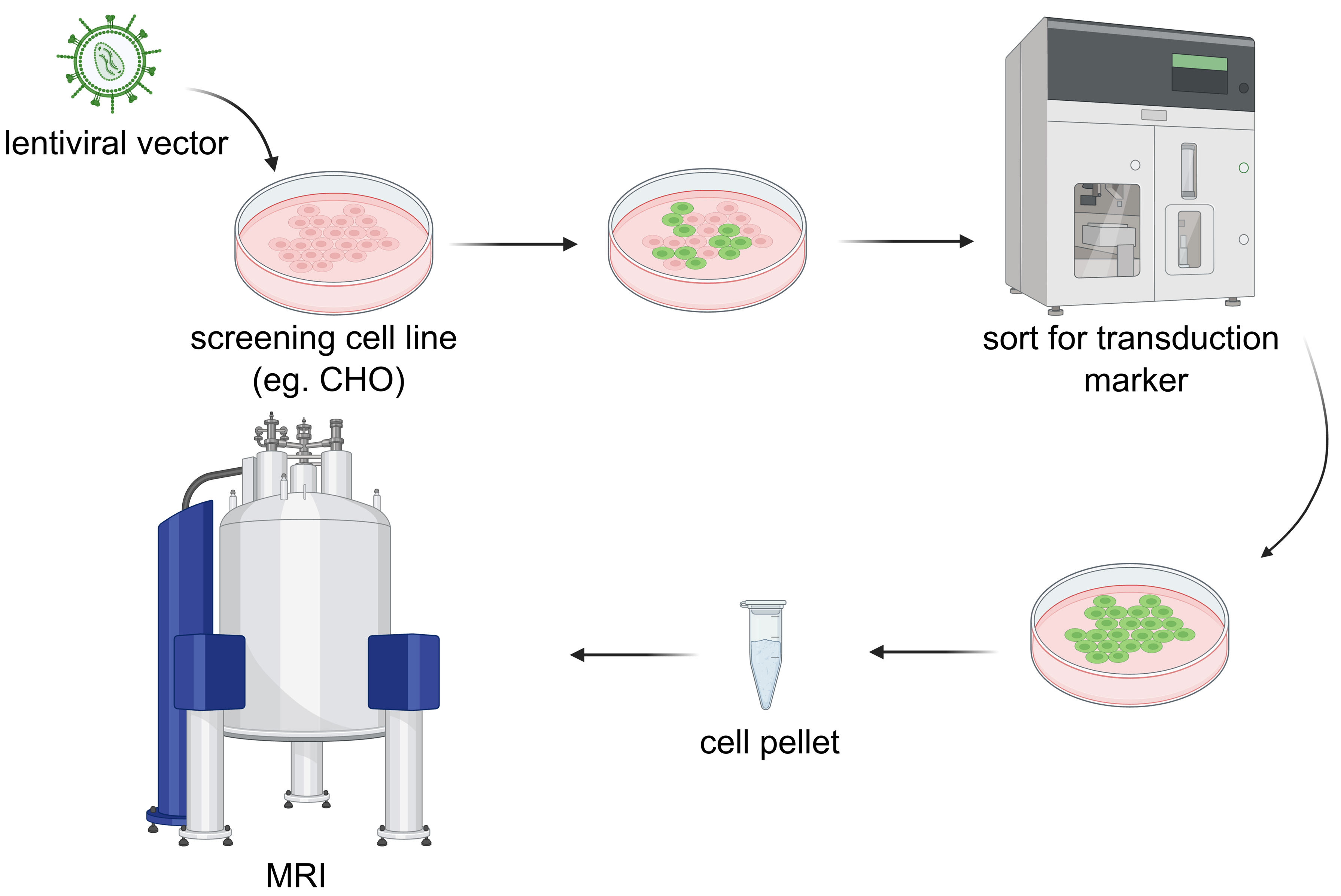
Workflow for generating and imaging MAPPER-expressing cells. CHO cells are stably transduced with lentiviral vectors encoding MAPPER constructs, sorted by fluorescence-activated cell sorting (FACS) to select successfully transduced cells, expanded to confluence, pelleted by centrifugation, and imaged by diffusion-weighted magnetic resonance imaging (MRI). MAPPER: modular aquaporin-based protease-activatable probe for enhanced reporting.
Background
The ability to monitor molecular events in living tissues noninvasively is a long-standing goal of biomedical research [1]. Understanding how diseases such as cancer, neurodegeneration, and inflammation develop at the molecular level requires tools that can report on biological events such as enzyme activation, protein–protein interactions, signaling, and drug action, within intact, optically opaque experimental models. Magnetic resonance imaging (MRI) is uniquely suited to this challenge: it generates high-resolution (from millimeters in humans to ~100 μm in rodents), three-dimensional images of soft tissue without utilizing ionizing radiation and has no depth constraint, making it one of the few modalities compatible with longitudinal molecular imaging in whole animals in both research and clinical settings.
Consequently, the development of genetically encoded reporters that produce detectable MRI signals has been an active area of research for over two decades [2–6]. Genetically encoded reporters for MRI can be broadly categorized by their contrast mechanism: metal-based reporters that alter T1 or T2/T2* relaxation times by accumulating paramagnetic ions, and water-exchange reporters that increase the apparent diffusion coefficient (ADC) of cellular water detectable by diffusion-weighted MRI. The latter is the foundation of the modular aquaporin-based protease-activatable probe for enhanced reporting (MAPPER) platform described in this protocol. Early approaches focused on metalloprotein overexpression by engineering cells to accumulate iron via ferritin or transferrin receptor overexpression, producing local perturbations in T2 or T2* relaxation rates that could be detected using standard T2-weighted imaging protocols similar to blood oxygenation level–dependent functional MRI (BOLD-fMRI) [7–9]. While these reporters have been used successfully in cell-tracking studies, they carry notable limitations. They are constitutively active, signal magnitude depends on expression level rather than on a specific molecular event, and, critically, require exogenous iron supplementation to generate sufficient MRI contrast. Together, these properties make them poorly suited to biosensing applications that require a switchable, activity-dependent readout. A second class of reporters exploits paramagnetic metal-binding proteins, including engineered variants of the dopamine- and serotonin-sensing MRI reporters developed by directed evolution [2], and calcium-responsive reporters based on calprotectin-driven chelation of manganese [10]. These provide genuine biosensing functionality but still require exogenous metal cofactors, limiting their applicability in vivo and raising translational concerns. A third class exploits chemical exchange saturation transfer (CEST), in which highly charged polypeptides, such as polylysine, exchange protons with bulk water at a distinct frequency offset, generating contrast without paramagnetic metals [11]. While promising, CEST reporters suffer from relatively low sensitivity compared to metal-based approaches. More recently, gas vesicle–based acoustic reporters derived from aquatic microorganisms have been developed and shown to function in mammalian cells, offering a distinct and genetically encodable contrast mechanism [12]. However, the large genetic payload required for gas vesicle biosynthesis, combined with the lower depth penetration of ultrasound compared to MRI, constrains their widespread practical deployment.
Aquaporin-based reporters represent a conceptually different and practically attractive strategy. Aquaporins are compact, single-gene transmembrane water channels that accelerate transcellular water exchange, producing a measurable increase in the ADC detectable by diffusion-weighted MRI (DW-MRI), a sequence available on virtually every clinical and preclinical MRI scanner with no exogenous contrast agent required. Human aquaporin-1 (hAqp1) was established as a genetically encoded MRI reporter by our lab in 2016 [5], and subsequent work demonstrated its safety and utility across diverse mammalian cell types [13] and in vivo. Notably, hAqp1 has been used as a gene reporter to track viral delivery across the blood–brain barrier (BBB), trace neural connectivity in the rodent brain, and monitor tumor progression in mice. Despite these advances, a key limitation remained: aquaporin-based reporters lacked a general, modular framework for coupling the reporter to arbitrary molecular inputs. Each new biosensor application required independent engineering of the aquaporin protein itself, with no guarantee that the sensing domain would modulate channel activity as intended.
The MAPPER platform described in this protocol addresses this limitation by decoupling the reporter (hAqp1) from the sensing module [14]. Rather than engineering the aquaporin directly, MAPPER places hAqp1 under the post-translational control of synthetic viral protease circuits, a strategy that exploits the well-characterized programmability of protease-based signaling architectures developed in the synthetic biology field [15,16]. Two complementary mechanisms are provided: DD-MAPPER, in which hAqp1 stability is controlled by a protease-cleavable destabilizing domain (DD) [17–19], and ER-MAPPER, in which hAqp1 membrane trafficking is regulated by a protease-cleavable endoplasmic reticulum retention signal (ER) [20]. Because the protease recognition sequence and not the aquaporin itself is the point of input integration, a new molecular target can be detected simply by rewiring the upstream protease circuit: substituting a different protease, splitting the protease across two interacting proteins to sense protein–protein interactions, or placing protease expression under ligand-dependent post-transcriptional control. This modularity sharply reduces the engineering burden compared with prior reporter strategies and, critically, has been validated across diverse mammalian cell types and multiple target classes without requiring optimization of the core reporter for each application.
One potential limitation of MAPPER-based sensors is the latency associated with protease-based regulation; signal changes reflect cumulative protease activity over several minutes to hours, rather than capturing instantaneous molecular events. This mechanism is similar to that of integral sensors in optical imaging and offers several practical benefits, such as a higher signal-to-noise ratio and applicability in awake, ambulatory animals. Another limitation of MAPPER is the larger genetic footprint of these sensors because of their circuit-based (rather than conventional single protein–based) architecture. Although most MAPPER constructs remain within the packaging limits of lentiviral vectors (~9.2 kb), delivering MAPPERs via adeno-associated viral vectors (AAVs) will likely require splitting the sensor across multiple AAVs for co-transduction. Within this context, it is worth noting that a distinct advantage of MAPPERs is their entirely post-transcriptional operational mechanism, which potentially allows for delivery as mRNA-encoded devices in lipid nanoparticles. This approach circumvents the packaging limitations inherent to viral gene delivery vectors and may be particularly beneficial for the in vivo delivery of gene-based diagnostics, which do not require sustained or permanent genetic modification of cells and tissues.
In summary, the primary advantage of MAPPER over existing MRI reporter technologies is its programmability, offering a sensor engineering platform vis-à-vis a single bespoke biosensor. Beyond the applications demonstrated in the original publication, we anticipate that MAPPER will be useful across a broad biomedical space, from capturing various stages of signal transduction in physiological and disease states to longitudinally tracking the functional outcomes of gene- and cell-based medicines.
Materials and reagents
Biological materials
1. NEB® stable competent E. coli (NEB, catalog number: C3040H)
2. CHO Tet-On® 3G cell line (Clontech, catalog number: 631195)
3. HEK293T (ATCC, catalog number: CRL-3216)
4. pPkg (second-generation lentiviral packaging plasmid, Addgene #12259)
5. pVSV-G (second-generation lentiviral envelope plasmid, Addgene #8454)
6. DD-MAPPER for TEV (Addgene #248354)
7. ER-MAPPER for TEV (Addgene #248355)
8. TEV on inducible CMV Tet ON promoter (Addgene #248360)
Reagents
Cell culture reagents
1. DMEM (4.5 g/L glucose, L-glutamine, sodium pyruvate, phenol red) (Corning, catalog number: MT10013CM)
2. Fetal bovine serum (FBS) (GenClone, catalog number: 25-550)
3. Penicillin-Streptomycin (Pen/Strep) (10,000 U/mL) (Gibco, catalog number: 15140-122)
4. TrypLETM Express Enzyme (1×), phenol red (Gibco, catalog number: 12605-010)
5. PBS (10×, pH 7.4, diluted to 1× in MilliQ water and autoclaved before use) (Apex BioResearch Products, catalog number: 18-244)
6. Sodium butyrate (Thermo Scientific, catalog number: A11079)
7. Polybrene (10 mg/mL, dilute to 8 mg/mL using autoclaved MilliQ water as a 1,000× stock) (Sigma-Aldrich, catalog number: TR-1003-G)
8. Lenti-XTM Concentrator (TaKaRa, catalog number: 631232)
Transfection and cloning reagents
9. LB agar, Miller (Fisher Bioreagents, catalog number: BP1425-500)
10. SOC medium (NEB, catalog number: B9035S)
11. Monarch DNA Gel Extraction kit (NEB, catalog number: T1120L)
12. Ampicillin (GOLDBIO, catalog number: A-301-100)
13. PEI (25 kDa, linear) (Polysciences, catalog number: 23966)
14. Q5® High-Fidelity 2× master mix (New England Biolabs, catalog number: M0492S)
15. PureYieldTM Plasmid Miniprep System (Promega, catalog number: A1223)
16. PureYieldTM Plasmid Midiprep System (Promega, catalog number: A2495)
17. 50× TAE buffer (Thermo Scientific, catalog number: B49)
18. Agarose LE (Goldbio, catalog number: A-201-100)
19. Doxycycline hyclate (Sigma-Aldrich, catalog number: D9891)
20. Gibson assembly master mix; prepared using the protocol described by Miller Lab (https://pengxulab.weebly.com/uploads/7/9/3/5/79359982/gibson_assembly_%E2%80%93_samuel_miller_lab_uw_seattle.pdf)
Chemical reagents
21. 12 M HCl (Sigma-Aldrich, catalog number: 320331-500ML)
22. NaOH (Sigma-Aldrich, catalog number: 881-500G)
23. CuSO4.5H2O (Sigma-Aldrich, catalog number: 939315-100G)
Solutions
1. DMEM complete medium (see Recipes)
2. PEI transfection stock (see Recipes)
3. Sodium butyrate 1 M (see Recipes)
4. Agarose gel (see Recipes)
5. 1% agarose phantom gel with 0.15 g/L CuSO4 (see Recipes)
Recipes
1. DMEM complete medium
| Reagent | Final concentration | Quantity or volume |
|---|---|---|
| DMEM (4.5 g/L glucose, L-glutamine, phenol red) | n/a | 450 mL |
| FBS | n/a | 50 mL |
| Pen/Strep | 10,000 U/mL | 5 mL (100 U/mL) |
Mix all reagents, aliquot into 50 mL tubes, and store at 4 °C until use.
2. PEI transfection stock (0.258 mg/mL, 500 mL)
| Reagent | Final concentration | Quantity or volume |
|---|---|---|
| PEI (25 kDa) | 0.258 mg/mL | 129 mg |
| MilliQ water | n/a | 450 mL |
| 12 M HCl | n/a | As required |
| 10 M NaOH | n/a | As required |
Pour ~450 mL of Milli-Q H2O into a 500 mL glass beaker with stirring. Add 129 mg of linear 25 kDa PEI. Add concentrated HCl dropwise until pH < 2.0 (~800 μL of 12 M HCl). Stir for 2–3 h until PEI is fully dissolved; a small amount of fine fiber particles that do not dissolve is normal and does not affect performance. Add concentrated NaOH dropwise until pH 7.0 (~500 μL of 10 M NaOH). Transfer to a 500 mL glass cylinder and adjust the final volume to 500 mL with MilliQ H2O. Filter-sterilize through a 0.22 μm membrane. Aliquot 10 mL per tube. Store at -20 °C for long-term storage and 4 °C for day-to-day use.
3. Sodium butyrate 1 M, 10 mL
| Reagent | Final concentration | Quantity or volume |
|---|---|---|
| Sodium butyrate | 1 M | 1.1 g |
| MilliQ water | n/a | 10 mL |
Filter-sterilize through a 0.22 μm membrane and store at 4 °C.
4. Agarose gel (for DNA gel electrophoresis)
| Reagent | Final concentration | Quantity or volume |
|---|---|---|
| Agarose | 0.9% w/v | 0.45 g |
| 50× TAE buffer | 1× | 1 mL |
| Deionized water | n/a | 49 mL |
Microwave in 30-s intervals with swirling between each interval, until the solution reaches a full boil, the agarose is completely dissolved, and the solution is clear. Pour into the casting tray with the combs inserted in place. Let the gel form by letting it sit undisturbed for approximately 15–20 min. Remove the comb before use.
5. 1% agarose phantom gel with 0.15 g/L CuSO4 (per 100 mL; adjust to phantom volume)
| Reagent | Final concentration | Quantity or volume |
|---|---|---|
| Agarose | 1% w/v | 1 g |
| CuSO4.5H2O | 0.15 g/L | 15 mg |
| Deionized water | 100 mL |
Combine agarose, CuSO4·5H2O, and deionized water in a heat-resistant flask. Microwave in 30-s intervals with swirling between each interval, until the solution reaches a full boil, the agarose is completely dissolved, and the solution is clear. Pour the solution into the phantom mold before it begins to gel. Once the solution has cooled sufficiently (surface is no longer steaming), place 200 μL PCR tubes into the phantom. Ensure all tubes remain upright throughout cooling.
Critical: Tube tilt at this stage will be permanent once the gel sets, causing systematic errors in MRI ROI placement. Use the weighted phantom stopper (see Equipment) to keep tubes fixed while the gel solidifies. Note that the MRI image will display the mirror image of the physical tube layout.
Pause point: Sealed phantoms can be stored at 4 °C for up to 4 weeks. Inspect for gel cracking, mold growth, or tube drift before each use.
Laboratory supplies
1. 200 μL PCR tubes (Olympus plastics, catalog number: 24-154)
2. 1.5 mL microcentrifuge tubes, sterilized (MBPS Inc., catalog number: MC15-S)
3. Syringe (10 mL) (BD Syringe, catalog number: 309604)
4. 0.22 μm syringe filter (GenClone, catalog number: 25-244)
5. Vacuum bottle top filter (Millipore, catalog number: S2GPT05RE)
6. Aspirating pipettes, 2 mL (Fisherbrand, catalog number: 14-955-135)
7. Serological pipettes 5, 10, and 25 mL (Fisherbrand, catalog numbers: 13-678-11D, 13-678-11E, 13-678-11)
8. 15 and 50 mL conical centrifuge tubes (Thermo Scientific, catalog numbers: 12-565-268, 12-565-270)
9. Tissue culture plates, 10 cm (Fisherbrand, catalog number: FB012924)
10. Tissue culture–treated 6-well plates (Corning, catalog number: 07-200-83)
Equipment
1. Class II Type A2 biosafety cabinet (ThermoFisher Scientific, 1300 series, model: 1377)
2. CO2 incubator, 37 °C, 5% CO2 (ThermoFisher Scientific, model: Forma steri-cycle i160 LK)
3. Refrigerated centrifuge with swing-bucket rotor (ThermoFisher Scientific, catalog number: 75004521)
4. Microcentrifuge/benchtop centrifuge, up to 16,000× g (ThermoFisher Scientific, catalog number: 75002431)
5. Thermocycler for PCR (Applied Biosystems SimpliAmp Thermal Cycler, catalog number: A24812)
6. NanoDrop OneC (Thermo Scientific, catalog number: 13-400-519)
7. Cell sorter (Sony, model: MA900)
8. Diffusion-weighted MRI scanner, Bruker 7 T vertically oriented, actively shielded super-wide bore scanner, 154 mm bore diameter
9. Radiofrequency (RF) probe, 66 mm diameter (MiniSWB90, Bruker, model: 1P T134743)
Software and datasets
1. ImageJ/Fiji, version 2.x (NIH, free; https://imagej.net/software/fiji), used for ROI selection, mean gray value extraction, and basic DW-MRI image processing
2. ParaVision 6.0.1 (Bruker BioSpin, commercial license required; https://www.bruker.com); used for DW-MRI data acquisition and raw image export
3. SnapGene or Benchling (SnapGene: DSBio, commercial; https://www.snapgene.com; Benchling: free for academic use; https://www.benchling.com); used for MAPPER construct design, primer design, and sequence verification
4. MATLAB R2022a Update 4 (version 9.12.0.200938, MathWorks, commercial license required; https://www.mathworks.com); used for ADC fitting and data visualization. The Matlab code used for generating ADC values from the mean grey value and effective b-values has been deposited to GitHub (https://github.com/asishninanchacko-cloud/Bio-Protocols) and archived on Zenodo (https://doi.org/10.5281/zenodo.21045388).
Procedure
登录/注册后免费查看全文
文章信息
稿件历史记录
提交日期: May 19, 2026
接收日期: Jul 14, 2026
在线发布日期: Aug 3, 2026
出版日期: Sep 5, 2026
版权信息
© 2026 The Author(s); This is an open access article under the CC BY-NC license (https://creativecommons.org/licenses/by-nc/4.0/).
如何引用
Readers should cite both the Bio-protocol article and the original research article where this protocol was used:
- Chacko, A., He, Y., Borg, R. E., Tang, T. and Mukherjee, A. (2026). Engineering MRI-Based Programmable Genetic Sensors Using the MAPPER Platform. Bio-protocol 16(17): e5795. DOI: 10.21769/BioProtoc.5795.
- Chacko, A. N., Dhanabalan, K. M., Wan, J., Chien, R., Anderson, N. T., Xu, B., Pham, K., Tiwari, R. and Mukherjee, A. (2026). A programmable genetic platform for engineering noninvasive biosensors. Sci Adv. 12(3): eaec1211. https://doi.org/10.1126/sciadv.aec1211

 Download Citation in RIS Format
分类
生物工程 > 合成生物学 > 基因修饰
分子生物学 > 蛋白质 > 活性
生物物理学 > 核磁共振波谱法 > NMR成像技术
您对这篇实验方案有问题吗?
在此处发布您的问题,我们将邀请本文作者来回答。同时,我们会将您的问题发布到Bio-protocol Exchange,以便寻求社区成员的帮助。
 发布问题
发布问题


