


Cryo-EM Pipeline for Actin Filament End Structures
肌动蛋白丝末端结构的冷冻电镜分析流程

发布: 2026年09月05日第16卷第17期 DOI: 10.21769/BioProtoc.5794 浏览次数: 706
评审: Gundeep KaurAnonymous reviewer(s)
 PDF
PDF  Q&A
Q&A
参见作者原研究论文
The authors used this protocol in:

Mar 2024





Abstract
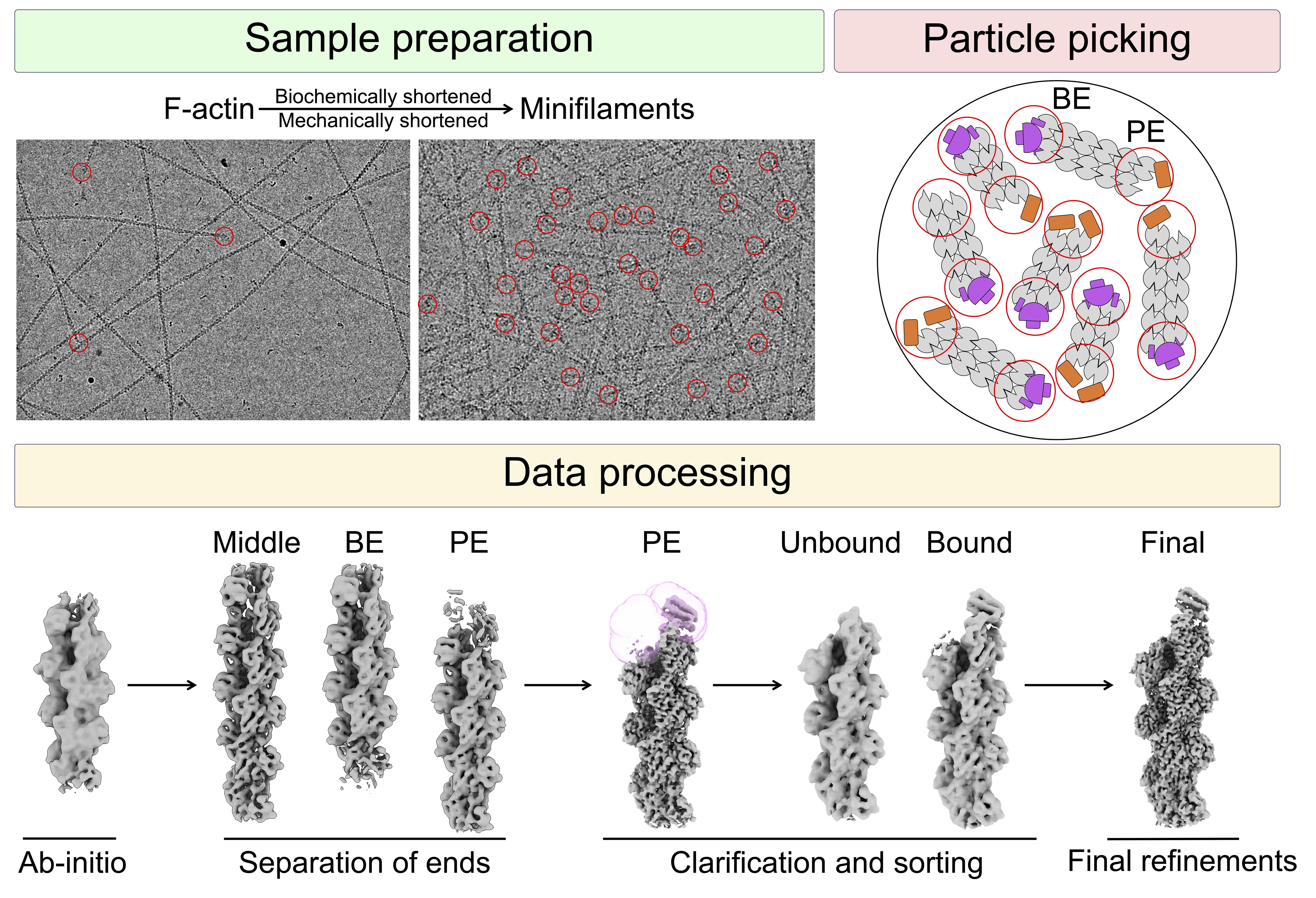
Actin filaments undergo dynamic growth and disassembly at their ends, regulated by many actin-binding proteins. However, structural analysis of filament end dynamics has been challenging due to the low abundance of filament ends in cryo-electron microscopy (cryo-EM) micrographs, their intrinsic polymorphisms, and the diversity and flexibility of end-binding proteins. Here, we describe a standardized cryo-EM protocol for determining actin filament end structures. First, short actin filaments are generated either biochemically using capping or severing proteins or mechanically through shearing. Filaments are then vitrified under conditions optimized for each specific end-binding protein. We describe data collection parameters using a 300 kV Titan Krios G3i microscope, including optimized grid preparation and imaging settings. Finally, we present a data processing pipeline for filament end structure determination based on machine learning–based particle picking, masking, and sorting strategies. This protocol has enabled the determination of multiple high-resolution structures of free, capped, elongating, and depolymerizing actin filament ends, and we further discuss considerations for extending this approach to other end-binding proteins.
Key features
• Generation of short actin filaments for cryo-EM using capping proteins, severing proteins, or mechanical shearing.
• Integrated data collection and image processing workflow for filament end structure determination.
• Common challenges, solutions, and experimental considerations for diverse actin filament end-binding complexes.
Keywords: Cryo-EM (冷冻电镜) Actin filament (肌动蛋白丝)
Filament ends (丝末端)
Machine learning (机器学习)
Vitrification (玻璃化冷冻)
Protein structure (蛋白质结构)
Cytoskeleton (细胞骨架)
Actin filament (肌动蛋白丝)
Filament ends (丝末端)
Machine learning (机器学习)
Vitrification (玻璃化冷冻)
Protein structure (蛋白质结构)
Cytoskeleton (细胞骨架)
Graphical overview
Background
Actin is a highly conserved and abundant protein that participates in numerous protein–protein interactions and plays crucial roles in cell motility, cytokinesis, and intracellular trafficking [1]. Central to actin’s functions is its ability to transition between monomeric (G-actin) and filamentous (F-actin) states in a highly dynamic and regulated manner, with an approximately 50:50 distribution between these two states in cells. F-actin is a polar filament with fast- and slow-growing ends, known as the barbed and pointed ends, respectively [2]. In cells, filament end dynamics are tightly regulated by a variety of barbed- and pointed-end binding proteins that control polymerization and depolymerization [1,3]; dysregulation of these processes is implicated in multiple human diseases [4,5].
High-resolution structures of filament ends are essential for understanding actin dynamics at the molecular level, but obtaining such structures has proven challenging. Cryo-electron microscopy (cryo-EM), the primary technique used, relies on averaging thousands of particles to reconstruct three-dimensional maps of proteins and complexes [6]. However, actin filaments can extend over micrometers, often spanning entire micrographs, resulting in very few filament ends per image. To overcome this limitation, filaments can be shortened either biochemically, using proteins that cap, slow filament growth, or sever filaments, or mechanically through shearing. Additional considerations include the activity of the end-binding protein under study, such as whether it interacts stably or transiently with filament ends or alters the polymerization and depolymerization rates. A further challenge is particle picking, as classical methods such as blob- or template-based approaches often fail to distinguish filament ends from filament middles. Machine learning–based picking methods implemented in software such as Topaz [7] and crYOLO [8] can be trained to address this limitation.
Here, we describe a step-by-step workflow for determining cryo-EM structures of actin filament ends using cryoSPARC [9,10] and highlight recently published examples in which this workflow has been applied successfully [11–15]. This framework may also be applicable to other classes of filamentous proteins and end-binding proteins.
Materials and reagents
Biological materials
1. Rabbit alpha skeletal muscle actin (UniProt P68135), purified in our laboratory from rabbit muscle using an established protocol [16]; alternatively, actin can be purified from commercially available muscle acetone powder from sources such as Cytoskeleton (catalog number: SKU: AKL99) or Pel-Freez Biologicals (catalog number: 41995-2)
Note: Actin preparations should be made fresh and stored in G-buffer (see Recipes) on ice or at 4 °C. A fresh preparation can last between 1 and 3 weeks in these conditions before a new preparation should be made.
2. Capping protein (CP) (UniProt: P52907 and P47756-2), purified as a heterodimer as described previously [11]
3. Cyclase associated protein-1 construct (UniProt: Q01518), purified as described previously [15]
Reagents
1. HEPES (Gold Biotechnology Inc, catalog number: H-400-1)
2. Potassium chloride (KCl) (Santa Cruz Biotechnology, catalog number: sc-203207A)
3. Ethylene glycol tetraacetic acid (EGTA) (Gold Biotechnology Inc, catalog number: E-217-100)
4. Magnesium chloride (MgCl2) (Fisher Scientific Company, catalog number: 442611500GM)
5. Dithiothreitol (DTT) (Gold Biotechnology Inc, catalog number: DTT100)
6. Adenosine 5′-triphosphate (ATP) (VWR International LLC, catalog number: 77877-060)
7. Tris base (Santa Cruz Biotechnology, catalog number: sc-3715C)
8. Calcium chloride dihydrate (CaCl2) (LabChem, catalog number: LC127251)
Note: It is important to store DTT and ATP as frozen aliquots at -20 °C and use freshly thawed aliquots for sample preparation. All buffers containing DTT and ATP should be made fresh.
Solutions
1. G-buffer in G-buffer (see Recipes)
2. Capped filaments in F-buffer (see Recipes)
3. F-actin in F-buffer (see Recipes)
Recipes
1. G-buffer
| Reagent | Final concentration | Quantity or volume |
|---|---|---|
| 1 M Tris, pH 8.0 | 5 mM | 2.5 μL |
| 20 mM CaCl2 | 0.2 mM | 5 μL |
| 20 mM ATP | 0.2 mM | 5 μL |
| 20 mM DTT | 0.2 mM | 5 μL |
| H2O | n/a | 482.5 μL |
| Total | n/a | 500 μL |
2. Capped filaments in F-buffer
| Reagent | Final concentration | Quantity or volume |
|---|---|---|
| 1 M HEPES pH 7.5 | 20 mM | 2 μL |
| 2.5 M KCl | 50 mM | 2 μL |
| 0.1 M EGTA | 1 mM | 1 μL |
| 0.1 M MgCl2 | 1 mM | 1 μL |
| 0.1 M ATP | 1 mM | 1 μL |
| 0.1 M DTT | 1 mM | 1 μL |
| H2O | n/a | 42 μL |
| 29.4 μM capping protein (CP) | 5 μM | 17 μL |
| 75.8 μM G-actin | 25 μM | 33 μL |
| Total | n/a | 100 μL |
3. F-actin in F-buffer
| Reagent | Final concentration | Quantity or volume |
|---|---|---|
| 1 M HEPES pH 7.5 | 20 mM | 2 μL |
| 2.5 M KCl | 50 mM | 2 μL |
| 0.1 M EGTA | 1 mM | 1 μL |
| 0.1 M MgCl2 | 1 mM | 1 μL |
| 0.1 M ATP | 1 mM | 1 μL |
| 0.1 M DTT | 1 mM | 1 μL |
| H2O | n/a | 72 μL |
| 75.8 μM G-actin | 15 μM | 20 μL |
| Total | n/a | 100 μL |
Laboratory supplies
1. Standard Vitrobot filter paper grade 595 (Ted Pella, Inc, catalog number: 47000-100)
2. Quantifoil R1.2/1.3 300 mesh, copper grids (Quantifoil, https://www.quantifoil.com/products/quantifoil/quantifoil-circular-holes)
3. Autogrid rings and C-clips (Electron Microscopy Sciences, catalog number: 71167-97)
Equipment
1. PELCO easiGlow Glow Discharge (PELCO, Ted Pella, Inc, catalog number: 91000S)
2. Ultrasonic Cleaner (Fisher Scientific, catalog number: FS20D)
3. Slide-Clamp Tweezers Assembly for FEI Vitrobot Mark IV-I (Ted Pella, Inc, catalog number: 47000-500)
4. Vitrobot Mark IV (Thermo Fisher)
5. Vitrobot dewar with metal parts (Ted Pella, Inc, catalog number: 47000-705)
6. Cryo grid boxes (Sub-Angstrom, catalog number: GBV01)
7. Krios G3i Cryo-Transmission Electron Microscope, 300 kV (Thermo Fisher)
8. K3-GIF Direct Electron Detector equipped with BioQuantum K3 imaging filter (Gatan Inc., AMETEK)
9. Customized 8 GPU Cryo-EM workstation (Single Particle)
Software and datasets
1. CryoSPARC (Structura Biotechnology, v4.7, dependencies and prerequisites can be found at https://guide.cryosparc.com/)
2. UCSF ChimeraX (https://www.cgl.ucsf.edu/chimerax, v1.10.1)
3. Topaz (https://github.com/3dem/topaz, v0.3.0, dependencies can be found in the GitHub repository)
4. EPU (Thermo Fisher, v3.12)
Procedure
登录/注册后免费查看全文
文章信息
稿件历史记录
提交日期: May 27, 2026
接收日期: Jul 19, 2026
在线发布日期: Aug 3, 2026
出版日期: Sep 5, 2026
版权信息
© 2026 The Author(s); This is an open access article under the CC BY license (https://creativecommons.org/licenses/by/4.0/).
如何引用
Readers should cite both the Bio-protocol article and the original research article where this protocol was used:
- Palmer, N. J. and Dominguez, R. (2026). Cryo-EM Pipeline for Actin Filament End Structures. Bio-protocol 16(17): e5794. DOI: 10.21769/BioProtoc.5794.
- Carman, P. J., Barrie, K. R., Rebowski, G. and Dominguez, R. (2023). Structures of the free and capped ends of the actin filament. Science. 380(6651): 1287–1292. https://doi.org/10.1126/science.adg6812

 Download Citation in RIS Format
分类
生物物理学 > 显微技术 > 低温显微镜技术
生物信息学与计算生物学
生物化学 > 蛋白质 > 结构
您对这篇实验方案有问题吗?
在此处发布您的问题,我们将邀请本文作者来回答。同时,我们会将您的问题发布到Bio-protocol Exchange,以便寻求社区成员的帮助。
 发布问题
发布问题


