


Efficient and Fast Site-Directed Mutagenesis via Partially or Completely Overlapping Primer Pairs
利用部分或完全重叠引物对实现快速高效的定点突变

发布: 2026年08月20日第16卷第16期 DOI: 10.21769/BioProtoc.5792 浏览次数: 454
评审: Hemant Kumar PrajapatiPriyanka MittalAnonymous reviewer(s)
 PDF
PDF  Q&A
Q&A
参见作者原研究论文
The authors used this protocol in:
Jan 2026





Abstract
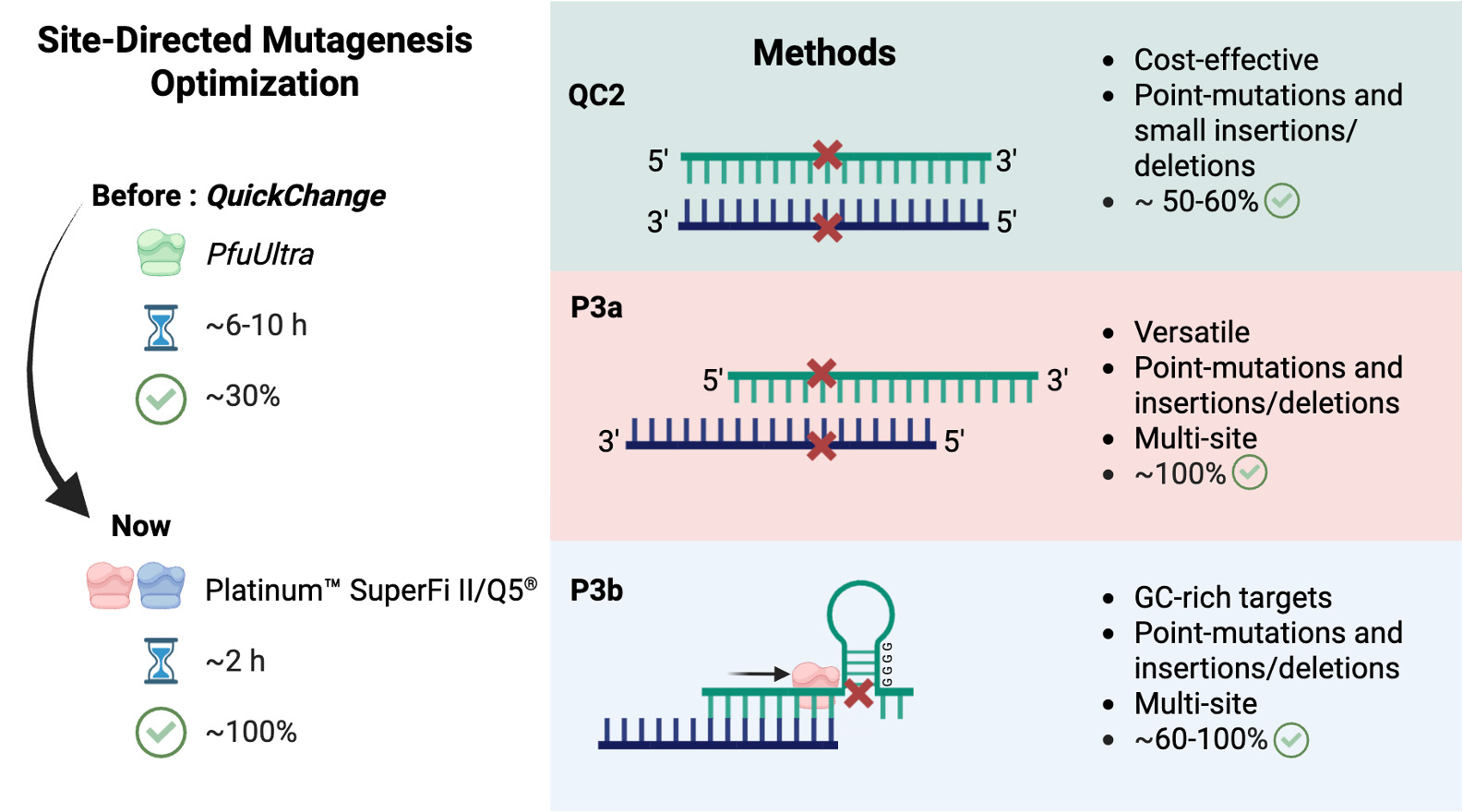
Site-directed mutagenesis is an indispensable molecular biology tool, but traditional methods often suffer from extended reaction time, structural limitations, and variable success rates. This article details three optimized protocols: P3a (primer pairs with 3′-overhangs, version a), P3b, and QuickChange 2.0, which rely on two highly processive DNA polymerases (Platinum SuperFi II and Q5) to accelerate and standardize plasmid engineering. The P3a method utilizes partially complementary primer pairs with distinct 3′-overhangs, achieving ~100% efficiency and enabling seamless cassette mutagenesis (insertion, deletion, and replacement). Building on this, the P3b method introduces specific thermal cycling modifications and a pre-denaturation step to overcome structural barriers resulting from GC-rich sequences. QuickChange 2.0 applies these two advanced polymerases to completely complementary primer pairs, even though the average efficiency decreases to 50%–60%. Replacing Pfu with the highly processive DNA polymerases also reduces PCR time to approximately 2 h. Thus, these new methods are more efficient and rapid than classical QuickChange mutagenesis based on Pfu polymerase.
Key features
• P3a uses 3′-overhang primers and superior polymerases for near-perfect efficiency in routine point mutations and seamless cassette mutagenesis.
• P3b adapts the 3′-overhang primer design with specialized thermal cycling to successfully overcome structural barriers in highly GC-rich templates.
• The QC2 protocol leverages completely complementary primers for fast and reliable introduction of point mutations and small insertions or deletions.
Keywords: Site-directed mutagenesis (定点突变) QuickChange method (QuickChange 方法)
PCR (PCR)
Epigenetic regulator (表观遗传调控因子)
Cas9 (Cas9)
Genome editing (基因组编辑)
G-quadruplex (G-四链体)
QuickChange method (QuickChange 方法)
PCR (PCR)
Epigenetic regulator (表观遗传调控因子)
Cas9 (Cas9)
Genome editing (基因组编辑)
G-quadruplex (G-四链体)
Graphical overview
Background
Since its initial report was made by the Nobel Laureate Michael Smith and his colleagues in 1978 [1], site-directed mutagenesis has become an essential molecular biology technique for engineering specific gene mutations in vitro [2]. Early approaches commonly used phage- or phagemid-based systems to generate single-stranded DNA templates, making the workflow labor-intensive and technically challenging [3–6]. The QuickChange method (or QuikChangeTM, as marketed initially by Stratagene and then by Agilent) is one of the most widely employed strategies in different laboratories, including our own, around the world [7–13]. The method relies on Pfu DNA polymerase-mediated PCR with pairs of completely complementary primers (Figure 1A), followed by DpnI restriction digestion to selectively degrade parental plasmids, which are methylated at the GATC sites and need to be isolated from Dam+ bacterial hosts. While the theoretical mutagenesis efficiency is 100%, the practical level varies widely, depending on the target plasmids and mutations. Furthermore, reliance on the slow, relatively low-fidelity Pfu DNA polymerase often results in extended PCR time and extensive troubleshooting, highly dependent on the plasmid backbones and the mutations to be engineered [14–16]. Another limitation of the QuickChange primer design is that the newly synthesized DNA strands are unsuitable as templates for subsequent PCR amplification cycles, at least as considered by many published reports [14–16], even though an alternative mechanism has also been identified [17]. Consequently, the reaction was considered to proceed linearly rather than exponentially, which, in combination with primer–primer dimer formation and unwanted insertions at primer sites, dramatically reduces the success rates [14–16].
To address these two limitations posed by the QuickChange method, an alternative strategy using partially complementary primers with 3′-protruding ends was initially developed in 2004 [14] and subsequently refined by four different laboratories in 2008 [15,18,19] and 2015 [17]. For convenience, we have referred to this strategy as P3 (primer pairs with 3′-protruding ends) site-directed mutagenesis [16]. Mechanistically, this approach was thought to allow newly synthesized strands to serve as templates for subsequent cycles, enabling exponential amplification [14–16], but it is noteworthy that two alternative mechanisms have been proposed to explain why such primers are advantageous over completely complementary primers [17,20]. Despite these refinements, the mutagenesis efficiency still varies substantially from gene to gene and from mutation to mutation. To investigate this, we systematically tested different Pfu-derived polymerases for generating over a hundred mutations on a dozen plasmids with different sizes and GC contents [16]. To improve the method further, we replaced PfuUltra (an improved version of Pfu) with two super-fidelity polymerases: PlatinumTM SuperFi II and Q5® DNA polymerases [21]. The resulting P3a method achieves a mutagenesis efficiency of near 100% for various plasmids and mutations [21]. These plasmids encode different epigenetic regulators, Cas9, and a protein kinase. The unique "handshaking" feature of P3 primer pairs also enables seamless cassette mutagenesis, allowing for efficient fragment deletions (up to 5 kb), insertions (up to 0.4 kb), and replacements [21]. Notably, Q5 DNA polymerase was found to be inactive for mutagenesis in an early study, likely due to the specific primers used and/or the specific mutation that was engineered [17]. Alternatively, new lots of Q5 polymerase are better than the early ones, likely due to improved purification procedures.
During development of the P3 and P3a methods [16,21], we faced difficulties with plasmids possessing GC-rich sequences, likely due to the formation of G-quadruplexes [22,23]. To resolve such structural barriers posed by high-GC regions, we developed the P3b method. By incorporating a pre-denaturation step prior to PCR and adjusting specific thermal cycling parameters, P3b successfully adapts the P3a framework for GC-rich templates [24]. Alternatively, because classical completely complementary primer pairs remain highly popular and cost approximately 50% less to synthesize than P3a and P3b primers, we applied these advanced polymerases to the traditional QuickChange primer design [20]. This updated protocol, detailed here as QuickChange 2.0 (QC2), successfully reduces the required PCR length from over 10 h to ~2 h. Thus, proper primer design and superior thermostable DNA polymerases are both key to successful PCR-based site-directed mutagenesis.
This article adds value to the related published research by translating these combined findings into a collection of actionable, step-by-step experimental protocols and by providing precise PCR thermal cycling parameters, reagent formulations, and template preparation steps required to execute the P3a, P3b, and QC2 methods. Furthermore, in light of the specific primer-site insertion phenomenon recently identified [20], this collection of protocols, supplemented by various practical tips, should help implement them in different laboratories, provide the context necessary to select the appropriate primer design strategy, and establish a concrete troubleshooting framework for mitigating failed mutagenesis reactions.
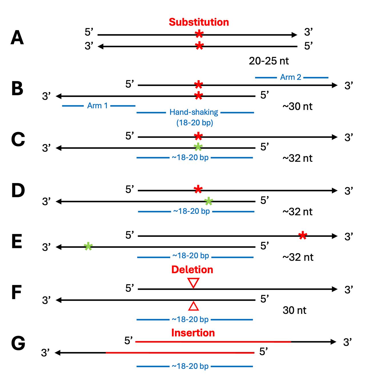
Figure 1. Schematic comparison of primer designs for site-specific mutagenesis. (A) The traditional QuickChange method utilizes completely complementary primer pairs, typically 20–25 nucleotides (nt) in length. Red asterisks indicate the targeted mutation site [16,21]. (B) The P3 method employs partially overlapping primer pairs that generate 3′-overhangs. This optimized P3 design utilizes simplified, shorter primers of approximately 30 nt. These feature 9-bp complementary regions flanking the single-nucleotide mutation (red asterisks). Utilizing shorter primers reduces synthesis costs and minimizes the risk of introducing unwanted mutations caused by impurities in chemical synthesis [16,21]. (C–E) Schematics demonstrating the simultaneous introduction of two distinct mutations (denoted respectively by the red and green asterisks) using a single partially overlapping primer pair. (F) Schematic representation of cassette deletion using P3a primers, where the 5′-arms are complementary to the regions immediately flanking the sequence to be deleted. (G) Schematic representation of cassette insertion using P3a primers, where the complementary arms correspond to the regions flanking the desired insertion site. The figure panels were adapted from [16,21].
Materials and reagents
Biological materials
1. Mammalian expression vectors
Note: As a reference, more than a dozen of plasmids have been tested for these methods, including mammalian expression plasmids for HA-tagged BRPF1 and the FLAG-tagged KAT2B, which have been deposited to Addgene (Addgene, catalog numbers: 250445 and 249547, respectively) [25,26]; expression plasmids for CDK13 and Cas9 [Addgene, catalog numbers: 135276 (deposited by Ben Major and colleagues) and 113096, respectively) [27]; and mammalian expression vectors for untagged D614G and Omicron spike proteins of SARS-COV-2 (SinoBiological, catalog numbers: VG40589-UT and VG40835-UT, respectively) [16,21].
All template plasmids were transformed into and isolated from Dam+ bacterial strains, such as DH5α. Competent cells were prepared as described previously [16,28].
2. 25-nmol scale, standard, and desalted DNA oligos
Note: All primers we have tested have been synthesized at Integrated DNA Technologies, Inc. No polyacrylamide gel or HPLC purification was used.
Reagents
1. 2× PlatinumTM SuperFi II PCR master mix (Thermo Fisher Scientific, catalog number: 12368010) or Q5® High-Fidelity DNA Polymerase (New England Biolabs, catalog number: M0494S)
Note: Notably, while we have used these two enzymes extensively, we have not tested their related preparations, such as the SuperFi PCR master mix and Q5 Ultra II.
2. DpnI restriction enzyme, 20 U/μL (New England Biolabs, catalog number: R0176)
3. Autoclaved Nanopure water
4. Plasmid Miniprep kit (Qiagen, catalog number: 27016)
5. Tryptone (BioShop, catalog number: TRP402)
6. Yeast extract (Gibco, catalog number: 212750)
7. NaCl (Wisent Inc., catalog number: 600-082-1K)
8. KCl (BioShop, catalog number: POC308)
9. NaOH (BioShop, catalog number: SHY700)
10. Glucose (Gibco, catalog number: 15023021)
11. MgCl2, hexahydrate (BioShop, catalog number: MAG510)
12. Ampicillin (BioShop, catalog number: AMP201)
Solutions
1. Super optimal broth with catabolite repression (SOC) medium (see Recipes)
2. Ampicillin stock solution (100 mg/mL) (see Recipes)
3. Luria–Bertani (LB) broth and agar plates (see Recipes)
Recipes
1. Super optimal broth with SOC medium
| Reagent | Quantity | Final concentration |
| Tryptone | 5 g | 2% w/v |
| Yeast extract | 1.25 g | 0.5% w/v |
| NaCl | 125 mg | 8.56 mM |
| KCl | 46.5 mg | 2.5 mM |
| 1 M NaOH | 0.5 mL | 2 mM |
| 1 M glucose | 5 mL | 20 mM |
| 2 M MgCl2 | 5 mL | 40 mM |
| Nanopure water | to 250 mL | — |
| Total | 250 mL | 100% |
Weigh and add tryptone, yeast extract, NaCl, and KCl to a glass bottle. Adjust pH to ~7.0 by adding 0.5 mL of 1 M NaOH. Adjust the volume to 250 mL with Nanopure water. Autoclave for 15 min in a liquid cycle. After autoclaving, cool to room temperature and then add 5 mL of sterile-filtered 1 M glucose and 5 mL of autoclaved 2 M MgCl2 while close to a flame or in a biosafety cabinet. Once opened, 1 mL aliquots should be prepared for storage at -20 °C.
2. Ampicillin stock solution (100 mg/mL)
Weigh 1 g of ampicillin powder into a 15 mL sterile Falcon tube. Add 10 mL of autoclaved Nanopure water and invert until completely dissolved. To prevent contamination, sterilize the solution by passing it through a sterile 0.22 or 0.45 µm syringe filter inside a biosafety cabinet or close to a flame, but this step is unnecessary and often omitted, without causing any problems. Prepare 1–1.5 mL aliquots for storage at -20 °C.
3. LB broth
Weigh and add 10 g of tryptone, 5 g of yeast extract, and 5 g of NaCl into a 1-L glass bottle. Adjust the volume to 1 L with deionized H2O. Add 2 mL of 1 M NaOH to adjust pH to ~7.0. Tightly close the bottle with a cap and shake the bottle several times to mix. Loosen the cap slightly in preparation for autoclaving in the next step. Sterilize by autoclaving according to a liquid sterilization protocol (a 15-min cycle is sufficient for 1 L; for larger volumes, a 30-min cycle is required). Take out the flask carefully from the autoclave machine, allow the media to cool down at room temperature, and tighten the lid. Store the sterilized LB medium at room temperature. Once opened, store it at 4 °C for up to 6 months.
4. LB agar plates
Weigh and add 10 g of tryptone, 5 g of yeast extract, 5 g of NaCl, and 15 g of agar into a 2-L Erlenmeyer flask. Adjust the volume to 1 L with deionized H2O. Add 2 mL of 1 M NaOH to adjust pH to ~7.0. Mix the solution carefully by gently swirling the flask in a circular motion. Sterilize by autoclaving according to a liquid sterilization protocol (a 15-min cycle is sufficient for 1 L; for larger volumes, a 30-min cycle is required). In the meantime, set up a water bath to 50 °C. Take out the flask carefully from the autoclave and place it in the water bath for ~60 min to bring the temperature down, while preventing it from solidifying. Add some water from the 50 °C water bath to a Styrofoam bucket and place the flask with the molten LB agar media inside (this will slow down the solidification of the media). Next to an open flame, add 1 mL of ampicillin (100 mg/mL) and mix thoroughly by gently swirling the flask to uniformly mix the ampicillin, avoiding the formation of air bubbles. With the flame still on, use a 25-mL sterile pipette to transfer ~20 mL of the molten LB media onto the bottom of a sterile Petri dish; then, place the lid on top.
Repeat this procedure quickly with the rest of the molten LB media. Plates can be stacked in 5–10 piles to make the pouring easier and quicker. Leave the dishes on top of an even bench overnight to solidify and reduce the condensation that forms on the lid. The next day, use a permanent marker to mark the sides of the plates with 2 or 3 parallel black bars or lines as the code to indicate that the plates contain ampicillin (other colors or markings can be used depending on the labelling system used in each laboratory). Stack the plates into plastic bags. Remove as much air as possible from the bags and seal them properly to prevent the plates from drying out. Store them at 4 °C. They can be kept there up to 4 months.
Laboratory supplies
1. PCR tubes (Diamed Lab Supplies Inc., catalog number: DIATEC420-1378)
2. Autoclaved Pipetman tips
3. 25 mL sterile serological pipette (Fisherbrand, catalog number: 13-678-11)
4. 100 × 15 mm stackable Petri dishes (Fisherbrand, catalog number: FB0875712)
6. 1.5 mL autoclaved Eppendorf tubes
7. 50 mL plastic centrifuge tubes (Fisherbrand, catalog number: 0644321)
8. Resealable plastic bag
9. LB agar plates with the appropriate antibiotic
10. 2 L Erlenmeyer flask
11. 1 L Graduated glass bottle
Equipment
1. P2, P20, P200 and P100 Pipetman
2. Thermal Cycler (e.g., Bio-Rad, model: PCR T100)
3. Nanodrop UV-Visible spectrophotometer (e.g., Thermo Fisher Scientific, model: NanoDrop 2000)
4. 37 °C bacterial incubator (e.g., Sanyo, model: MIR-153)
5. 37 °C bacterial shaker (e.g., Infors AG, catalog number: 111096)
6. 42 °C water bath
7. Bunsen burner
8. Pipette controller (e.g., Drummond, model: Pipet-Aid)
9. Autoclave machine (e.g., STERIS Life Sciences, model: AMCO Lab 250 Steam Sterilizer)
10. 50 °C water bath (e.g., Precision Scientific, 66800)
11. Analytical scale (e.g., Mettler Toledo, 4200 g scale)
12. Styrofoam bucket
13. Centrifuge (e.g., Beckman Coulter, model: AllegraTM 6R Centrifuge)
14. Benchtop centrifuge (e.g., Eppendorf, model: Centrifuge 5425)
Software and datasets
1. SnapGene software package (v.8.2): https://www.snapgene.com/
Procedure
登录/注册后免费查看全文
文章信息
稿件历史记录
提交日期: Apr 22, 2026
接收日期: Jun 19, 2026
在线发布日期: Jul 29, 2026
出版日期: Aug 20, 2026
版权信息
© 2026 The Author(s); This is an open access article under the CC BY-NC license (https://creativecommons.org/licenses/by-nc/4.0/).
如何引用
Varela-Castillo, P., Razavi, A., Zhao, C., Geng, M. M., Nour, A. and Yang, X. (2026). Efficient and Fast Site-Directed Mutagenesis via Partially or Completely Overlapping Primer Pairs. Bio-protocol 16(16): e5792. DOI: 10.21769/BioProtoc.5792.

 Download Citation in RIS Format
分类
分子生物学 > DNA > 诱/突变
分子生物学 > DNA > PCR
分子生物学 > DNA > DNA 克隆
您对这篇实验方案有问题吗?
在此处发布您的问题,我们将邀请本文作者来回答。同时,我们会将您的问题发布到Bio-protocol Exchange,以便寻求社区成员的帮助。
 发布问题
发布问题


