


Optical Tissue Clearing and Small-Molecule Labeling of Paraffin-Embedded Breast Cancer and Axillary Lymph Node Human Tissue Samples
石蜡包埋乳腺癌及腋窝淋巴结人体组织样本的光学透明化与小分子标记

(*contributed equally to this work) 发布: 2026年08月20日第16卷第16期 DOI: 10.21769/BioProtoc.5790 浏览次数: 618
评审: Kirill AgashkovAnonymous reviewer(s)
 PDF
PDF  Q&A
Q&A





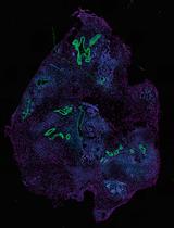
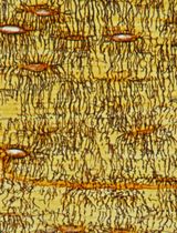
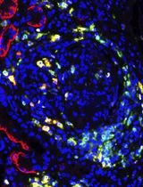
Abstract
Breast cancer is the most frequently diagnosed cancer in women, representing approximately 25% of all cancers in women worldwide. Both breast cancer research and histopathological diagnostics mainly show a two-dimensional planar view of the three-dimensional breast cancerous architecture. Recently, the application of optical tissue clearing, together with 3D microscopy, has been applied to visualize the complexity of whole tumor samples. Preliminary studies on whole-organ mouse mammary glands and tissues from human breast cancer patients subjected to optical tissue clearing and volumetric imaging have enabled the detection of previously unrecognized spatial cellular interactions and structural features within intact breast tissue. There is currently no standardized clearing workflow for breast and lymph node tissues. In this protocol, we optimized and validated the MASH (multiscale architectonic staining of human cortex) immunolabeling-enabled three-dimensional imaging of solvent-cleared organs (iDISCO)-like clearing and labeling pipeline for the investigation of formalin-fixed and paraffin-embedded (FFPE) breast tissue and lymph nodes obtained from breast cancer patients. This illustrates the application of the protocol in a new biological and clinical context, as human breast and lymph node tissues differ substantially from brain tissues in their composition, architecture, and optical properties. Whole FFPE tissue blocks are deparaffinized in liquid paraffin and xylene, bleached through methanol dehydration and a subsequent hydrogen peroxide incubation, and stained with a diverse set of small molecule dyes. As a next step, the tissues are delipidated and subjected to refractive index matching with ethyl cinnamate to reach optimal tissue transparency. Importantly, the applied dehydration and delipidation nicely preserve the morphology of the tissue, and the shrinkage is minimal. This allows reliable 3D imaging of large tissue samples within a timeframe of 10 days, providing clinicians and biomedical researchers with a more holistic view of the FFPE tissue sample and its spatial organization.
Key features
• Optimized, low-cost approach for optical tissue clearing and labeling of whole human breast and lymph node FFPE tissue samples ≥30 × 20 × 3 mm.
• Prepared tissue samples can be assessed by means of advanced 3D volume microscope modalities, such as twophoton microscopy (TPM) or light-sheet fluorescence microscopy (LSFM).
• The clearing protocol is compatible with a variety of small-molecule dyes, such as neutral red (cell body), eosin Y (cytoplasm, collagen), and methyl green (nucleus).
Keywords: Optical tissue clearing (组织光学透明化) Breast cancer (乳腺癌)
Lymph nodes (淋巴结)
Human cancer tissue samples (人体肿瘤组织样本)
FFPE tissue samples (福尔马林固定石蜡包埋组织样本)
Breast cancer (乳腺癌)
Lymph nodes (淋巴结)
Human cancer tissue samples (人体肿瘤组织样本)
FFPE tissue samples (福尔马林固定石蜡包埋组织样本)
Graphical overview
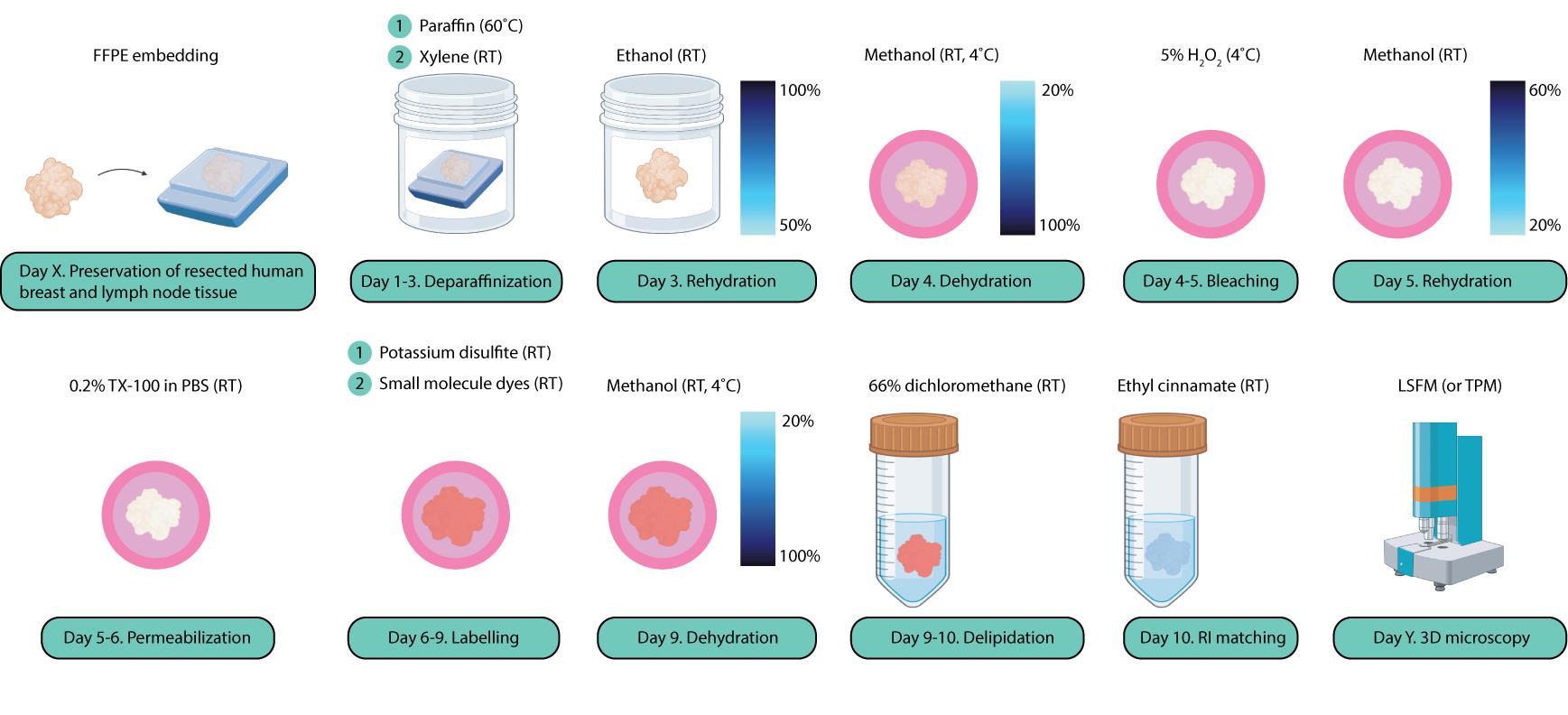
Schematic overview of the optical tissue clearing and small-molecule labeling steps of human breast cancer and lymph node tissue samples. This illustration was created using Adobe Illustrator and https://BioRender.com.
Background
With approximately 2.3 million new cases and over 670,000 deaths annually, breast cancer remains the leading type of cancer in women worldwide [1]. There are several distinct subtypes, with invasive breast carcinoma of no special type (NST) and invasive lobular carcinoma (ILC) being the most common [2]. To date, in the field of histopathology, clinicians and pathologists have relied on histological examinations of 3–5 μm-thin formalin-fixed and paraffin-embedded (FFPE) 2D tissue slices subjected to hematoxylin and eosin (H&E) staining. Despite recent advances in the level of early detection and precision medicine, patient numbers are increasing; therefore, new methodologies to improve diagnostics and patient therapy are essential [1]. At the same time, most breast cancer research is primarily focused on two-dimensional models, which only enable a limited, planar representation of the large-scale 3D breast cancer tissue architecture. As a result, key characteristics of large-scale human cancer tissue samples, such as the tumor volume, the high-resolution 3D tissue architecture, and the spatial organization of distinct cell populations, have remained largely unexplored due to the lack of suitable tools and methodologies.
Recent advances in optical tissue clearing, paired with light-sheet fluorescence microscopy (LSFM), are beginning to overcome the limitations of conventional slide-based breast cancer studies, enabling three-dimensional volume assessment of breast tissue samples [3]. Optical tissue clearing renders several millimeter thick tissues samples transparent, which circumvents scattering during image acquisition, resulting in deep microscopic imaging [4]. Barner et al. demonstrated that cleared, FFPE breast cancer lymph nodes imaged with LSFM offer a non-destructive, 3D alternative to traditional 2D histology with classic imaging modalities [5]. Cleared-tissue LSFM has also been applied to examine the terminal ductal lobular unit (TDLU) architecture and cell phenotypes in healthy breast tissue, contributing to our understanding of normal breast histology and its alterations in disease [6]. Despite these advances, workflows that combine optical tissue clearing and non-destructive 3D imaging of large FFPE human breast cancer tissues and their associated axillary lymph nodes remain underdeveloped. Robust and reproducible protocols for such tissues, both malignant and non-malignant, are still limited.
Here, we adapted and refined the MASH (multiscale architectonic staining of human cortex) immunolabeling-enabled three-dimensional imaging of solvent-cleared organs (iDISCO)-like optical tissue clearing and labeling protocol for human breast and lymph node tissues [7–9]. Our protocol demonstrates optical tissue clearing of complete, unsectioned FFPE tissue blocks of both breast and lymph nodes from breast cancer patients with dimensions of at least 30 × 20 × 3 mm. As the first step describes the deparaffinization of FFPE material, the protocol is applicable and suitable for archival tissue samples and their retrospective analyses. The combination of small molecule dyes with this optical tissue clearing protocol allows the specific visualization of subcellular compartments such as the nucleus, the lysosomes, and the cytoplasm. This allows for the establishment of a robust and broadly applicable 3D imaging workflow, which is less expensive and time-consuming than the use of immunohistochemistry. Antibody-based labeling has already been successfully combined with other iDISCO-based clearing protocols, and the list of verified antibodies is growing [10]. However, it has to be noted that other antibodies still have to be tested and verified; hence, here we focus on a universally transferable method. The cleared and labeled tissue specimen can eventually be examined with various microscope modalities, such as multiphoton or light-sheet fluorescence microscopy. This way, the potential for 3D analysis is harnessed and combined with LSFM advantages, including low levels of photobleaching, high imaging speed, and an exceptional signal-to-noise ratio [11]. In the future, this technique has the potential to aid in the investigation of immune cell infiltration, intratumoral heterogeneity, vascularization, and the spatial relations between malignant, stromal, and immune cell populations for both research and clinical investigations [12].
Materials and reagents
Biological materials
1. Human breast cancer samples of surgical specimens of the primary tumor site within the breast, preserved as FFPE tissue blocks, obtained from the Pathology Department of Maastricht University Medical Center+
2. Human lymph node samples of surgical specimens from breast cancer patients undergoing axillary surgery, preserved as FFPE tissue blocks, obtained from the Pathology Department of Maastricht University Medical Center+
Note: After the clearing process, tissue samples are stored in ethyl cinnamate in Falcon tubes at room temperature (RT) until imaging or in the cold room at 4 °C for longer storage.
Reagents
1. Liquid paraffin (histological grade) (Carl Roth, catalog number: 9190.1)
2. Xylene (Laboratorium Discounter, catalog number: XY834.1)
3. Ethanol, absolute (100%) (Carl Roth, catalog number: t913.2)
4. Methanol, absolute (100%) (Carl Roth, catalog number: 4627.4)
5. Milli-Q water
6. Phosphate-buffered saline (PBS), 0.1 M (Carl Roth, catalog number: 9143.2)
7. Thymol (Carl Roth, catalog number: 5391.1)
8. TritonTM X-100 (octylphenol ethoxylate) (Sigma-Aldrich, catalog number: X100)
9. 30% hydrogen peroxide (H2O2) (Carl Roth, catalog number: cp26.1)
10. Potassium disulfite (K2S2O5) (Carl Roth, catalog number: 7995.1)
11. Disodium hydrogen phosphate dihydrate (Na2HPO4·2H2O) (Carl Roth, catalog number: 27ke.1)
12. Citric acid monohydrate (C6H8O7·H2O) (Carl Roth, catalog number: 3958.2)
13. Neutral Red (NR) (Carl Roth, catalog number: T122.3)
14. Methyl Green (MG) (Sigma-Aldrich, catalog number: M8884)
15. Eosin Y (Morphisto, catalog number: 12199.00100)
16. 4′,6-Diamidino-2-phenylindole (DAPI) stock (1 mg/mL) (Carl Roth, catalog number: 6843.3)
17. Dichloromethane (DCM) (Carl Roth, catalog number: 8424.2)
18. Ethyl cinnamate (ECi) (Sigma-Aldrich, catalog number: 112372)
19. Chloroform (trichloromethane) (Carl Roth, catalog number: 6340.1)
Solutions
1. 50% (v/v) ethanol (see Recipes)
2. 70% (v/v) ethanol (see Recipes)
3. 20% (v/v) methanol (see Recipes)
4. 40% (v/v) methanol (see Recipes)
5. 50% (v/v) methanol (see Recipes)
6. 60% (v/v) methanol (see Recipes)
7. 80% (v/v) methanol (see Recipes)
8. 5% (v/v) hydrogen peroxide (H2O2) (see Recipes)
9. 50% (w/w) potassium disulfite solution (see Recipes)
10. McIlvaine buffer (phosphate-citrate buffer), pH 4.0 (see Recipes)
11. NR stock solution, 0.1% (w/v) (see Recipes)
12. MG stock solution, 4% (w/v) (see Recipes)
13. DAPI stock solution, 1 mg/mL (v/v) (see Recipes)
14. Dichloromethane:methanol (DCM) (2:1, v/v) (see Recipes)
15. 0.1 M PBS + 0.2% (v/v) Triton X-100 (see Recipes)
Recipes
See General note 1.
1. 50% (v/v) ethanol
| Reagent | Final concentration | Quantity or volume |
|---|---|---|
| Ethanol, absolute | 50% (v/v) | 50 mL |
| Milli-Q water | / | 50 mL |
| Total | / | 100 mL |
First, measure absolute ethanol (100%) using a graduated cylinder and dilute with Milli-Q water to the final volume. Then, mix thoroughly by gentle agitation. Prepare at the laboratory bench, away from ignition sources, due to ethanol flammability. Store absolute ethanol and all ethanol dilutions at RT in tightly closed, clearly labeled containers in a flammable-liquids storage cabinet, according to institutional safety regulations. Keep containers away from heat sources and open flames.
2. 70% (v/v) ethanol
| Reagent | Final concentration | Quantity or volume |
|---|---|---|
| Ethanol, absolute | 70% (v/v) | 70 mL |
| Milli-Q water | / | 30 mL |
| Total | / | 100 mL |
3. 20% (v/v) methanol
| Reagent | Final concentration | Quantity or volume |
|---|---|---|
| Methanol, absolute | 20% (v/v) | 20 mL |
| Milli-Q water | / | 80 mL |
| Total | / | 100 mL |
First, measure absolute methanol (100%) using a graduated cylinder and dilute with Milli-Q water to the final volume. Then, mix thoroughly by gentle agitation. Prepare in a chemical fume hood due to methanol toxicity and volatility. Store absolute methanol and all methanol dilutions at RT in tightly closed, clearly labeled containers in a flammable-liquids storage cabinet, according to institutional safety regulations. Due to methanol toxicity and volatility, ensure containers are clearly labeled and minimize opening time during use.
4. 40% (v/v) methanol
| Reagent | Final concentration | Quantity or volume |
|---|---|---|
| Methanol, absolute | 40% (v/v) | 40 mL |
| Milli-Q water | / | 60 mL |
| Total | / | 100 mL |
5. 50% (v/v) methanol
| Reagent | Final concentration | Quantity or volume |
|---|---|---|
| Methanol, absolute | 50% (v/v) | 50 mL |
| Milli-Q water | / | 50 mL |
| Total | / | 100 mL |
6. 60% (v/v) methanol
| Reagent | Final concentration | Quantity or volume |
|---|---|---|
| Methanol, absolute | 60% (v/v) | 60 mL |
| Milli-Q water | / | 40 mL (≈40 g) |
| Total | / | 100 mL |
7. 80% (v/v) methanol
| Reagent | Final concentration | Quantity or volume |
|---|---|---|
| Methanol, absolute | 80% (v/v) | 80 mL |
| Milli-Q water | / | 20 mL |
| Total | / | 100 mL |
8. 5% (v/v) H2O2
| Reagent | Final concentration | Quantity or volume |
|---|---|---|
| H2O2 stock | 30% (v/v) | 16.7 mL |
| Methanol | / | 83.3 mL |
| Total | 5% (v/v) | 100 mL |
To prepare a 5% (v/v) hydrogen peroxide solution, measure the required volume of 30% stock using a volumetric pipette and dilute in pre-cooled methanol. Add hydrogen peroxide slowly to the methanol while gently mixing by agitation. Prepare the solution in a chemical fume hood due to the strong oxidative properties of hydrogen peroxide and the potential release of oxygen gas. Protect the solution from light at all times. Prepare the diluted hydrogen peroxide solution immediately prior to use. It is not advised to store diluted solutions, as hydrogen peroxide rapidly decomposes upon dilution and light exposure. Store 30% (v/v) hydrogen peroxide stock at 4 °C in its original, vented, light-protected container, according to the manufacturer’s instructions.
9. 50% (w/v) potassium disulfite solution
| Reagent | Final concentration | Quantity or volume |
|---|---|---|
| K2S2O5 stock | 50% (w/w) | 50 g |
| Milli-Q water | / | 50 mL |
| Total | / | 100 mL |
First, weigh K2S2O5 using an analytical balance and then add Milli-Q water until just before the final volume. Stir with a magnetic stir while heating the solution up to approximately 60–70 °C. When dissolved, add the remaining Milli-Q to the final volume. Please note that the solution is oversaturated and the salt will precipitate again over time. Avoid inhalation of dust. Store at RT in a tightly sealed glass container. Directly before use, it is advised to stir the solution and heat it up on a magnet stirrer to dissolve the precipitate. This is followed by filtering the clear dissolved solution.
10. McIlvaine buffer (phosphate-citrate buffer), pH 4.0
Prepare 0.2 M Na2HPO4 and 0.1 M citric acid stock solutions separately by weighing salts on an analytical balance and dissolving them in Milli-Q water to the required volumes by mixing on a magnetic stir. Combine the two solutions in the specified ratio to achieve pH 4.0 and mix thoroughly. Prepare at the laboratory bench. Store the buffer at 4 °C.
See General note 2.
Step 1. 0.2 M Na2HPO4 stock solution
| Reagent | Final concentration | Quantity or volume |
|---|---|---|
| Na2HPO4·2H2O | 0.2 M | 35.447 g |
| Milli-Q water | / | 1,000 mL |
| Total | / | 1,000 mL |
Step 2. 0.1 M C6H8O7·H2O stock solution
| Reagent | Final concentration | Quantity or volume |
|---|---|---|
| C6H8O7·H2O | 0.1 M | 21.017 g |
| Milli-Q water | / | 1,000 mL |
| Total | / | 1,000 mL |
Step 3. Final phosphate-citrate buffer
| Reagent | Final concentration | Quantity or volume |
|---|---|---|
| 0.2 M Na2HPO4 solution | / | 40 mL |
| 0.1 M citric acid solution | / | 60 mL |
| Total | / | 100 mL |
11. NR stock solution, 0.1% (w/v)
| Reagent | Final concentration | Quantity or volume |
|---|---|---|
| NR | 0.1 mg/mL (0.1%) | 1 mg |
| McIlvaine buffer (pH 4.0) | / | 10 mL |
| Total | / | 10 mL |
First, prepare a 0.1% (0.1 mg/mL) stock solution by weighing NR using an analytical balance and dissolving it in McIlvaine buffer (pH 4.0). Mix gently until fully dissolved. Dilute the stock dilution 1:100 in McIlvaine buffer (pH 4.0) to obtain the final 0.001% working concentration. Prepare at the laboratory bench under reduced light conditions. Protect from light and store at 4 °C. Store NR powder at RT in a tightly closed container, protected from light and moisture. See General note 3.
12. MG stock solution, 4% (w/v)
| Reagent | Final concentration | Quantity or volume |
|---|---|---|
| MG | 40 mg/mL (4%) | 40 mg |
| McIlvaine buffer (pH 4.0) | / | 1 mL |
| Total | / | 1 mL |
First, prepare a highly concentrated 4% MG aqueous stock solution according to [13]. Remove crystal violet impurities by extractions with chloroform in a separation funnel, thereby discarding the lower (violet) phase until the lower chloroform phase no longer contains the violet color. Dilute the stock solution 1:5,000 in McIlvaine buffer (pH 4.0). Prepare at the laboratory bench under reduced light conditions. Protect from light and store at 4 °C. Store MG powder at RT in a tightly sealed container, protected from light and humidity. See General note 3.
13. DAPI stock solution, 1 mg/mL (v/v)
| Reagent | Final concentration | Quantity or volume |
|---|---|---|
| DAPI | 1 mg/mL | 10 mg |
| McIlvaine buffer (pH 4.0) | / | 10 mL |
| Total | / | 10 mL |
First, prepare a 1 mg/mL stock solution by weighing DAPI using an analytical balance and dissolving it in McIlvaine buffer (pH 4.0). Mix gently until fully dissolved and minimize light exposure. Store DAPI stock solutions at -20 °C, protected from light, according to the manufacturer’s instructions. Dilute the DAPI stock solution 1:1,000 in McIlvaine buffer (pH 4.0) to obtain the final 1 μg/mL working solution. Prepare at the laboratory bench immediately prior to use. See General note 3.
14. DCM (2:1, v/v)
| Reagent | Final concentration | Quantity or volume |
|---|---|---|
| DCM | 66% (v/v) | 66.7 mL |
| Methanol, absolute | 33% | 33.3 mL |
| Total | / | 100 mL |
Measure DCM and methanol using glass graduated cylinders and combine in a 2:1 (v/v) ratio. It is advised to prepare this strictly in a chemical fume hood due to the high volatility and toxicity of both dichloromethane and methanol. Stir until the inhomogeneities disappear. Store dichloromethane in tightly closed, chemically compatible containers at RT in a dedicated halogenated-solvent storage cabinet, separate from flammable solvents. Ensure containers are clearly labeled and kept away from heat sources. See General notes 4 and 5.
15. 0.1 M PBS + 0.2% (v/v) Triton X-100
| Reagent | Final concentration | Quantity or volume |
|---|---|---|
| Triton X-100 | 0.2% (v/v) | 0.2 mL |
| PBS | 0.1 M | 99.8 mL |
| Total | / | 100 mL |
Prepare 0.1 M PBS using MilliQ water. Triton X-100 is highly viscous, which can complicate pipetting. To facilitate handling, the end of the pipette tip may be cut. Solutions containing Triton X-100 should not be shaken; instead, gently stir and allow the detergent to dissolve passively, which may take up to 1 h.
Laboratory supplies
1. Glass containers, 100, 250, and 500 mL
2. Glass Petri dish
3. Beakers, 50 and 200 mL
4. Erlenmeyer flask, 50 mL
5. Graduated cylinders, 25, 50, 100, 250, and 500 mL
6. 50 mL Falcon tubes
7. Six-well cell culture plates
8. Pipettes, 0.2, 1, and 5 mL, with compatible pipette tips
9. Forceps
10. Filter paper
11. Funnel
12. Millimeter paper
13. Nitrile gloves
Equipment
1. Water bath, set to 60 °C (Gesellschaft für Labortechnik mbH, model: 1002)
2. Fume hood (Vinitex Laboratoriuminrichtingen B.V., model: L1K1-GR+51)
3. Chemical waste disposal system (institutional facility)
4. Orbital shaker (Polymax, model: 1040)
5. Cold room or refrigerated storage, 4 °C
6. Analytical balance (Mettler Toledo, model: PG2002-S DeltaRange)
7. Magnetic stirrer (Framo®-Gerätetechnik, model: M21/1)
8. Led-light pad (optional) (Crafts & Co, Karsten International, catalog number: 973)
9. Two-photon (2P) laser scanning microscope (Leica TCS SP5 MP, Leica Mikrosysteme Vertrieb GmbH)
10. UltraMicroscope II light-sheet microscope (La Vision Biotech, Miltenyi)
Software and datasets
1. Leica Application Suite Advanced Fluorescence (Leica Microsystems), for Leica 2-photon microscopy data
2. Imaris Biplane 11.0, for 2-photon microscopy and LSFM 3D volume data
3. FIJI (open-source Bio-image analysis software)
Procedure
登录/注册后免费查看全文
文章信息
稿件历史记录
提交日期: Apr 1, 2026
接收日期: Jul 2, 2026
在线发布日期: Jul 24, 2026
出版日期: Aug 20, 2026
版权信息
© 2026 The Author(s); This is an open access article under the CC BY license (https://creativecommons.org/licenses/by/4.0/).
如何引用
Schiffelers, L. D. J., Pisarović, U., Bitorina, A., Hildebrand, S., Walravens, B., van Nijnatten, T. J. A., Kooreman, L. F. S. and Schueth, A. (2026). Optical Tissue Clearing and Small-Molecule Labeling of Paraffin-Embedded Breast Cancer and Axillary Lymph Node Human Tissue Samples. Bio-protocol 16(16): e5790. DOI: 10.21769/BioProtoc.5790.

 Download Citation in RIS Format
分类
癌症生物学 > 通用技术 > 肿瘤微环境
细胞生物学 > 组织分析 > 组织成像
细胞生物学 > 组织分析 > 组织染色
您对这篇实验方案有问题吗?
在此处发布您的问题,我们将邀请本文作者来回答。同时,我们会将您的问题发布到Bio-protocol Exchange,以便寻求社区成员的帮助。
 发布问题
发布问题


