


A Simple and Reproducible ImageJ Workflow for Measuring Areas of Irregularly Shaped Necrotic Lesions on Plant Leaves
利用 ImageJ 测量植物叶片不规则坏死病斑面积的简便可重复流程

发布: 2026年08月20日第16卷第16期 DOI: 10.21769/BioProtoc.5788 浏览次数: 410
评审: Shweta PanchalPawan KumarAnonymous reviewer(s)
 PDF
PDF  Q&A
Q&A





Abstract
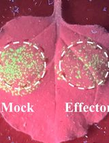
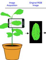
Accurately quantifying the areas of necrotic lesions on plant leaves is essential for evaluating plant–pathogen interactions and disease resistance. Although digital image analysis methods using ImageJ are widely employed, they often require case-specific optimization and may not be readily applicable across different experimental conditions. Furthermore, many studies have used ImageJ for lesion measurement without providing methodological details, which limits reproducibility. Here, we present a simple, step-by-step ImageJ workflow for measuring irregular necrotic lesions using a standard personal computer and mouse. The procedure relies on manual lesion selection using the freehand selection tool, followed by Gaussian smoothing, binarization, and automated particle analysis to extract lesion area measurements. By balancing manual isolation with computational thresholding, this protocol eliminates the need for extensive parameter tuning. This approach provides an accessible, reliable alternative to time-consuming color thresholding methods, thereby improving transparency and reproducibility in lesion quantification. The workflow's reproducibility has been confirmed through both intra-user and inter-user analyses.
Key features
• Relies on simple manual freehand selection combined with minimal image processing, requiring only a standard computer and mouse without specialized software or advanced training.
• Enables accurate quantification of irregular necrotic lesions in conditions where automated thresholding methods require time-consuming optimization.
• Provides a fully detailed, reproducible ImageJ workflow addressing common gaps in published methods, facilitating direct implementation.
• Demonstrates high reproducibility, validated by intra-user and inter-user statistical analyses, ensuring reliable lesion quantification regardless of the operator.
Keywords: ImageJ/Fiji (ImageJ/Fiji) Necrotic lesion quantification (坏死病斑定量)
Lesion area measurement (病斑面积测量)
Leaf disease analysis (叶片病害分析)
Plant phenotyping (植物表型分析)
Plant pathology (植物病理学)
Necrotic lesion quantification (坏死病斑定量)
Lesion area measurement (病斑面积测量)
Leaf disease analysis (叶片病害分析)
Plant phenotyping (植物表型分析)
Plant pathology (植物病理学)
Background
Quantification of lesion area is widely used in plant pathology to assess disease severity, compare pathogen virulence, and evaluate host resistance in plant–pathogen interaction studies [1]. Necrotic lesions on leaves are common readouts in experimental systems involving viral, bacterial, and fungal pathogens, making accurate and reproducible lesion measurement important for comparative analyses across genotypes, treatments, and time points [2].
Several approaches have been used to quantify lesion areas, including visual scoring, manual tracing, and, more recently, digital image analysis, which has become the conventional method [3]. Among digital tools, ImageJ has become widely adopted because it is easy to use, freely available, and frequently used for color-based segmentation approaches such as global thresholding [4]. However, color-based threshold methods can perform poorly when lesions are heterogeneous in color, have diffuse or irregular borders, or are captured under non-uniform lighting conditions. Under such conditions, lesions may be incompletely detected, adjacent lesions may be merged, or healthy tissues with similar color may be misclassified as diseased areas. While advanced machine learning and automated segmentation tools offer powerful alternatives to thresholding [5], they frequently require specialized computational expertise, extensive training datasets, or high-end hardware. Furthermore, studies reporting ImageJ-based lesion quantification often describe the software without specifying workflows or parameters, hampering reproduction by readers.
The protocol described here addresses these gaps by providing a highly controlled and standardized manual ImageJ workflow for quantifying irregular necrotic lesions on plant leaves. It requires no specialized equipment beyond standard image acquisition and freely available software and is well-suited for experiments involving individual lesions or a moderate number of lesions per sample. Compared with automated color thresholding–based methods, this approach offers controlled precision in lesion boundary selection, particularly when lesion shape, color, or contrast complicates segmentation. Although the workflow becomes labor-intensive when lesions are extremely numerous, it remains well-suited to datasets with several tens of lesions per leaf. This manual ImageJ-based approach can be adapted to quantify other visible, localized plant symptoms on the leaves of different plant species. For example, the workflow can measure symptoms caused by diverse pathogens that induce localized tissue damage, necrosis, or pathogen-associated mycelial growth. It is also applicable to distinct discolorations on leaves of varying colors (such as chlorotic spots), as well as physical deformations like hypertrophic regions or insect herbivory (chewing damage). However, the suitability of this approach depends on how clearly symptom boundaries can be defined.
Materials and reagents
Biological materials
1. Nicotiana tabacum plants harboring the N′ resistance gene [6]
2. Tomato mosaic virus (ToMV).
Note: Necrotic lesions were generated by mechanical inoculation using carborundum as an abrasive and applying 20 μL of ToMV solution at 1 μg/mL on the fourth true leaves of Nicotiana tabacum. Plants were maintained at 25 °C under a 16 h light/8 h dark photoperiod with a light intensity of 60–100 μmol/m2/s. Leaves were scanned at 7 days post-inoculation, when necrotic lesions were clearly visible.
Equipment
1. CCD-type flatbed scanner (e.g., Epson, model: GT-X820)
2. Standard metric ruler
3. Computer systems: User A, CPU: Intel® CoreTM i7-8650U @ 1.90 GHz; RAM: 16 GB; GPU: Intel® UHD Graphics 620; OS: Windows 11 Pro (64-bit); monitor: Acer KA242Y, 23.8-inch (connected via HDMI); and User B, CPU: Intel® CoreTM i7-11800H @ 2.30 GHz; RAM: 16 GB; GPU: NVIDIA® GeForce RTX 3050 Laptop GPU (4 GB); OS: Windows 11 Pro (64-bit); monitor: Philips 234E SoftBlue, 23-inch (connected via HDMI).
Note: Use of a large display screen is recommended to facilitate accurate visualization and manual tracing of lesion boundaries. Zooming in during selection improves precision, particularly for irregular lesion edges.
4. Standard computer mouse (highly recommended over a laptop trackpad for accurate freehand selection).
Note: Use of a mouse pad is recommended to improve cursor stability during manual lesion tracing.
Software and datasets
1. ImageJ or Fiji, a package of ImageJ; Available for free download at https://imagej.net/ij/ or https://imagej.net/software/fiji/ (v1.54s)
Procedure
登录/注册后免费查看全文
文章信息
稿件历史记录
提交日期: May 2, 2026
接收日期: Jul 8, 2026
在线发布日期: Jul 24, 2026
出版日期: Aug 20, 2026
版权信息
© 2026 The Author(s); This is an open access article under the CC BY-NC license (https://creativecommons.org/licenses/by-nc/4.0/).
如何引用
Bensedira, H. E. S. and Chehaba, O. K. (2026). A Simple and Reproducible ImageJ Workflow for Measuring Areas of Irregularly Shaped Necrotic Lesions on Plant Leaves. Bio-protocol 16(16): e5788. DOI: 10.21769/BioProtoc.5788.

 Download Citation in RIS Format
分类
植物科学 > 植物免疫 > 病害症状
植物科学 > 植物免疫 > 病害生物测定
植物科学 > 植物生理学
您对这篇实验方案有问题吗?
在此处发布您的问题,我们将邀请本文作者来回答。同时,我们会将您的问题发布到Bio-protocol Exchange,以便寻求社区成员的帮助。
 发布问题
发布问题


