


Calcium Imaging in H4IIE Liver Cells and Primary Rodent Hepatocytes: A Cost-Effective Protocol for Use With Fura-2 AM Ca2+ Indicator
H4IIE肝细胞和啮齿类原代肝细胞的钙成像:一种基于Fura-2 AM Ca²⁺指示剂的低成本实验方法

(*contributed equally to this work) 发布: 2026年08月20日第16卷第16期 DOI: 10.21769/BioProtoc.5783 浏览次数: 760
评审: Swetha MurthygowdaAnonymous reviewer(s)
 PDF
PDF  Q&A
Q&A
参见作者原研究论文
The authors used this protocol in:
Jul 2021





Abstract
Calcium signaling is a universal, versatile process in which ionized or free calcium (Ca2+) acts as a second messenger to regulate various cellular activities, including hormone secretion, contraction, proliferation, gene expression, and apoptosis. Changes in the cytoplasmic free Ca2+ concentration ([Ca2+]cyt) in hepatocytes play a central role in mediating the actions of insulin, glucagon, catecholamines, and other hormones on carbohydrate, lipid, and protein metabolism in the liver. Ratiometric chemical Ca2+ indicators are fluorescent dyes that change their emission or excitation spectrum upon binding to calcium, allowing for precise, quantitative measurements of changes in the intracellular Ca2+ concentration. They enable calibration by calculating the ratio of two fluorescence intensities, correcting for artifacts such as uneven dye loading, photobleaching, and cell volume variations. Fura-2 acetoxymethyl ester (AM) (hereinafter referred to as Fura-2), a ratiometric and sensitive indicator dye, is a popular fluorescent Ca2+ reporter for measuring intracellular calcium. Here, we describe a comprehensive and detailed protocol for Ca2+ imaging of the H4IIE cell line and primary rodent hepatocytes in vitro via the chemical reporter Fura-2, which can also be employed on a wide variety of cell types. Unlike previously published protocols, this protocol addresses the challenge of facilitating the attachment of liver cell lines and primary hepatocytes to glass coverslips for imaging using an inverted fluorescence microscope. Our protocol describes two different loading/labeling strategies for Fura-2 dye: one is cost-effective but requires skillful pipettor handling, and the second one is easy but expensive as it needs a large volume of Krebs-Ringer HEPES (KRH)-Fura-2 solution. If the coverslips are handled properly, the cost-effective coverslip-only loading approach produces similar quality results as the large volume method. Finally, we describe a simple and user-friendly procedure to analyze Ca2+ signals over time using Microsoft Excel’s functional equations.
Key features
• This protocol enables real-time monitoring of intracellular Ca2+ in living hepatocytes.
• The method is based on fluorescence microscopy of hepatocytes loaded with Fura-2 fluorescent dye; moreover, the protocol can be extended to other mammalian cell lines.
• We used this protocol to monitor the activity of store-operated Ca2+ channels or SERCA pumps, but it could also be extended to other intracellular phenomena.
• If the Fura-2 loading strategy and incubation time are followed properly, this protocol produces highly reproducible imaging results.
Keywords: Calcium signaling (钙信号) Single-cell imaging (单细胞成像)
H4IIE liver cells (H4IIE肝细胞)
Hepatocytes (肝细胞)
Store-operated Ca2+ entry (储存操纵性Ca²⁺内流)
Fura-2 AM (Fura-2 AM)
Single-cell imaging (单细胞成像)
H4IIE liver cells (H4IIE肝细胞)
Hepatocytes (肝细胞)
Store-operated Ca2+ entry (储存操纵性Ca²⁺内流)
Fura-2 AM (Fura-2 AM)
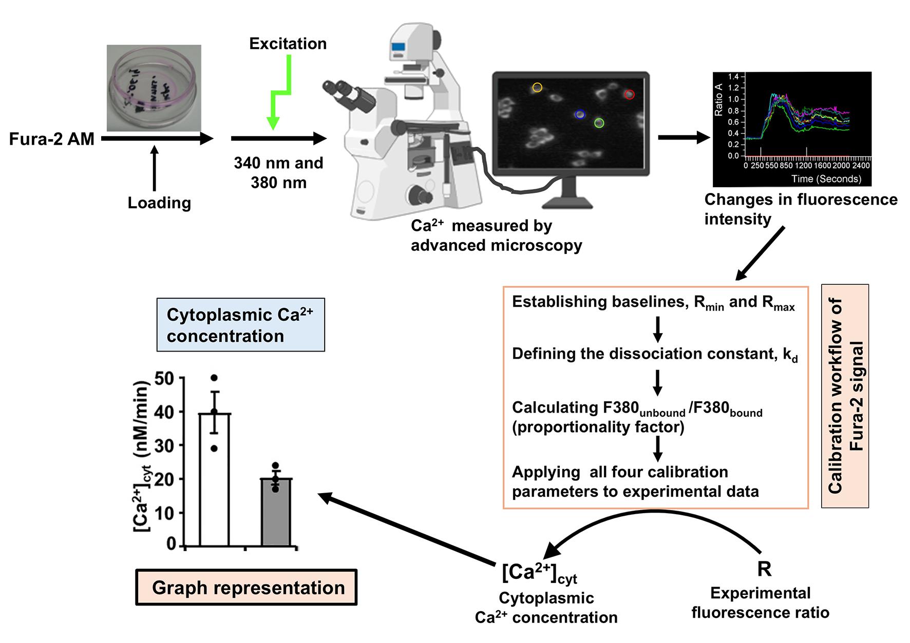
Graphical overview
Background
Intracellular calcium plays a central role in regulating key cellular processes, including transcription, signal transduction, excitability, motility, and apoptosis in hepatocytes [1,2]. The concentration of free ionized Ca2+ in the cytoplasmic space of hepatocytes is approximately 10-7 M (0.1 μM) at rest, whereas a large proportion (99%) of total Ca2+ in the cytoplasmic space is bound to proteins and metabolites [3]. Ca2+ is stored (accumulated) in the endoplasmic reticulum (ER), sarcoplasmic reticulum, mitochondria, and other intracellular organelles. For example, the total Ca2+ concentration in the ER can be 10 mM, with a free Ca2+ concentration of 10 μM. Hormones, neurotransmitters, and other extracellular signals increase cytoplasmic free Ca2+ concentration ([Ca2+]cyt) from 0.1 to 1–5 μM [4]. Since the hormonal and metabolic cues are associated with dynamic fluctuations of intracellular Ca2+, accurate quantification of cytosolic Ca2+ dynamics has been central to interpreting liver-specific signaling pathways. Intracellular fluorescent Ca2+ reporters provide one of the best techniques to measure the free Ca2+ concentration in the cytoplasmic space and organelles. Fura-2 acetoxymethyl ester (Fura-2 AM), which enables calibrated measurements of absolute intracellular Ca2+ concentrations in isolated cells, is one of the most widely used fluorescent indicators for intracellular Ca2+ imaging.
With a variety of calcium indicators available, such as fluorescence dyes including Fluo-3, Fluo-4, Fura-2, and Indo-1, or genetically encoded calcium indicators (e.g., GCaMP), it is important to consider the specific requirements of the experiment, including the type of cells being investigated, the desired emission wavelengths, and the sensitivity needed for detecting calcium binding events. Since chemical Ca2+ indicators are not required to be transfected or expressed into cells, they provide an unparalleled flexibility in experimental design for tracking intracellular calcium dynamics, largely outperforming genetically encoded indicators in terms of commercial availability and customization. The main advantage of using ratiometric dyes (e.g., Fura-2) compared to single-wavelength probes (e.g., Fluo-3) is that the ratio signal remains unchanged by variations in dye concentration, optical path length, and/or light intensity. This property of ratiometric dyes permits investigators to determine the intracellular calcium concentration with greater accuracy. The primary advantage of Fura-2 over other calcium reporters is its ratiometric capability, which calculates calcium concentration by taking the ratio of fluorescence emitted at 340 and 380 nm excitation [5–7]. This mathematically cancels out experimental artifacts like uneven dye loading, cell thickness variations, and photobleaching, allowing for absolute calcium quantification [4]. While both Fura-2 and Indo-1 are ratiometric indicators, Fura-2 remains the superior choice for fluorescence imaging because of its high resistance to photobleaching compared to the Indo-1 reporter [8]. The principle for the measurement of cytoplasmic Ca2+ using this Ca2+ fluorescent dye is that the acetoxymethyl ester (AM) form of the fluorescent dyes can diffuse passively across the cell membrane (and intracellular membranes); once the dye is inside the cell, esterases cleave off the AM groups [5,6]. Intracellular or cytoplasmic Ca2+ binds to Fura-2 free acid and provides fluorescence intensity (Figure 1).
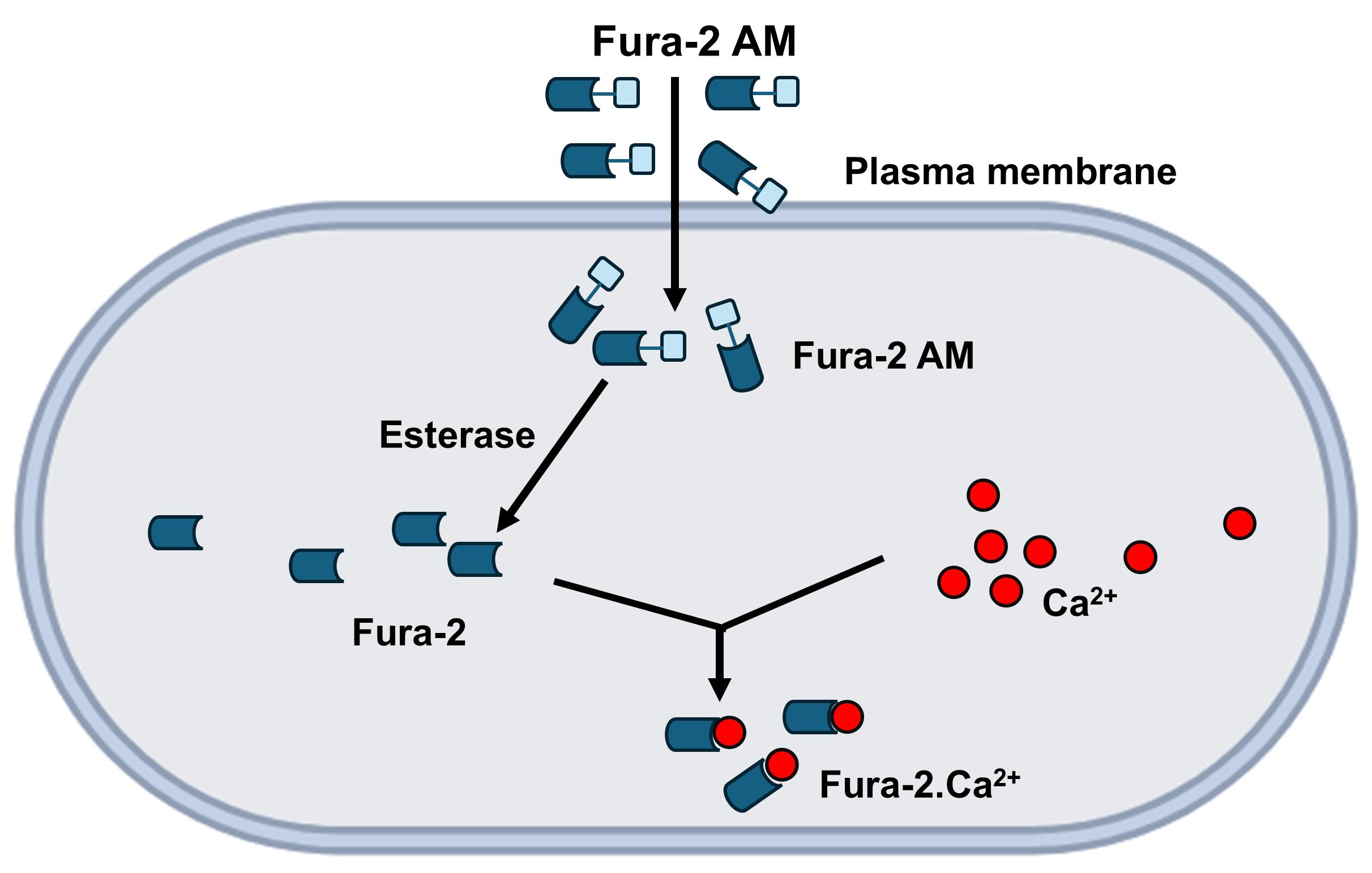
Figure 1. Principles of Fura-2 AM calcium imaging. Once the Fura-2 AM dye is inside the cell, intracellular esterases cleave the AM groups [5,6]. Intracellular or cytoplasmic Ca2+ can bind to Fura-2 free acid, providing fluorescence intensity.
One important methodological gap in existing Fura-2 imaging protocols and tutorials is the lack of a framework for relating image-based measurements to quantitative interpretation. The Grynkiewicz formula (for details, see section E of this protocol) is used to accurately calculate intracellular free Ca2+ concentrations using ratiometric fluorescent indicators like Fura-2 [9]. Eventually, the formula translates background-corrected fluorescence ratios into a reliable Ca2+ concentration. In this protocol, to successfully use the formula in calibration-based analysis, we have followed a specific protocol (ionomycin-EGTA assay) to acquire the values of different variables and parameters used in the formula. In the case of the image-based implementation workflow, before calculating the ratio, we have subtracted background noise (from optics or autofluorescence) for each excitation wavelength to prevent large calculation errors. With the imaging software, we also computed the pixel-by-pixel intensity ratio 340 nm/380 nm across the entire image through the selection of 10–15 regions of interest. This ratiometric approach inherently corrects for uneven dye loading, minor dye leakage, and photobleaching. Finally, we performed the calibration assay to convert the Fura-2 dye fluorescence ratio into the cytoplasmic Ca2+ concentration.
Our group used Fura-2 dye to examine Ca2+ signaling by fluorescence microscopy in hepatocytes isolated from Alms1 mutant mice [10], hepatocytes from obese Zucker rats and Hooded Wistar rats [11], and H4IIE rat liver cell lines [12]. Hepatocytes can be easily isolated and purified from rat or mouse liver tissue. Freshly isolated primary hepatocytes, however, only survive for a few hours in non-adherent suspension culture [13,14] and are not amenable to attaching onto commercially available coverslips. This protocol addresses the well-known challenge of cell adhesion by employing an acid (hydrochloric acid, 1 M) washing strategy. As attachment-dependent cells, acid washing provides conditions on the coverslips in which primary hepatocytes can survive. Hydrochloric acid (1 M) washing removes microscopic debris, dust, and toxic manufacturing residues from the glass surface of coverslips, ensuring a chemically sterile environment. The acid treatment hydroxylates the glass surface and yields a highly charged, hydrophilic surface that facilitates robust bonding of extracellular matrix (ECM) proteins of hepatocytes [15]. From our experience, if the acid washing protocol for coverslip is appropriately followed, freshly isolated hepatocytes are shown to adhere and survive up to 36–40 h. It is worth noting that the acid washing strategy is reasonably cost-effective compared to the collagen- or Polylysine-coating method. Furthermore, in this protocol, we have adopted a new and cost-effective Fura-2 dye loading approach, which produces similar quality results as the traditional method using a large dye volume.
The detailed step-by-step protocol described below, using chemical Ca2+ indicators, can also be readily adapted to various suspensions and other adherent cell types.
Materials and reagents
Biological materials
1. H4IIE rat hepatoma cells (ATCC CRL-1548)
Note: The H4IIE is an epithelial rat hepatoma cell line widely used in toxicology, cancer research, and pharmacology. Derived from the Reuber H-35 rat hepatoma, it serves as a critical in vitro model for evaluating how the liver responds to toxins, metabolic regulation, and drug-induced enzymatic activities [11,12].
2. Freshly isolated primary hepatocytes (isolated in-house from C57BL/6J Alms1 mouse; Hooded Wistar rat, Obese Zucker rat [10–12])
Reagents
1. Dulbecco's modified Eagle medium (DMEM) (Thermo Fisher Scientific, catalog number: 11-965-092)
2. F-12 (Thermo Fisher Scientific, catalog number: 11-765-054)
3. Fetal bovine serum (FBS) (Bovogen Biologicals, Keilor East VIC, catalog number: SFBS-AU)
4. Glucose (Millipore Sigma, catalog number: G7021)
5. Dimethyl sulfoxide (DMSO) (Thermo Fisher Scientific, catalog number: D12345)
6. Penicillin-Streptomycin (Gibco, catalog number: 15140122)
7. Phosphate-buffered saline (PBS), cell culture grade (Corning, catalog number: 21-040-CV)
8. Trypsin–ethylenediaminetetraacetic acid (Trypsin-EDTA) (Millipore Sigma, catalog number: T4049)
9. Pluronic acid, F-127 (Thermo Fisher Scientific, catalog number: P3000MP)
10. Fura-2 AM (Thermo Fisher Scientific, catalog number: F3021)
11. Ethylene glycol-bis(2-aminoethylether)-N,N,N′,N′-tetraacetic acid (EGTA) (Sigma-Aldrich, catalog number: E8145)
12. 4-(2-hydroxyethyl)-1-piperazineethanesulfonic acid (HEPES); chemical formula: C8H18N2O4S (EMD Millipore, catalog number: 391338)
13. Dexamethasone (Sigma-Aldrich, catalog number: D4902)
14. Insulin (Millipore Sigma, catalog number: 91077C)
15. Sodium chloride (NaCl) (Sigma-Aldrich, catalog number: S9888)
16. Potassium chloride (KCl) (Millipore Sigma, catalog number: P5405)
17. Calcium chloride (CaCl2) (Millipore Sigma, catalog number: C5670)
18. Magnesium chloride (MgCl2) (Millipore Sigma, catalog number: M4880)
19. Sodium hydroxide (NaOH) (Sigma-Aldrich, catalog number: S2770)
20. Hydrochloric acid (HCl) (Sigma-Aldrich, catalog number: 320331)
21. Ethanol 200-proof (Westlab, catalog number: AL048X)
22. Collagenase Type IV (working concentration: 0.2 mg/mL) (Worthington Biochemical Corporation, catalog number: CLS-4)
23. Digitonin (Sigma-Aldrich, catalog number: D141)
24. Tris(hydroxymethyl)aminomethane (Tris base) (Sigma-Aldrich, catalog number: 252859)
25. Ionomycin (Sigma-Aldrich, catalog number: AABH9A95673F)
26. 2,5-di-tert-butylhydroquinone (DBHQ) (Sigma-Aldrich, catalog number: 112976)
Solutions
1. Krebs Ringer’s (KRH) solution (see Recipes)
2. Calcium-free KRH solution (see Recipes)
3. Pluronic acid 20% (w/v) in DMSO (see Recipes)
4. Complete DMEM medium (see Recipes)
5. Growth medium (see Recipes)
6. Attachment medium (see Recipes)
Recipes
1. KRH solution
| Reagent | Final concentration | Quantity or volume (for 500 mL) |
|---|---|---|
| NaCl | 136 mM | 3.97 g |
| KCl | 4.7 mM | 0.175 g |
| CaCl2 | 2.4 mM | 0.133 g |
| MgCl2 | 1.25 mM | 59.51 mg |
| HEPES (stock 1 M) | 10 mM | 5 mL |
| Glucose | 10 mM | See note |
Prepare the KRH solution in ddH2O and adjust to pH 7.4 with NaOH. Store at room temperature.
Note: Prepare KRH without glucose and add the required amount of glucose on the day of the experiment. For example, to prepare 50 mL of KRH working buffer, add 90.1 mg of glucose to the tube containing 50 mL of KRH. For the calibration experiment, 5 mM CaCl2 is used instead of 2.4 mM CaCl2.
2. Calcium-free KRH solution
| Reagent | Final concentration | Quantity or volume (for 500 mL) |
|---|---|---|
| NaCl | 136 mM | 3.97 g |
| KCl | 4.7 mM | 0.175 g |
| MgCl2 | 1.25 mM | 59.51 mg |
| HEPES (stock 1 M) | 10 mM | 5 mL |
| EGTA | 1 mM | 0.19018 g |
| Glucose | 10 mM | See note |
Prepare the Ca2+-free KRH solution in ddH2O and adjust to pH 7.4 with NaOH. Store at room temperature.
Note: KRH buffer is prepared with or without Ca2+ (CaCl2) according to the experimental need. Prepare KRH without glucose and add the required amount of glucose on the day of the experiment.
3. Pluronic acid 20% (w/v) in DMSO
Add 0.2 g of Pluronic acid to 1 mL of DMSO and vortex until dissolved. Pluronic acid is difficult to dissolve, so vortex continuously for ~30 min or until dissolved. Keep at room temperature.
4. Complete DMEM medium
| Reagent | Final concentration | Volume (for ~500 mL) |
|---|---|---|
| DMEM | 89% | 445 mL |
| FBS | 10% (v/v) | 50 mL |
| Penicillin/Streptomycin (Penicillin 10,000 units/mL and Streptomycin 10,000 μg/mL) | 100 units/mL (penicillin), 0.1 mg/mL streptomycin | 5 mL |
5. Growth medium
| Reagent | Final concentration | Volume (for ~500 mL) |
|---|---|---|
| DMEM | 44% | 220 mL |
| F-12 | 44% | 220 mL |
| FBS | 10% (v/v) | 50 mL |
| Penicillin/Streptomycin (Penicillin 10,000 units/mL and Streptomycin 10,000 μg/mL) | 100 units/mL (penicillin), 0.1 mg/mL streptomycin | 5 mL |
| HEPES (stock 1 M) | 10 mM | 5 mL |
For preparing FBS aliquots, thaw frozen FBS (500 mL bottle) in a 37 °C water bath. In the laminar flow hood, prepare 10× 50 mL aliquots. For penicillin/streptomycin (concentration 100×), after thawing, prepare 5 mL aliquots. Additionally, prepare HEPES stock buffer 1 M by dissolving 47.66 g of HEPES in 200 mL of ddH2O, and adjust the pH to 7.4. Then, filter-sterilize in a laminar flow hood. Finally, prepare 500 mL of growth medium containing DMEM and F-12 (1:1), supplemented with 10% (v/v) FBS, penicillin (100 units/mL), streptomycin (0.1 mg/mL), and 10 mM HEPES.
6. Attachment medium
| Reagent | Final concentration | Volume (for ~500 mL) |
|---|---|---|
| DMEM | 44% | 220 mL |
| F-12 | 44% | 220 mL |
| FBS | 10% (v/v) | 50 mL |
| Penicillin/Streptomycin (Penicillin 10,000 units/mL and Streptomycin 10,000 μg/mL) | 100 units/mL (penicillin), 0.1 mg/mL streptomycin | 5 mL |
| HEPES (1 M stock) | 10 mM | 5 mL |
| Dexamethasone (100 μM stock) | 100 nM | 500 μL |
| Insulin (100 μM stock) | 100 nM | 500 μL |
Prepare 500 mL of attachment medium containing DMEM and F-12 (1:1), supplemented with 10% (v/v) FBS, penicillin (100 units/mL), streptomycin (0.1 mg/mL), 10 mM HEPES, and the required amount of dexamethasone (final concentration 100 nM) and insulin (final concentration 100 nM).
Laboratory supplies
1. 35 mm TC-treated culture dish (Corning, catalog number: 430165)
2. 22 mm diameter, 1-mm thick glass coverslip (Oxford Instruments, catalog number: 51-1625-0129)
3. Gloves (Westlab, catalog number: 100714-3007)
4. Forceps (Westlab, catalog number: 663-938)
5. Cell scrapers (TH Geyer, catalog number: 7696760)
6. 15 mL conical tubes (TH Geyer, catalog number: 7696714)
7. 50 mL conical tubes (Greiner Bio-One, catalog number: 227261)
8. 5 mL serological pipettes (Greiner Bio-One, catalog number: 606180)
9. 10 mL serological pipettes (Greiner Bio-One, catalog number: 607180)
10. 20 μL pipette tips (TH Geyer, catalog number: 7695882)
11. 200 μL pipette tips (TH Geyer, catalog number: 7695884)
12. 1,250 μL pipette tips (TH Geyer, catalog number: 7695887)
13. Racks (John Morris Scientific, catalog number: 0030119819)
14. Glass beaker (John Morris Scientific, catalog number: 632417010400)
15. Aluminum foil (John Morris Scientific/LabFriend, catalog number: 60032)
16. Silicon grease (Molykote/Dow Corning, catalog number: 408524)
17. 75 cm2 sterile flasks (Greiner Bio-One, catalog number: 658175)
Equipment
1. Nikon TE300 Eclipse microscope equipped with a Sutter DG-4 (this Sutter DG4 also combines a high-intensity xenon light source) wavelength switcher (to generate alternating excitation light 340 and 380 nm), Omega XF04 filter set for Fura-2 Photonic Science ISIS-3 ICCD camera
2. CO2 incubator
3. Computer system equipped with MetaFluor software
4. Freezer (-20 °C)
5. Refrigerator (2–8 °C)
6. Liquid nitrogen (N2) tank
7. Water bath (Westlab, catalog number: 663-311)
8. Tissue culture hood (NuAire, catalog number: NU-978-002)
9. Fume hood (Westlab, catalog number: 665-846)
10. Oven
11. Centrifuge (Eppendorf, model: Centrifuge 5420, catalog number: 5420000245D)
Software and datasets
1. MetaFluor suite (https://imagxcell.com/metafluor)
2. Microsoft Excel (https://www.microsoft.com/en-us/microsoft-365/excel)
3. GraphPad Prism (https://www.graphpad.com/)
Procedure
登录/注册后免费查看全文
文章信息
稿件历史记录
提交日期: May 15, 2026
接收日期: Jul 5, 2026
在线发布日期: Jul 22, 2026
出版日期: Aug 20, 2026
版权信息
© 2026 The Author(s); This is an open access article under the CC BY-NC license (https://creativecommons.org/licenses/by-nc/4.0/).
如何引用
Fang, L., Habib, A. G. K., Harris, M. D., Begley, A. J. and Ali, E. S. (2026). Calcium Imaging in H4IIE Liver Cells and Primary Rodent Hepatocytes: A Cost-Effective Protocol for Use With Fura-2 AM Ca2+ Indicator. Bio-protocol 16(16): e5783. DOI: 10.21769/BioProtoc.5783.

 Download Citation in RIS Format
分类
细胞生物学 > 基于细胞的分析方法 > 钙离子稳态
细胞生物学 > 细胞成像 > 活细胞成像
细胞生物学 > 细胞成像 > 荧光
您对这篇实验方案有问题吗?
在此处发布您的问题,我们将邀请本文作者来回答。同时,我们会将您的问题发布到Bio-protocol Exchange,以便寻求社区成员的帮助。
 发布问题
发布问题


