


Liposome-based Expression of the PIEZO1 Sensor GenEPi in Hippocampal Neurons in Organotypic Slices
脂质体介导的PIEZO1传感器GenEPi在器官型海马切片神经元中的表达


发布: 2026年08月05日第16卷第15期 DOI: 10.21769/BioProtoc.5780 浏览次数: 192
评审: Anonymous reviewer(s)
 PDF
PDF  Q&A
Q&A





Abstract
Expressing large DNA constructs in the native three-dimensional brain microenvironment remains technically challenging. Although viral vectors provide high transduction efficiency and cell-type selectivity, their genetic payload capacity is limited. Various non-viral approaches have been used in brain tissue, but they may compromise tissue viability or require specialised equipment, such as biolistic delivery or electroporation. We present an adapted protocol for delivering the large DNA vector encoding the optical PIEZO1 sensor GenEPi into brain tissue to enable sensor expression in pyramidal neurons. By applying DNA–Lipofectamine liposomes directly to the slice surface, we achieved efficient, minimally invasive transfection of pyramidal neurons in the CA1 and CA3 regions of organotypic hippocampal slices. PIEZO1 sensor expression was detectable as early as 7 days after transfection, increased with longer tissue maintenance, and was sustained for 3–4 weeks in vitro. This protocol describes a cost-effective, non-invasive approach that preserves cell viability and enables investigation of PIEZO1-mediated mechanotransduction in a native brain microenvironment.
Key features
• DNA–Lipofectamine liposomes are applied directly to slice surface, enabling efficient transfection of superficial hippocampal neurons, important for imaging experiments performed using upright microscope systems.
• The protocol provides a cost-effective gene delivery approach that requires only small volumes of DNA and transfection reagent.
• Robust expression of the PIEZO1 sensor GenEPi is achieved within a relatively short time (approximately 1 week after transfection).
• The method is compatible with long-term tissue maintenance, with neuronal viability and GenEPi expression maintained for up to 3–4 weeks after transfection.
Keywords: Organotypic hippocampal slices (器官型海马切片) PIEZO1 (PIEZO1)
Optical sensor (光学传感器)
Pyramidal neurons (锥体神经元)
Lipofectamine transfection (Lipofectamine转染)
Large DNA constructs (大型DNA构建体)
Fluorescence imaging (荧光成像)
PIEZO1 (PIEZO1)
Optical sensor (光学传感器)
Pyramidal neurons (锥体神经元)
Lipofectamine transfection (Lipofectamine转染)
Large DNA constructs (大型DNA构建体)
Fluorescence imaging (荧光成像)
Graphical overview
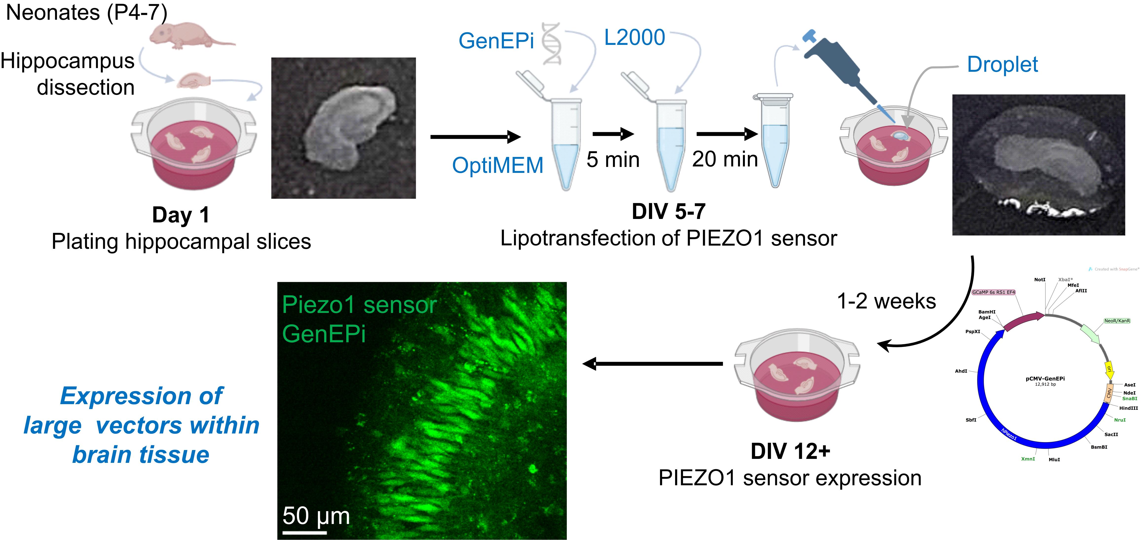
Schematic illustration of lipotransfection of the PIEZO1 sensor GenEPi, a large DNA construct, in organotypic hippocampal slices. A freshly prepared droplet of DNA–Lipofectamine 2000 complex is applied to the surface of individual hippocampal slices approximately 1 week after plating. This approach enables expression of the PIEZO1 sensor GenEPi as early as 1 week post-transfection, corresponding to approximately 12 days in vitro (DIV).
Background
Genetically encoded tools have become essential for probing protein function in complex biological systems. However, many receptors and ion channels have multi-domain configurations encoded by genes whose expression constructs exceed the packaging capacity of commonly used viral vectors. As a result, expressing large DNA constructs remains a major technical challenge, and gene delivery methods are often limited in their ability to achieve robust and sustained expression in brain cell types. Several delivery approaches have been developed for this purpose, including viral vectors, biolistics (gene gun), electroporation, and lipotransfection [1]. Viral vectors, such as adeno-associated viruses, offer stable long-term expression with high transduction efficiency and relatively low cytotoxicity [2], yet their packaging capacity is limited to ~5 kb, restricting their use for larger constructs [3]. By contrast, non-viral methods can support the delivery of larger constructs and may enable access to deeper tissue layers, but they are often more invasive, may disrupt tissue integrity, and typically require specialised equipment [1,4]. Inefficient delivery, often associated with low expression levels and reagent-induced toxicity, therefore restricts the ability to achieve robust and sustained expression of large constructs when studying large, multi-domain channels.
This limitation is critically relevant for mechanosensitive ion channels of the PIEZO family, which form large complexes within the membrane. PIEZO1 represents a large homo-trimeric protein [5] and is a challenging target for genetic manipulations, particularly because of the technical constraints of current gene delivery approaches. Existing genetically encoded optical PIEZO1 sensors, such as GenEPi and Ca2+-sensitive HaloTag ligands, have been designed to report PIEZO1 conformational changes by detecting mechanosensitive Ca2+ influx [6,7]. While these sensors have been tested in cell models and in zebrafish, there are no reports of their successful implementation in brain neurons, either in primary cultures or in organised brain tissue preparations. The PIEZO1-specific sensor GenEPi has been validated in HEK293T, HFF, and HeLa cells, cardiomyocytes, and zebrafish [6]; however, its large vector size (12,912 bp) and structural complexity further exacerbate the limitations of classical viral and non-viral delivery methods. Despite the growing interest in PIEZO1-mediated mechanosensation within the brain, approaches that enable successful expression of the PIEZO1 sensor in brain cell types remain to be developed.
Organotypic hippocampal slices provide a powerful model for studying neuronal function in a preserved 3D environment. Unlike monolayer cultures, organotypic slices retain the layered architecture, synaptic circuitry, and cell-type diversity of the hippocampus, together with native cell–cell interactions [1,4,8], while offering experimental access comparable to in vivo settings. This combination of structure and accessibility makes organotypic slices particularly well-suited for investigating channels and receptors that operate within a native environment, such as mechanosensitive PIEZO1 channels. When combined with genetic manipulation, organotypic brain slices provide a valuable platform for studying protein and/or channel function within a native tissue structure that is essential for mechanobiology in a physiologically relevant context.
Here, we describe a modified, minimally invasive protocol for expressing large DNA constructs in organotypic hippocampal slices. Lipid-based transfection, such as Lipofectamine 2000–mediated delivery, is a well-established non-viral method for DNA transfer that is relatively simple, cost-effective, and less invasive than mechanical delivery techniques [9,10]. Lipid–nucleic acid complexes enter cells primarily via endocytosis; however, in thick tissue, diffusion barriers within the extracellular matrix can limit reagent penetration and reduce transfection efficiency. Existing protocols often rely on introducing large volumes of DNA–Lipofectamine complexes into the culture at very early stages, with short incubation periods [1,11]. This may dilute the transfection reagent, reduce targeting efficiency, or compromise tissue integrity. We provide a step-by-step description for applying DNA–Lipofectamine 2000 complexes directly onto the tissue surface, thereby maximising reagent access to the superficial layers while minimising physical stress to the slices. Using this approach, we enable efficient expression of the genetically encoded PIEZO1 sensor GenEPi in hippocampal pyramidal neurons as early as 7 days post-transfection, with expression maintained for 3–4 weeks. The protocol combines cost-effectiveness, straightforward implementation, and compatibility with standard imaging setups, making it suitable for functional studies of PIEZO1-mediated mechanotransduction that require rapid and sustained expression of large DNA constructs in intact 3D brain tissue.
Materials and reagents
Biological materials
1. Sprague-Dawley rats (Rattus norvegicus) from Charles River Laboratories, strain code: 001 (neonates, P4–P6, of both sexes)
2. Alternatively, C57BL/6 J mice (Mus musculus) from Charles River Laboratories, strain code: 632 (neonates, P4–P7, of both sexes)
3. pCMV-GenEPi (Addgene plasmid #140236; http://n2t.net/addgene:140236; RRID: Addgene_140236)
Note: For long-term storage, plasmid DNA was maintained at -20 °C with minimal freeze/thaw cycles. We successfully expressed GenEPi in organotypic slices using DNA concentrations of 400–1,000 ng/μL. DNA purity was within the range of A260/A280 ratio 1.8–2.0 and A260/A230 ratio 2.0–2.2.
Reagents
1. Glucose (Sigma-Aldrich, catalog number: G8270)
2. Sodium chloride (Sigma-Aldrich, catalog number: S9888)
3. Sodium bicarbonate (Sigma-Aldrich, catalog number: S0751)
4. Sodium monophosphate (NaH2PO4) (Sigma-Aldrich, catalog number: S6040)
5. Potassium chloride (Sigma-Aldrich, catalog number: P9333)
6. Magnesium chloride hexahydrate (Sigma-Aldrich, catalog number: M2670)
7. Calcium chloride dihydrate (Sigma-Aldrich, catalog number: C7902)
8. Sucrose (Sigma-Aldrich, catalog number: S0389, CAS number: 57-50-1)
9. MEM (with Earl’s salts, without L-glutamine) (Gibco, catalog number: 21090-022)
10. HBSS 10× (without CaCl2/MgCl2) (Gibco, catalog number: 14185-045)
11. Horse serum, heat inactivated (Thermo Fisher Scientific, catalog number: 26050070)
12. Pen/Strep 10,000 units/mL (Gibco, catalog number: 15140-122)
13. B27 supplement (Gibco, catalog number: 17504-044)
14. Ara-C (Sigma-Aldrich, catalog number: C1768)
15. Ascorbic acid (Sigma-Aldrich, catalog number: A92902)
16. HEPES buffer (Gibco, catalog number: 15630-049)
17. Sterile water (Sigma-Aldrich, catalog number: W3500)
18. Lipofectamine 2000 (Invitrogen, catalog number: 11668030)
19. OptiMEM (Gibco, catalog number: 31985-062)
20. Paraformaldehyde solution (PFA), 4% in phosphate buffer solution (Thermo Fisher Scientific, catalog number: 15670799)
21. Phosphate buffered solution (PBS) (Sigma-Aldrich, catalog number: P4417)
22. Antibodies for neuronal staining (NeuN) (Abcam, catalog number: ab104224) and astrocyte labelling (GFAP) (Abcam, catalog number: ab134436)
23. Milli-Q water (prepared using a Milli-Q purification system available in the laboratory)
Note: Reagents 11–14 and antibodies should be stored at -20 °C. We suggest aliquoting reagents to minimise freeze/thaw cycles. PFA stocks should be stored within a designated flammable storage cabinet and used inside a fume hood.
Solutions
1. Sucrose-rich Ringer solution (see Recipes)
2. Culture medium (see Recipes)
3. DNA–Lipofectamine mixture (see Recipes)
4. Ringer solution (see Recipes)
Recipes
1. Sucrose-rich Ringer solution (for hippocampal slicing)
| Reagent | Molecular weight (g/mol) | Final concentration (mM) | Quantity (mg) |
|---|---|---|---|
| Sucrose | 342.3 | 119.8 | 41,000 |
| Sodium chloride | 58.44 | 64 | 3,738 |
| Potassium chloride | 74.55 | 2.5 | 186 |
| Calcium chloride dihydrate | 147.02 | 0.5 | 73.5 |
| Magnesium chloride hexahydrate | 203.3 | 7 | 1,423 |
| Sodium bicarbonate | 84.01 | 25 | 2,100 |
| Glucose | 180.2 | 10 | 1,802 |
| Sodium monophosphate | 120 | 1.25 | 150 |
| Total | - | - | 1 L |
Reagents should be dissolved in 1 L of Milli-Q water; pH = 7.4; osmolarity ~300 mOsm/kg.
To avoid precipitation, the solution should be bubbled with 95% O2 and 5% CO2 before adding calcium chloride. To maintain sterility, decant into a 50 mL syringe fitted with a 0.22 μm filter. Pass the solution through the filter into a sterile container. We recommend always using a freshly prepared solution.
2. Culture medium
| Reagent | Stock concentration | Final concentration | Volume (mL) |
|---|---|---|---|
| MEM (with Earl’s salts, without L-glutamine) | 1× | 0.50× | 25 |
| HBSS (without CaCl2/MgCl2) | 10× | 0.25× | 1.25 |
| Horse serum | 100% | 25% (v/v) | 12.5 |
| Pen/Strep | 10,000 U/mL | 100 U/mL | 0.5 |
| Glucose | 1 M | 16 mM | 0.8 |
| Ascorbic acid | 250 mM | 0.5 mM | 0.1 |
| HEPES buffer | 1 M | 8 mM | 0.4 |
| B27 supplement | 50× | 0.50× | 0.5 |
| Sterile water | - | - | 8.95 |
| Total | - | - | 50 mL |
Note: Culture medium should be prepared under sterile conditions in a tissue culture hood. The medium can be stored at 4 °C for the duration of tissue maintenance.
3. DNA–Lipofectamine mixture (amount per one slice)
| Reagent | Quantity or volume | |
| DNA:Lipofectamine ratio 1:2 | DNA:Lipofectamine ratio 1:1 | |
| DNA | 2.5 μg | 3.5 μg |
| Lipofectamine 2000 | 5 μL | 3.5 μL |
| OptiMEM | up to 10 μL | up to 10 μL |
This recipe is for the transfection of a single organotypic hippocampal slice with the PIEZO1 sensor GenEPi. For further guidance, please see General note 3. In our experiments, the concentration of DNA ranged from 400 to 1,000 ng/μL; we used a mass of 2.5 or 3.5 μg.
4. Ringer solution [12]
| Reagent | Molecular weight (g/mol) | Final concentration (mM) | Quantity (mg) |
|---|---|---|---|
| Sodium chloride | 58.44 | 126 | 7,358 |
| Potassium chloride | 74.55 | 3 | 224 |
| Calcium chloride dihydrate | 147.02 | 1.25 | 150 |
| Magnesium chloride hexahydrate | 203.3 | 2 | 294 |
| Sodium bicarbonate | 84.01 | 2 | 493 |
| Sodium monophosphate | 120 | 26 | 2,184 |
| Glucose | 180.2 | 10 | 1,802 |
| Total | - | - | 1 L |
Reagents should be dissolved in 1 L of Milli-Q water; pH = 7.4; osmolarity ~300 mOsm/kg. Solutions can be stored at 4 °C for up to a week. To avoid precipitation, the solution should be bubbled with 95% O2 and 5% CO2 before adding calcium chloride.
Laboratory supplies
1. Isoflurane (Abbvie, catalog number: B506, or Henry Schein, catalog number: 1182097, or any available)
2. Carbogen cylinder (95% O2 and 5% CO2 gas mixture)
3. Spray bottle with 70% ethanol (Fisher Scientific, catalog number: BP82031GAL)
4. Petri dish 100 mm × 15 mm (Corning, catalog number: 351029)
5. Fine forceps (Fine Science Tools, catalog number: 11412-11 or any similar fine forceps)
6. 6-well plate (Thermo Fisher Scientific, catalog number:140675)
7. Millicel membrane inserts (Millipore, catalog number: PICM0RG50)
8. Disposable Pasteur pipette (Kimblex, catalog number: DWK883350-0575)
9. 1.5 mL Eppendorf tubes (Sigma-Aldrich, catalog number: T9661)
10. Corning Falcon 50 mL tubes (Sigma-Aldrich, catalog number: CLS352070)
11. P20 pipette (Gilson, catalog number: F144056M)
12. P200 pipette (Gilson, catalog number: F144058M)
13. P1000 pipette (Gilson, catalog number: F144059M)
14. Sterile pipette tips 1,000 μL (Gilson, catalog number: F171703)
15. Sterile pipette tips 200 μL (Gilson, catalog number: F171503)
16. Sterile pipette tips 10 μL (Gilson, catalog number: F171203)
17. Plastic 50 mL syringe (Merck, catalog number: XX1105005)
18. 0.22 μm syringe filters (Millex, catalog number: SLMPR25SS)
Equipment
Dissection and preparation
1. Anaesthesia induction chamber (VetEquip, US) or any other available
2. Standard scissors (Fine Science Tools, catalog number: 14002-12)
3. Coarse forceps (Fine Science Tools, catalog number: 11652-10)
4. Spring scissors (Fine Science Tools, catalog number: 15025-10)
5. Fine forceps (Fine Science Tools, catalog number: 11412-11)
6. Vibratome (Leica, model: VT1200 S)
7. Double-edge prep blades (AccuThrive, catalog number: AVBL-3002-0000)
8. Bucket or plastic box for ice (any available)
Tissue culturing and transfection
1. Tissue culture hood
2. Humidified incubator (set at 37 °C with 5% CO2)
3. Bead heater or water bath
Electrophysiology and visualisation
1. Two-photon upright microscope (Femtonics, Budapest), connected to a tuneable femtosecond pulsed laser (Insight X3, Spectra-Physics/Newport); alternatively, a confocal microscope (Leica, model: DM2500)
2. XLPlan N 25× water-immersion objective (NA 1.05, Olympus)
3. Camera (any equipped with the microscope)
4. PC, workstation
Software and datasets
1. Imaging software (MES, Femtonics; LAS X, Leica, either requires a license)
2. ImageJ/Fiji (ImageJ 1.54f or g, or any other version)
3. Microsoft Excel (freely available)
4. GraphPad Prism 10
Procedure
登录/注册后免费查看全文
文章信息
稿件历史记录
提交日期: May 13, 2026
接收日期: Jun 28, 2026
在线发布日期: Jul 16, 2026
出版日期: Aug 5, 2026
版权信息
© 2026 The Author(s); This is an open access article under the CC BY license (https://creativecommons.org/licenses/by/4.0/).
如何引用
Bhavnani, A. and Kopach, O. (2026). Liposome-based Expression of the PIEZO1 Sensor GenEPi in Hippocampal Neurons in Organotypic Slices. Bio-protocol 16(15): e5780. DOI: 10.21769/BioProtoc.5780.

 Download Citation in RIS Format
分类
神经科学 > 细胞机理 > 组织分离与培养
分子生物学 > DNA > 转染
您对这篇实验方案有问题吗?
在此处发布您的问题,我们将邀请本文作者来回答。同时,我们会将您的问题发布到Bio-protocol Exchange,以便寻求社区成员的帮助。
 发布问题
发布问题


