


Sample Preparation for Imaging-Based Spatial Transcriptomics in Rigid Plant Tissues (Roots, Shoots)
硬质植物组织(根和地上部)成像型空间转录组学的样品制备

发布: 2026年08月05日第16卷第15期 DOI: 10.21769/BioProtoc.5779 浏览次数: 337
评审: Samik BhattacharyaAnonymous reviewer(s)
 PDF
PDF  Q&A
Q&A
参见作者原研究论文
The authors used this protocol in:

Jun 2025
Advertisement






Abstract
Plant roots dynamically respond to environmental changes and serve as an ideal system for studying cell development and gene regulation. Recent advances in imaging-based spatial transcriptomics have enabled high-resolution mapping of gene expression while preserving spatial context. However, existing sample preparation techniques remain inadequate for handling rigid plant tissues such as crop roots. Here, we present a detailed and practical protocol for preparing rigid plant tissue samples for imaging-based spatial transcriptomics. The workflow ensures effective tissue handling while maintaining RNA integrity and spatial organization. Within approximately eight days, samples can be processed and mounted onto commercial slides, making them ready for subsequent probe hybridization and imaging. This protocol also includes an integrated sample attachment test performed to assess slide quality. It has been optimized to produce consistent and reliable results across experiments. Overall, our method provides a robust solution for spatial transcriptomic analysis in rigid plant tissues, facilitating broader application of these technologies in plant research.
Key features
• Builds upon the method developed by Zhu et al. [1] and introduces an optimized sample preparation protocol for imaging-based spatial transcriptomics in rigid rice roots.
• Ensures effective tissue fixation and sectioning, while preserving RNA integrity and spatial organization.
• Includes an integrated sample attachment test to assess the adhesion of tissue sections to commercial slides.
• Requires approximately 8 days to complete the sample preparation, with another 6 days for the attachment test.
Keywords: Imaging-based spatial transcriptomics (成像型空间转录组学) Rigid plant tissues (硬质植物组织)
Crop roots (作物根系)
PFA-based fixation (多聚甲醛(PFA)固定)
Paraffin-embedded tissue sectioning (石蜡包埋组织切片)
Section adhesion test (切片附着性测试)
Rigid plant tissues (硬质植物组织)
Crop roots (作物根系)
PFA-based fixation (多聚甲醛(PFA)固定)
Paraffin-embedded tissue sectioning (石蜡包埋组织切片)
Section adhesion test (切片附着性测试)
Graphical overview
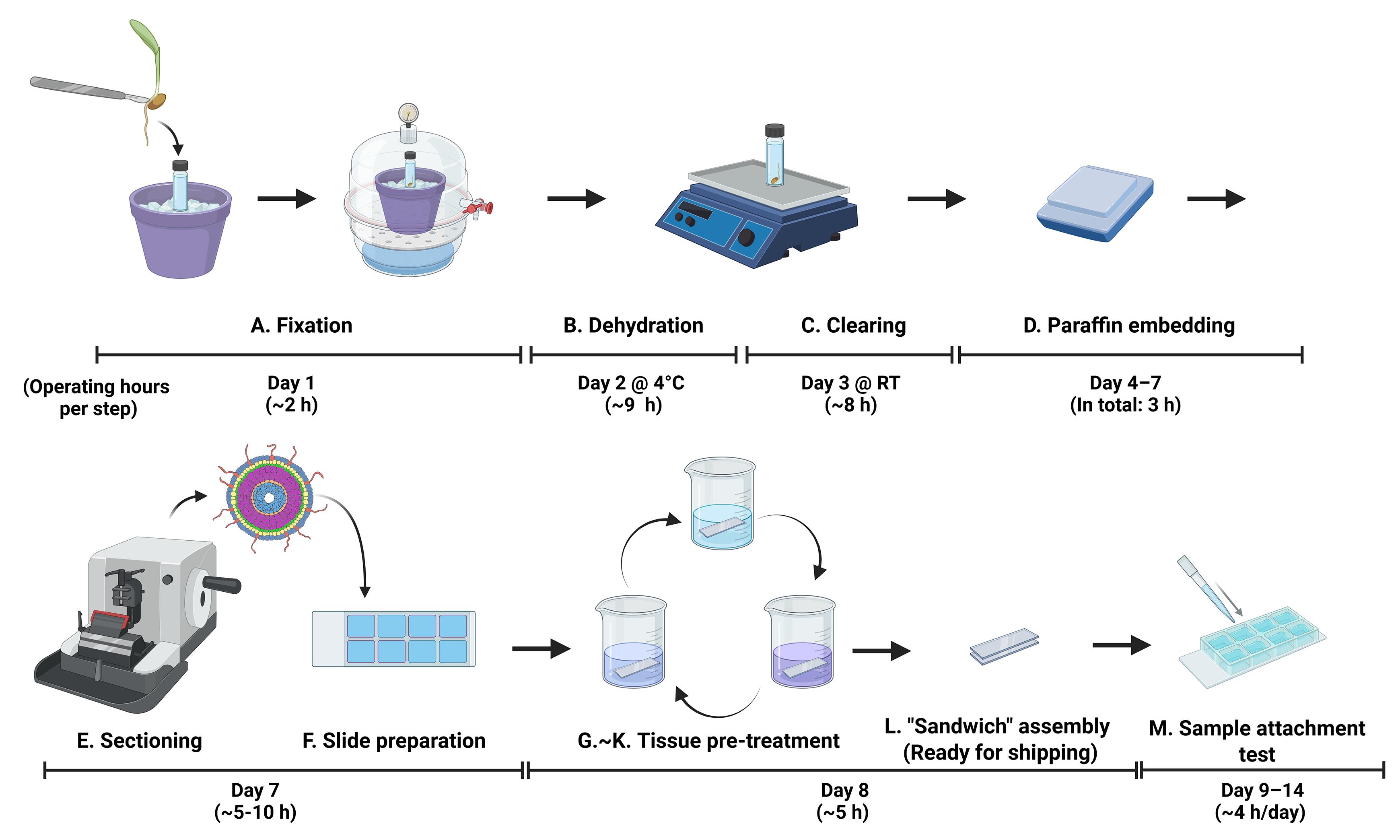
Overview of sample preparation for imaging-based spatial transcriptomics in rice roots
Background
The rapid development of single-cell and spatial transcriptomic technologies has revolutionized our understanding of gene expression at the cellular level and its spatial organization within tissues and organs [2]. In plant science research, various single cell–based approaches have been widely adopted and further developed, enabling insights into diverse biological processes, including cell differentiation trajectories, hormone signaling, and plant responses to biotic and abiotic stresses [3–6].
However, most studies rely on single-cell or single-nucleus RNA sequencing, which requires dissociation of cells from tissues and can lead to the loss of spatial information [7]. Traditional approaches, such as in situ hybridization and confocal imaging with fluorescent reporters, have been used to recover spatial context [8,9], but these methods are limited by low throughput (restricted number of detectable genes) and resolution and are not fully compatible with high-throughput single-cell technologies.
To overcome these limitations, we collaborated with Resolve Biosciences to implement a multiplexed, imaging-based spatial transcriptomics approach that enables high-throughput visualization of gene expression while preserving spatial context [1]. Our sample preparation workflow is specifically optimized for rice roots. This protocol can be readily adapted to other rigid plant tissues, such as developing maize ears [10], maize roots [11], soybean roots and mature nodules [12], and barley shoot meristem [13], facilitating robust imaging-based spatial transcriptomic analyses across diverse plant systems. With further optimization, the protocol may also be extended to support additional omics applications.
Materials and reagents
Biological materials
1. Rice primary roots, 4–6 days old
Note: Rice seeds were dehulled and surface-sterilized using 50% bleach for 30 min, followed by five rinses with sterile water. The seeds were then placed in Yoshida’s medium solidified with 0.15% gellan gum (Gelzan, Caisson), with the embryos oriented upward. Seeds were incubated at 30 °C in darkness for 2–3 days to allow germination. Germinated seedlings were either kept in a gel system or transferred to soil conditions. For soil condition, soils (Wedowee sandy loam soils, Johnston County, NC, USA) were air dried, crushed, and then passed through a sieve with a 2-mm mesh size, then lightly sprayed with sterilized water and mixed thoroughly. Non-compacted soil condition was packed up to 1.2 g/cm3, and compacted soil was pressed to make 1.6 g/cm3. Seedlings were grown in a growth chamber maintained at 28 °C under continuous light (45 μmol/m2/s) for an additional 2–3 days prior to harvesting.
Reagents
1. Paraformaldehyde (PFA) (Sigma-Aldrich, catalog number: 158127)
2. Triton X-100 (Thermo Fisher Scientific, catalog number: A16046)
3. Phosphate-buffered saline (PBS) (10×) pH 7.4, RNase-free (Thermo Fisher Scientific, Invitrogen, catalog number: AM9625)
4. Sodium hydroxide (NaOH) (Sigma-Aldrich, catalog number: S5881)
5. DEPC-treated water (Thermo Fisher Scientific, Invitrogen, catalog number: 4387937)
6. Absolute ethanol, 200 proof, molecular biology grade (Thermo Fisher Scientific, Invitrogen, catalog number: T038181000)
7. Histo-Clear®/Histo-Clear II®, Electron Microscopy Sciences (VWR, catalog number: 101412-876)
8. SurgipathTM ParaplastTM tissue embedding medium (Leica Biosystems, catalog number: 39601006)
9. Proteinase K solution (20 mg/mL), RNA grade (Thermo Fisher Scientific, Invitrogen, catalog number: 25530049)
10. Tris (1 M), pH 8.0, RNase-free (Thermo Fisher Scientific, Invitrogen, catalog number: AM9855G)
11. EDTA (0.5 M), pH 8.0, RNase-free (Thermo Fisher Scientific, Invitrogen, catalog number: AM9260G)
12. Glycine, molecular biology grade (Promega, catalog number: H5073)
13. Triethanolamine (Sigma-Aldrich, catalog number: 90279)
14. Hydrochloric acid (HCl), 36.5%–38% (MilliporeSigma, catalog number: HX06034)
15. SlowFadeTM Diamond antifade mountant (Thermo Fisher Scientific, Invitrogen, catalog number: S36967)
16. Isopropanol (Sigma-Aldrich, catalog number: I9030)
17. Wash buffer (provided by Resolve Biosciences)
18. RNaseZapTM RNase decontamination solution (Thermo Fisher Scientific, Invitrogen, catalog number: AM9780)
19. Ammonium nitrate (NH4NO3) (Thermo Fisher Scientific, catalog number: A676-500)
20. Sodium dihydrogen phosphate dihydrate (NaH2PO4·2H2O) (Thermo Fisher Scientific, catalog number: AAA1131636)
21. Potassium sulfate (K2SO4) (Thermo Fisher Scientific, catalog number: AAA139750B)
22. Calcium chloride (CaCl2) (Thermo Fisher Scientific, catalog number: AAL131910B)
23. Magnesium sulfate heptahydrate (MgSO4·7H2O) (Thermo Fisher Scientific, catalog number: M63-500)
24. Manganese(II) chloride tetrahydrate (MnCl2·4H2O) (Thermo Fisher Scientific, catalog number: AC193451000)
25. Ammonium molybdate (para) tetrahydrate [(NH4)6Mo7O24·4H2O] (Thermo Fisher Scientific, catalog number: AAA1376618)
26. Boric acid (H3BO3) (Thermo Fisher Scientific, catalog number: A74-500)
27. Iron(III) chloride hexahydrate (FeCl3·6H2O) (Thermo Fisher Scientific, catalog number: AC217091000)
28. Citric acid monohydrate (Thermo Fisher Scientific, catalog number: A104-500)
29. Zinc sulfate heptahydrate (ZnSO4·7H2O) (Thermo Fisher Scientific, catalog number: Z68-500)
30. Copper(II) sulfate pentahydrate (CuSO4·5H2O) (Thermo Fisher Scientific, catalog number: AAA112620B)
31. Sulfuric acid (H2SO4) (Thermo Fisher Scientific, catalog number: MSX12446)
32. Gelzan™ (Cassion Labs, catalog number: G024)
33. Acetic anhydride (Sigma-Aldrich, catalog number: 320102)
Solutions
1. 1× PBS (see Recipes)
2. 30% Triton X-100 solution (see Recipes)
3. Ethanol gradient solutions (see Recipes)
4. Fixative (see Recipes)
5. Histo-Clear/ethanol gradient solutions (see Recipes)
6. Proteinase K buffer (see Recipes)
7. 0.2% (w/v) glycine solution (see Recipes)
8. Fixative without Triton (see Recipes)
9. 0.1 M triethanolamine (see Recipes)
10. Yoshida’s medium (see Recipes)
Recipes
1. 1× PBS
| Reagent | Final concentration | Quantity or volume |
| 10× PBS | 1× | 40 mL |
| DEPC-treated water | n/a | 360 mL |
| Total | n/a | 400 mL |
2. 30% Triton X-100 solution
| Reagent | Final concentration | Quantity or volume |
| Triton X-100 | 30% (v/v) | 3 mL |
| 1× PBS | n/a | 7 mL |
| Total | n/a | 10 mL |
3. Ethanol gradient solutions
| Reagent | Final concentration | Quantity or volume |
| Ethanol (absolute) | X% (v/v) | (X/10) mL |
| DEPC-treated water | n/a | 10 - (X/10) mL |
| Total | n/a | 10 mL |
Make fresh.
4. Fixative
| Reagent | Final concentration | Quantity or volume |
| PFA | 4% (w/v) | 0.8 g |
| 30% Triton X-100 solution | 0.03% | 20 μL |
| NaOH | n/a | 0.15 g |
| HCl | n/a | Adjust pH to 7.0 |
| 1× PBS | 1× | Make up to 20 mL |
| Total | n/a | 20 mL |
Add 0.15 g of NaOH pellets to dissolve PFA in 15 mL of 1× PBS, then adjust pH to 7.0 with HCl. Top up with 1× PBS to 20 mL. Always use freshly made fixative.
5. Histo-Clear/ethanol gradient solutions
| Reagent | Final concentration | Quantity or volume |
| Histo-Clear | Y% | (Y/2) mL |
| Ethanol (absolute) | (100 - Y)% | 50 - (Y/2) mL |
| Total | n/a | 50 mL |
Solutions cannot be stored for a long time.
6. Proteinase K buffer
| Reagent | Final concentration | Quantity or volume |
| 1 M Tris-HCl, pH 8.0 | 100 mM | 20 mL |
| 0.5 M EDTA, pH 8.0 | 50 mM | 10 mL |
| DEPC-treated water | n/a | 170 mL |
| Total | n/a | 200 mL |
7. 0.2% (w/v) glycine solution
| Reagent | Final concentration | Quantity or volume |
| Glycine | 0.2% (w/v) | 0.4 g |
| 1× PBS | n/a | 200 mL |
| Total | n/a | 200 mL |
Make fresh.
8. Fixative without Triton
| Reagent | Final concentration | Quantity or volume |
| PFA | 4% (w/v) | 8 g |
| NaOH | n/a | 1.5 g |
| HCl | n/a | Adjust pH to 7.0 |
| 1× PBS | 1× | Make up to 200 mL |
| Total | n/a | 200 mL |
Add 1.5 g of NaOH pellets to dissolve PFA in 150 mL of 1× PBS, then adjust pH to 7.0 with HCl. Top up with 1× PBS to 200 mL. Always use freshly made fixative.
9. 0.1 M triethanolamine
| Reagent | Final concentration | Quantity or volume |
| Triethanolamine | 0.1 M | 2.68 mL |
| HCl | 0.15% (v/v) | 0.8 mL |
| 1× PBS | n/a | 197 mL |
| Total | n/a | 200 mL |
10. Yoshida’s medium
| Reagent | Final concentration | Quantity or volume |
| NH4NO3 (800×) | 1.43 mM | 1.25 mL |
| NaH2PO4·2H2O (800×) | 0.32 mM | 1.25 mL |
| K2SO4 (800×) | 0.51 mM | 1.25 mL |
| CaCl2 (800×) | 1 mM | 1.25 mL |
| MgSO4·7H2O (800×) | 1.64 mM | 1.25 mL |
| Yoshida’s microstock (800×) | 1× | 1.25 mL |
| MES hydrate | 2.8 mM | 0.546 g |
| dH2O | n/a | Make up to 1 L |
| Total | n/a | 1 L |
Prepare 800× stock solution of NH4NO3 (1.14 M), NaH2PO4·2H2O (0.25 M), K2SO4 (0.41 M), CaCl2 (0.80 M), and MgSO4·7H2O (1.31 M).
Then, prepare Yoshida’s microstock (800×). Dissolve 1.5 g of MnCl2·4H2O in 50 mL of ddH2O. Dissolve 0.074 g of (NH4)6Mo7O24·4H2O in 50 mL of ddH2O. Dissolve 0.934 g of H3BO3 in 50 mL of ddH2O. Dissolve 7.7 g of FeCl3·6H2O in 50 mL of ddH2O. Dissolve 11.9 g of citric acid monohydrate in 50 mL of ddH2O. Also, prepare 50 mM ZnSO4·7H2O and 50 mM CuSO4·5H2O solutions. In a fume hood, slowly add 50 mL of H2SO4 to 550 mL of ddH2O while stirring, then add 50 mL of MnCl2·4H2O, 50 mL of (NH4)6Mo7O24·4H2O, 50 mL of H3BO3, 2.435 mL of 50 mM ZnSO4·7H2O, 2.483 mL of 50 mM CuSO4·5H2O, 50 mL of FeCl3·6H2O, and 50 mL of citric acid monohydrate. Make up the volume to 1 L.
Add all components into a beaker containing 800 mL of dH2O with a magnetic stir bar. Adjust pH to 5.8 with NaOH before making up the final volume to 1 L. Add 1.5 g of Gelzan and then autoclave.
Laboratory supplies
1. DWK Life Sciences WheatonTM glass 20 mL scintillation vials: polypropylene caps (Thermo Fisher Scientific, catalog number: 03-341-25D)
2. VWR® razor blades, 0.22 mm (VWR, catalog number: 55411-050)
3. Disposable scalpel blades, sterile, IntegraTM Miltex®, carbon steel blade, #21 (VWR, catalog number: 21909-624)
4. FisherbrandTM fine precision medium tipped tweezers/forceps (Thermo Fisher Scientific, catalog number: 12-000-157)
5. Olympus conical polypropylene centrifuge tubes, 50 mL (Genesee Scientific, catalog number: 28-108)
6. Olympus conical polypropylene centrifuge tubes, 15 mL (Genesee Scientific, catalog number: 28-103)
7. Eisco alcohol lamp (VWR, catalog number: 47036-104)
8. EprediaTM ultra disposable microtome blades (Thermo Fisher Scientific, catalog number: 31-537-35)
9. Princeton Artist Brush, Neptune Series 4750, Script, synthetic squirrel, Size 1 (Princeton Artist Brush Company, catalog number: P4750SC1)
10. VWR® Premium Superfrost® Plus microscope slides (VWR, catalog number: 48311-703)
11. VWR® micro cover glasses, rectangular, No. 2, 50 × 24 mm (VWR, catalog number: 48382-136)
12. Slide (sample coverslip) for sample mounting with regions drawn (provided by Resolve Biosciences)
13. Kimberly-Clark ProfessionalTM Kimtech ScienceTM KimwipesTM delicate task wipers, 1-Ply (Thermo Fisher Scientific, catalog number: 06-666)
14. VWR® transfer pipets, sterile, bulb draw: 3.5 mL (VWR, catalog number: 78062-460)
15. CorningTM PYREXTM low form Griffin beakers, 250 mL (Thermo Fisher Scientific, catalog number: 02-540K)
16. VWR® disposable Petri dishes, 100 × 15 mm (VWR, catalog number: 89022-320)
17. EprediaTM ShandonTM slide holder, 10 slide capacity (Thermo Fisher Scientific, catalog number: 14-7)
18. Mold, Peel-A-Way embedding; truncated, T8; size: 22 × 22 sq. mm truncated to 8 × 8 mm (Thermo Fisher Scientific, catalog number: NC9991740)
19. Wooden holder for ultramicrotome (Smallest pieces in Ward's® Essentials Magic Blocks Kit)
20. Sticky slide (provided by Resolve Biosciences)
21. FisherbrandTM 5-place slide mailer, end opening (Thermo Fisher Scientific, catalog number: HS15986)
Equipment
1. Thermo ScientificTM PH111 pH/mV Bench Meter Easy-to-Clean Bio Kit (Thermo Fisher Scientific, catalog number: 15200969PM)
2. Bel ArtTM Space Saver Vacuum Desiccator (Thermo Fisher Scientific, catalog number: 08-594-16A)
3. Dual-Stage HVAC Vacuum Pump, 5.0 CFM (Amazon)
4. FisherbrandTM Multi-Platform Shaker (Thermo Fisher Scientific, catalog number: 88-861-021)
5. VWR® VWB2 Unstirred Water Baths (VWR, catalog number: 77587-158)
6. Spencer 820 Precision Rotary Microtome (Spencer Lens Company, model: 820)
7. Microscope (Leica, model: DM5500 B)
8. Covered and uncovered flat-bed slide warmers (VWR, catalog number: 470303-828)
9. Water Jacket Incubator (VWR, model: 3015)
10. Cimarec+TM Stirring Hotplates (Thermo Fisher Scientific, catalog number: SP88857100)
11. Thermal Cycler (Bio-Rad, model: C1000)
Procedure
登录/注册后免费查看全文
文章信息
稿件历史记录
提交日期: Apr 21, 2026
接收日期: Jun 28, 2026
在线发布日期: Jul 12, 2026
出版日期: Aug 5, 2026
版权信息
© 2026 The Author(s); This is an open access article under the CC BY-NC license (https://creativecommons.org/licenses/by-nc/4.0/).
如何引用
Liu, H., Zhang, J. and Zhu, M. (2026). Sample Preparation for Imaging-Based Spatial Transcriptomics in Rigid Plant Tissues (Roots, Shoots). Bio-protocol 16(15): e5779. DOI: 10.21769/BioProtoc.5779.

 Download Citation in RIS Format
分类
植物科学 > 植物分子生物学 > RNA > RNA 检测
分子生物学 > RNA > 转录
系统生物学 > 空间转录组学
您对这篇实验方案有问题吗?
在此处发布您的问题,我们将邀请本文作者来回答。同时,我们会将您的问题发布到Bio-protocol Exchange,以便寻求社区成员的帮助。
 发布问题
发布问题


