


Fluorogenic Tissue-Based Assessment of Acid Ceramidase Activity
基于组织的酸性神经酰胺酶活性荧光检测

发布: 2026年08月20日第16卷第16期 DOI: 10.21769/BioProtoc.5778 浏览次数: 431
评审: Komuraiah MyakalaBhavneet KaurElena A. OstrakhovitchAnonymous reviewer(s)
 PDF
PDF  Q&A
Q&A





Abstract
Acid ceramidase (aCDase) is a lysosomal amidase that catalyzes the hydrolysis of sphingolipids (SphL), including ceramides and glucosylceramides. Altered expressions of aCDase are associated with several pathological conditions, such as cancer, inflammation, pain, and pulmonary disorders. aCDase activity is reduced in Farber disease, spinal muscular atrophy with progressive myoclonic epilepsy, diabetes, and cardiovascular disease. Recent reports suggest that aCDase inhibition may be an emerging strategy for treating several SphL-related neurodegenerative conditions, such as Krabbe, Gaucher, and Parkinson’s disease, due to its role in the accumulation of glycosphingolipids. Therefore, the development of a tissue-based aCDase activity assay has potential applications in clinical diagnostics and drug discovery, enabling the evaluation of the onset and progression of disease from biological samples of patients, drug-target engagement analysis, and identification of biomarkers. Here, we report a detailed protocol for detecting aCDase activity in tissue lysates, using Rbm14-12 as a specific fluorogenic substrate for aCDase. Assay protocol optimization, including a procedure for the preparation and storage of tissue lysates and the identification of optimal protein tissue lysate amounts and substrate concentrations based on kinetic enzymatic parameter analyses, is described.
Key features
• This protocol relies on the use of the Rbm14-12 fluorogenic substrate.
• This protocol was developed out of a need to measure the target engagement of an aCDase-targeting therapeutic in a Parkinson’s disease-related animal model.
• This protocol can be broadly useful for sensitively measuring aCDase activity in central and peripheral organ tissues.
Keywords: ASAH1 (ASAH1) Acid ceramidase (酸性神经酰胺酶)
Ceramides (神经酰胺)
Sphingolipids (鞘脂)
Neurodegeneration (神经退行性疾病)
Farber disease (法伯病)
Acid ceramidase (酸性神经酰胺酶)
Ceramides (神经酰胺)
Sphingolipids (鞘脂)
Neurodegeneration (神经退行性疾病)
Farber disease (法伯病)
Graphical overview
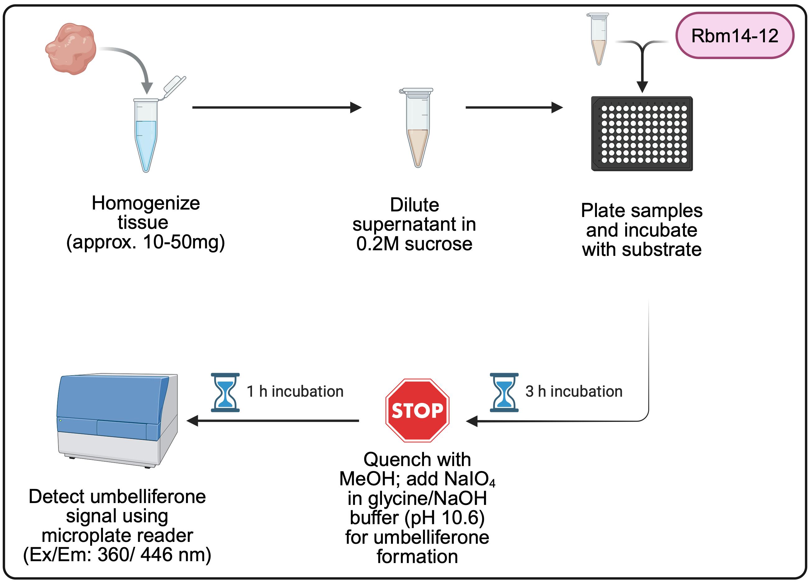
Overview of the fluorogenic tissue-based assessment of acid ceramidase activity methodology. MeOH, methanol; NaIO4, sodium periodate; NaOH, sodium hydroxide. NaIO4 solution is prepared in glycine/NaOH buffer (pH 10.6). Created with BioRender.com.
Background
Acid ceramidase (aCDase, encoded by the ASAH1 gene) is a cysteine amidase that hydrolyzes ceramides (Cer) to generate sphingosine and fatty acids. aCDase can also deacylate glucosylceramides (GlcCer) to form glucosylsphingosine (GlcSph) [1–5]. aCDase is localized within the lysosomal compartments and has a pH optimum of 4.5 to 5.0 [6–8]. Mutations in the ASAH1 gene associate with lysosomal storage disorder, Farber disease (FD), and spinal muscular atrophy with progressive myoclonic epilepsy (SMA-PME) [9]. In the presence of mutant ASAH1, aCDase activity is reduced, leading to the buildup of lysosomal Cer in cells throughout the body [10].
Diagnosing FD based on assays that determine aCDase activity has been challenging, often requiring the use of radiolabeled substrates, specialized equipment, non-selectivity, and significant amounts of biological material, which is not always feasible [11,12]. A cell-based aCDase activity assay was previously developed using the fluorogenic Rbm14–12 substrate and a series of FD patient-derived cell lines, demonstrating its usefulness for diagnosing FD [13]. According to this previous protocol, aCDase hydrolyzes the amide bond of Rbm14-12 to form an aminodiol, which is oxidized by sodium periodate; the oxidized product (aldehyde) then undergoes β-elimination at basic pH, leading to the release of umbelliferone, a fluorescent coumarin-derived compound that can be measured with a microplate reader [13].
Evaluating aCDase activity has implications beyond FD and SMA-PME. In recent years, ASAH1 expression and aCDase activity have been studied in the context of oncology, notably due to the overexpression of ASAH1 in various tumor types [14–16]. aCDase activity has also been studied in the context of cardiovascular disease, as well as in aging, immunity, diabetes, and inflammation [17–20]. Most recently, aCDase has also been investigated for its role in some neurological disorders, such as Gaucher disease, Krabbe disease, Alzheimer’s disease, and Parkinson’s disease, due to its potential role in the accumulation of neurotoxic glycosphingolipids [21–24]. With the increasing relevance of aCDase in a variety of diseases, being able to accurately detect its activity in tissue lysates is fundamental. aCDase activity measurements are particularly valuable in preclinical validation studies in a variety of disease animal models to predict the clinical translation of targeting aCDase as a valuable therapeutic strategy.
Here, we describe a protocol for a tissue-based aCDase activity assay using the Rbm14-12 fluorogenic substrate. We optimized this protocol to measure aCDase activity in the brain of a Parkinson’s disease mouse model. This cost-effective, not laborious, and efficient aCDase activity assay has broad applicability for a variety of tissue types and animal models. The specific reagents needed for tissue homogenization, the optimal protein tissue lysate, and Rbm14-12 substrate concentrations were all determined in brain tissue lysates of GBA1+/+ wild-type (WT) mice. Key considerations for optimizing the assay for other tissue types are included.
Materials and reagents
Biological materials
1. GBA1+/+ mouse brain tissue (WT mice, offspring of GBA1+/L444P heterozygotes originally obtained from the University of North Carolina at Chapel Hill Mutant Mouse Resource and Research Center, catalog number/origin: MMRRC-000117)
Reagents
1. Dimethyl sulfoxide (DMSO) (Fisher Scientific, catalog number: D12345)
2. D-Sucrose (Fisher Scientific, catalog number: BP220-10)
3. EDTA (0.5 M, pH 8.0) (Fisher Scientific, catalog number: 15575020)
4. Glycine (Fisher Scientific, catalog number: BP381-500)
5. MeOH (Fisher Scientific, catalog number: A411-4)
6. Sodium periodate (NaIO4) (Millipore Sigma, catalog number: 311448-5G)
7. Pierce BCA Protein Assay kit (Fisher Scientific, catalog number: 23225)
8. Rbm14-12 substrate (Avanti Polar Lipids, catalog number: A86855)
9. Sodium acetate (1 M, pH 4.5) (Fisher Scientific, catalog number: J63669.AE)
10. Sodium chloride (NaCl) (Fisher Scientific, catalog number: BP358-212)
11. Sodium hydroxide (NaOH) (1 M) (Fisher Scientific, catalog number: SS266-1)
12. Tris base (Fisher Scientific, catalog number: BP152-500)
13. Umbelliferone (Millipore Sigma, catalog number: H24003)
Solutions
1. Assay solution (see Recipes)
2. Detergent-free lysis buffer (see Recipes)
3. 100 mM glycine/NaOH buffer (see Recipes)
4. 4 mM Rbm14-12 substrate stock solution (see Recipes)
5. 25 mM Sodium acetate buffer (see Recipes)
6. 2.5 mg/mL NaIO4 solution (see Recipes)
7. 0.2 M sucrose solution (see Recipes)
8. 5 mM umbelliferone stock solution (see Recipes)
Recipes
1. Assay solution (per well)
| Reagent | Final concentration | Volume |
|---|---|---|
| 25 mM sodium acetate | 18.6 mM | 74.875 μL |
| Rbm14-12 substrate stock solution | 5 μM* | 0.125 μL |
| Fixed amount of protein | 10 μg* | 25 μL |
| Total | n/a | 100 μL |
*Conditions are to be optimized by the experimenter based on tissue type and animal model. See the aCDase activity assay reagent calculator in Supplementary information, Template 1, to easily calculate the amount of 4 mM Rbm14-12 substrate and the volume of 25 mM sodium acetate required to prepare the desired substrate concentration. For example, for a desired Rbm14-12 substrate concentration of 5 μM, 0.125 μL of 4 mM Rbm14-12 stock solution would be required per 100 μL of total volume. This can be scaled up using the aCDase activity assay reagent calculator in Template 1, so that a multi-channel pipette can be used to add 75 μL of assay solution to each necessary well.
The assay solution should be made fresh the day of the experiment.
2. Detergent-free lysis buffer
| Reagent | Final concentration | Volume |
|---|---|---|
| Tris, pH 8.0 | 50 mM | 2.5 mL |
| 5 M NaCl | 150 mM | 1.5 mL |
| 0.5 M EDTA, pH 8.0 | 5 mM | 0.5 mL |
| MilliQ water | n/a | 45.5 mL |
| Total | n/a | 50 mL |
The detergent-free lysis buffer can be stored at room temperature (20–25 °C) for long-term storage. Collected protein supernatants using the detergent-free lysis buffer can be stored at -80 °C for long-term storage. Please note that protease and phosphatase inhibitors are omitted from the lysis buffer. To prevent any changes in enzyme stability, it is recommended to use chilled buffers, keep samples on ice, and work as quickly as possible while maintaining accuracy.
3. 100 mM glycine/NaOH buffer
| Reagent | Final concentration | Amount or volume |
|---|---|---|
| Glycine | n/a | 1.875 g |
| MilliQ water | n/a | 200 mL |
| 1 M NaOH | n/a | to adjust pH to 10.6 |
| MilliQ water | n/a | Bring to 250 mL |
| Total | 100 mM | 250 mL |
The 100 mM glycine/NaOH buffer should be stored at 4 °C for up to two weeks, as long as the pH remains stable and unchanged. Please note that it is recommended to add 1.875 g of glycine to 200 mL of MilliQ water. Adjust the pH to 10.6 by slowly adding 1 M NaOH while stirring. After the solution reaches a pH of 10.6 based on the pH meter, add enough MilliQ water to bring the final volume to 250 mL. Mix thoroughly and verify the pH, adjusting if necessary.
4. 4 mM Rbm14-12 substrate stock solution
| Reagent | Final concentration | Amount or volume |
|---|---|---|
| Rbm14-12 | 4 mM | 1 mg |
| DMSO | n/a | 541.6 μL |
Due to the volatility of ethanol (EtOH), it is recommended to dissolve Rbm14-12 in DMSO, rather than EtOH. The Rbm14-12 substrate stock solution in DMSO (4 mM) should be stored at -20 °C for <1 month in an amber glass vial wrapped in foil. It is recommended to aliquot the DMSO stock solutions of Rbm14-12 prior to storage to minimize freeze-thaw cycles.
5. 25 mM sodium acetate buffer
| Reagent | Final concentration | Amount or volume |
|---|---|---|
| 1 M sodium acetate, pH 4.5 | 25 mM | 1.25 mL |
| MilliQ water | n/a | 48.75 mL |
| Total | 25 mM | 50 mL |
The 25 mM sodium acetate buffer can either be stored at 4 °C or room temperature (20–25 °C) for up to two weeks, as long as the pH (4.5) remains stable and unchanged.
6. 2.5 mg/mL NaIO4 solution
| Reagent | Final concentration | Amount or volume |
|---|---|---|
| NaIO4 | 2.5 mg/mL | 2.5 mg |
| 100 mM glycine/NaOH buffer | 1 mL | 1 mL |
The 2.5 mg/mL NaIO4 solution should be made fresh the day of the experiment.
7. 0.2 M sucrose solution
| Reagent | Final concentration | Volume |
|---|---|---|
| 1 M sucrose | 0.2 M | 12.5 mL |
| MilliQ water | n/a | 50 mL |
| Total | n/a | 62.5 mL |
The 0.2 M sucrose solution can be stored at 4 °C for up to two weeks, as long as the pH (6.0–6.5) remains stable and unchanged. Please note: to prepare the 1 M sucrose used for the final 0.2 M sucrose solution, 17.12 g of sucrose is dissolved in 250 mL of MilliQ water. The remaining 1 M sucrose solution that is not used can either be discarded or stored at 4 °C for up to two weeks.
8. 5 mM umbelliferone stock solution
| Reagent | Final concentration | Amount or volume |
|---|---|---|
| Umbelliferone | 5 mM | 0.81 mg |
| DMSO | n/a | 1 mL |
Due to the volatility of EtOH, it is recommended to dissolve umbelliferone in DMSO, rather than EtOH. The umbelliferone stock solution in DMSO (5 mM) should be stored at -20 °C for long-term storage. It is recommended to aliquot the DMSO stock solutions of umbelliferone prior to storage at -20 °C to minimize freeze-thaw cycles.
Laboratory supplies
1. Amber glass vial (Millipore Sigma, catalog number: 27046-U)
2. Black, flat-bottomed 96-well plate (Fisher Scientific, catalog number: M33090)
3. Microcentrifuge tubes (1.5 mL) (Fisher Scientific, catalog number: 05-408-130)
4. Microcentrifuge tubes (0.6 mL) (Fisher Scientific, catalog number: 05-408-120)
5. Motorized tissue grinder (Fisher Scientific, catalog number: 12-141-361)
6. Pellet pestles (Fisher Scientific, catalog number: 749521-1500)
Equipment
1. Microcentrifuge (Fisher Scientific, catalog number: 75002446, model: Sorvall Legend Micro 21R)
2. Microplate reader for BCA (Agilent Technologies, catalog number: 209924, model: BioTek Synergy 2 SL)
3. Microplate reader for umbelliferone detection (Molecular Devices, model: SpectraMax iD3)
Procedure
登录/注册后免费查看全文
文章信息
稿件历史记录
提交日期: Apr 1, 2026
接收日期: Jul 2, 2026
在线发布日期: Jul 14, 2026
出版日期: Aug 20, 2026
版权信息
© 2026 The Author(s); This is an open access article under the CC BY license (https://creativecommons.org/licenses/by/4.0/).
如何引用
Manabat, A., Russo, D., Penna, I., Scarpelli, R. and Volpicelli-Daley, L. (2026). Fluorogenic Tissue-Based Assessment of Acid Ceramidase Activity. Bio-protocol 16(16): e5778. DOI: 10.21769/BioProtoc.5778.

 Download Citation in RIS Format
分类
神经科学 > 神经系统疾病 > 帕金森氏症
生物化学 > 蛋白质 > 活性
细胞生物学 > 基于细胞的分析方法 > 酶学测定
您对这篇实验方案有问题吗?
在此处发布您的问题,我们将邀请本文作者来回答。同时,我们会将您的问题发布到Bio-protocol Exchange,以便寻求社区成员的帮助。
 发布问题
发布问题


