


A Novel Plate Reader–Based Protocol for Measurement of DNAJB6 Dimerization Activity
基于酶标仪测定DNAJB6二聚化活性的新方法

发布: 2026年08月05日第16卷第15期 DOI: 10.21769/BioProtoc.5776 浏览次数: 208
评审: Elena A. OstrakhovitchManasa VL ChanduriRichard Cardoso da Silva
 PDF
PDF  Q&A
Q&A
参见作者原研究论文
The authors used this protocol in:

Feb 2026
Advertisement






Abstract
Progressive neurodegeneration linked to the accumulation of misfolded proteins is a hallmark of several neurodegenerative disorders, including Parkinson’s disease, Huntington’s disease, and Alzheimer’s disease. Dysfunction in the protein homeostasis machinery correlates with pathology. The chaperone protein DNAJB6 is expressed in neurons and oligodendrocytes and has been shown to play a key role in preventing amyloid aggregation by binding to amyloidogenic proteins and facilitating their refolding or degradation, in cooperation with other chaperones. Here, we describe a simple and feasible assay that enables high-throughput screening for DNAJB6 activity in a plate reader format. We use genetically engineered HEK293 cells that stably express DNAJB6 fused to either CFP or YFP. These cells can be plated into multi-well plates, and the fluorescence resonance energy transfer (FRET) signal can be measured for analysis of DNAJB6 dimerization, which is linked to DNAJB6 activity. The protocol can be used for drug screening and to identify compounds that increase DNAJB6 dimerization, and can serve as a starting point for finding new medicines that act through modulating DNAJB6 activity.
Key features
• The protocol requires a plate reader capable of FRET analysis and bandwidth adjustment for CFP/YFP separation. It was developed using a CLARIOstar plate reader.
• The protocol requires access to the authors’ FRET DNAJB6 cell line or equivalent cells with stable expression of CFP/YFP-DNAJB6.
• The assay measures DNAJB6 dimerization and can potentially be adapted to other proteins whose functional state is linked to dimerization activity.
• The protocol is useful for compound screening purposes and requires pre-existing knowledge of basic cell culture.
Keywords: FRET (FRET) DNAJB6 (DNAJB6)
Plate reader assay (酶标仪检测)
Drug screening (药物筛选)
Cell-based assay (基于细胞的检测)
DNAJB6 (DNAJB6)
Plate reader assay (酶标仪检测)
Drug screening (药物筛选)
Cell-based assay (基于细胞的检测)
Graphical overview
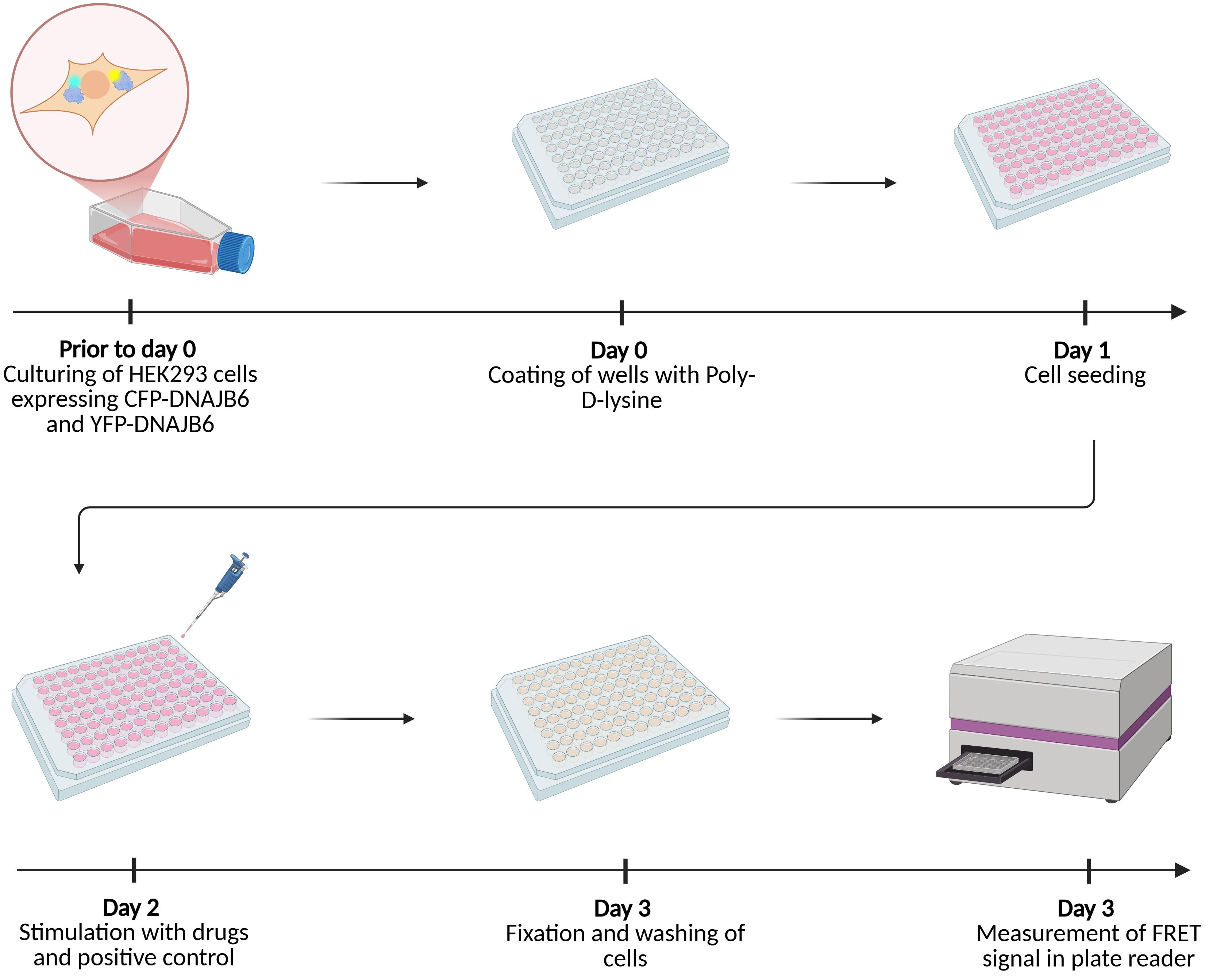
Graphical abstract of the cell-based fluorescence resonance energy transfer (FRET) assay protocol
Background
Neurodegenerative diseases, such as Alzheimer's disease (AD), Huntington’s disease (HD), and Parkinson’s disease (PD), are characterized by the accumulation of misfolded amyloid proteins, which are implicated in disease progression. There are currently no curative treatments for these diseases; consequently, there is a need to find new ways for drug targeting. Preventing misfolding and clearing out aggregates of misfolded protein are central processes in maintaining protein homeostasis in all cells. This is particularly important in post-mitotic cells, such as neurons. Chaperone proteins are key players in the process of preventing and clearing misfolded proteins.
The chaperone DNAJB6 is a member of the Heat Shock Protein 40 (HSP40) family, which is involved in maintaining protein homeostasis. DNAJB6 is highly expressed in the brain, primarily in neurons and oligodendrocytes [1]. Studies have shown that DNAJB6 suppresses aggregation of the amyloid protein alpha-synuclein (α-syn) in vitro, in cells, and in animal models [2–4]. Research also shows that DNAJB6 is dysregulated in synucleinopathies, such as PD and multiple system atrophy (MSA) [5]. Altogether, these data suggest that DNAJB6 is an important endogenous inhibitor of toxic aggregation of the amyloid protein α-syn. Moreover, accumulating data suggest that DNAJB6 may be protective across multiple neurodegenerative diseases, as it has been shown to prevent the formation of amyloid protein aggregates and pathology in animal and cellular models of HD and AD [6–8].
Structural studies suggest that the dimer of DNAJB6 is primarily the active form; an S/T-rich cleft in the DNAJB6 dimer has been identified as the main binding site for amyloid proteins [6,9–11]. While previous studies have established how DNAJB6 expression levels affect amyloid protein aggregation, no assay has previously been developed to study the activity of DNAJB6. We have generated cells that stably express DNAJB6 fused to CFP or YFP, which enables the use of fluorescence resonance energy transfer (FRET) measurements to assess the dimerization status of DNAJB6. In a previous study, we showed that upon treatment with tunicamycin (an antibiotic often used experimentally to activate the unfolded protein response in cells) or pre-formed α-syn fibrils (but not monomeric α-syn), the FRET signal in these cells is elevated, indicating that the activity of DNAJB6 correlates with an increase in misfolded proteins [12]. Here, we describe how these cells can be used in a plate reader–based study to measure fluorescent signal as a proxy for DNAJB6 activity. Tunicamycin was used as a positive control since it is a well-established inducer of the unfolded protein response and produced a robust and reproducible increase in FRET signal. This assay can be used for high-throughput screening for compounds that may alter the activity of DNAJB6. Depending on the intended screening application, assay conditions such as treatment duration and compound concentration may be further optimized to improve sensitivity for detecting weaker compound effects. The protocol is set up for one 96-well plate and can easily be scaled up to suit specific needs.
Materials and reagents
Biological materials
1. FRET DNAJB6 cells (HEK293 cells stably expressing CFP-DNAJB6 and YFP-DNAJB6, generated in-house) [12]
Note: The cell line can be obtained by contacting the corresponding author.
Reagents
1. Dulbecco's modified Eagle medium (DMEM) (Thermo Fisher Scientific, Gibco, catalog number: 61965026)
2. Penicillin/streptomycin (Pen/Strep) (Thermo Fisher Scientific, Gibco, catalog number: 15140122)
3. Amphotericin B 250 μg/mL (Thermo Fisher Scientific, Gibco, catalog number: 15290026)
Note: In the protocol, amphotericin is used directly from a stock solution, but since small quantities are used, it is recommended to aliquot it into, e.g., 2 mL Eppendorf tubes that can be stored for up to 4 weeks at 4 °C or for up to 12 months at -20 °C.
4. Fetal bovine serum (FBS) (Thermo Fisher Scientific, Gibco, catalog number: A5256701)
5. Dimethyl sulfoxide (DMSO) (Merck, Sigma-Aldrich, catalog number: D2438)
6. Phosphate buffered saline (PBS) pH 7.4 (Thermo Fisher Scientific, Gibco, catalog number: 10010015)
7. Paraformaldehyde 4% in PBS (PFA) (Thermo Fisher Scientific, catalog number: J61899)
8. Ethanol anhydrous 99.9% (KiiltoClean A/S, CAS number: 64-17-5)
9. 0.5% trypsin EDTA (Thermo Fisher Scientific, Gibco, catalog number: 15400054)
10. Poly-D-lysine 1 mg/mL (Merck, Sigma-Aldrich, catalog number: A003E)
11. Tunicamycin 1 mg (Sigma-Aldrich, catalog number: T7765-1MG)
Solutions
1. Culture medium (see Recipes)
2. Poly-D-lysine coating solution 100 μg/mL (see Recipes)
3. Trypsin working solution 0.05% (see Recipes)
4. Tunicamycin stock solution 5 mM (see Recipes)
5. Tunicamycin working solution 50 μM (see Recipes)
6. DMSO working solution 1% (see Recipes)
Recipes
Note: All solutions should be prepared aseptically in a biosafety class II cabinet.
1. Culture medium
| Reagent | Final concentration | Quantity or volume |
|---|---|---|
| DMEM | 90% | 500 mL |
| FBS | 9% | 50 mL |
| Pen/Strep | 1% | 5 mL |
| Total | n/a | 555 mL |
Thaw FBS and heat it at 65 °C for 20 min to inactivate proteins. Thaw Pen/Strep in a water bath at 37 °C. Transfer the reagents aseptically to the DMEM bottle.
2. Poly-D-lysine coating solution 100 μg/mL
| Reagent | Final concentration | Quantity or volume |
|---|---|---|
| Poly-D-Lysine 1 mg/mL | 100 μg/mL | 500 μL |
| PBS | n/a | 4.5 mL |
| Total | n/a | 5 mL |
Thaw Poly-D-lysine and dilute it 1:10 according to the table above. The solution can be saved after coating and used up to 10 times. Store at -20 °C and discard after 10 uses or freeze-thaw cycles.
3. Trypsin working solution 0.05%
| Reagent | Final concentration | Quantity or volume |
|---|---|---|
| Trypsin EDTA 0.5% | 0.05% | 100 μL |
| PBS | n/a | 900 μL |
| Total | n/a | 1 mL |
Thaw trypsin EDTA and dilute it 1:10 according to the table above. The trypsin working solution can be prepared in larger volumes and aliquoted into, e.g., 15 mL tubes. It can be stored at 4 °C for a couple of weeks or at -20 °C for long-term storage.
4. Tunicamycin stock solution 5 mM
| Reagent | Final concentration | Quantity or volume |
|---|---|---|
| Tunicamycin 1 mg (844.95 g/mol) | 5 mM | 1 mg |
| DMSO | n/a | 237 μL |
| Total | n/a | 237 μL |
Use the tunicamycin vial to prepare the solution. Pipette up and down and along the sides of the vial to ensure that all the powder is dissolved. Aliquot into PCR tubes. Store the solution at -20 °C and avoid more than five freeze-thaw cycles.
5. Tunicamycin working solution 50 μM
| Reagent | Final concentration | Quantity or volume |
|---|---|---|
| Tunicamycin stock solution (Recipe 4) | 50 μM | 1 μL |
| Culture medium (Recipe 1) | n/a | 99 μL |
| Total | n/a | 100 μL |
Prepare the working solution just before use.
6. DMSO working solution 1%
| Reagent | Final concentration | Quantity or volume |
|---|---|---|
| DMSO | 1% | 1 μL |
| Culture medium (Recipe 1) | n/a | 99 μL |
| Total | n/a | 100 μL |
Prepare the working solution just before use.
Laboratory supplies
1. Cell culture flasks Nunc EasYFlask 75 cm2 sterile (Thermo Fisher Scientific, catalog number: 156499)
2. Pipette tips 1,000 μL (Thermo Fisher Scientific, Sartorius, catalog number: 791000)
3. Pipette tips 300 μL (Thermo Fisher Scientific, Finntip Flex, catalog number: 94060513)
4. Pipette tips 10 μL (Merck, Maxymum Recovery Pipette Tips, catalog number: AXYT300LR)
5. Nunclon Delta Surface 96-well plates (Thermo Fisher Scientific, catalog number: 167008)
6. 15 mL conical tubes (MikroLab Frisenette, Nerbe Plus, catalog number: 02-502-8001)
7. Microcentrifuge tubes (Thermo Fisher Scientific, catalog number: 3404-DLBPK)
8. PCR tubes (Hounisen, Sarstedt, catalog number: 72990002)
9. Cell counting chamber (Marienfeld, Bürker, catalog number: n/a)
10. Matrix reagent reservoirs (Thermo Fisher Scientific, catalog number: 8093)
11. Serological pipette tips 5 mL (Thermo Fisher Scientific, Nunc, catalog number: 170355N)
12. Serological pipette tips 10 mL (Thermo Fisher Scientific, Nunc, catalog number: 170356N)
13. Parafilm (Merck, Sigma-Aldrich, catalog number: P7793-1EA)
Equipment
1. Biosafety cabinet class II (Thermo Scientific, model: SAFE2020)
2. Cell culture incubator (Thermo Scientific, model: Heracell Vios 160i)
3. Water bath (PolyScience, model: WB20)
4. Microscope (Carl Zeiss, model: Primovert)
5. Chemical fume hood
6. Plate reader CLARIOstar (BMG Labtech, model: CLARIOstar)
7. Centrifuge (Thermo Fisher Scientific, model: Sorvall X4RF Pro)
8. FinnPipette F2 2 μL (Thermo Fisher Scientific, catalog number: 4642010)
9. FinnPipette F2 20 μL (Thermo Fisher Scientific, catalog number: 4642060)
10. FinnPipette F2 200 μL (Thermo Fisher Scientific, catalog number: 4642080)
11. Multichannel FinnPipette F2 300 μL (Thermo Fisher Scientific, catalog number: 4662030)
12. FinnPipette F2 1,000 μL (Thermo Fisher Scientific, catalog number: 4642090)
13. Pipette controller (TH Geyer, Labsolute, catalog number: 7696030)
14. Freezer, -20 °C
15. Refrigerator, 4 °C
Software and datasets
The following software is used for initial analysis of the plate reader data, and a license is provided with the purchase of the plate reader.
1. CLARIOstar (BMG Labtech, software version 5.40.R3)
2. CLARIOstar MARS Data Analysis Software (BMG Labtech, software version 3.32)
Procedure
登录/注册后免费查看全文
文章信息
稿件历史记录
提交日期: May 28, 2026
接收日期: Jun 28, 2026
在线发布日期: Jul 9, 2026
出版日期: Aug 5, 2026
版权信息
© 2026 The Author(s); This is an open access article under the CC BY-NC license (https://creativecommons.org/licenses/by-nc/4.0/).
如何引用
Gelman, A., Nielsen, L. K. and Hansen, C. (2026). A Novel Plate Reader–Based Protocol for Measurement of DNAJB6 Dimerization Activity. Bio-protocol 16(15): e5776. DOI: 10.21769/BioProtoc.5776.

 Download Citation in RIS Format
分类
生物化学 > 蛋白质 > 相互作用 > 蛋白质-蛋白质相互作用
细胞生物学 > 基于细胞的分析方法 > 蛋白互作
神经科学 > 神经系统疾病 > 神经退行性病变
您对这篇实验方案有问题吗?
在此处发布您的问题,我们将邀请本文作者来回答。同时,我们会将您的问题发布到Bio-protocol Exchange,以便寻求社区成员的帮助。
 发布问题
发布问题


