


Mouse and Rat Oxygen-Induced Retinopathy Models to Study Vascular Features Seen in Retinopathy of Prematurity
用于研究早产儿视网膜病变血管特征的小鼠和大鼠氧诱导视网膜病变模型

(*contributed equally to this work) 发布: 2026年08月05日第16卷第15期 DOI: 10.21769/BioProtoc.5771 浏览次数: 255
评审: Sébastien GillotinSushma KalmodiaAbhishek VatsMarquis Walker
 PDF
PDF  Q&A
Q&A
Advertisement






Abstract
Retinopathy of prematurity (ROP), a retinovascular disease, is a leading cause of childhood blindness worldwide. Given the constraints of studying molecular mechanisms in preterm infants, reproducible animal models are important to understand ROP pathophysiology. Mouse and rat oxygen-induced retinopathy (OIR) models are the most commonly used and recapitulate key vascular features seen in ROP. However, these models are susceptible to inherent variability that limits reproducibility, including inter-litter variability, consistency of oxygen delivery across experiments, retinal dissection technique, and immunohistochemistry. Here, we describe a comprehensive protocol for performing the most common mouse and rat OIR models, and procedures such as eye enucleation, retinal dissection and flat mounting, isolectin GS-IB4 staining, whole retina stitched fluorescence imaging from Z-stacks, and quantification of vascular features. This protocol provides important materials and procedural details to increase the reproducibility of the mouse and rat OIR models.
Key features
• Rat and mouse oxygen-induced retinopathy (OIR) models.
• Eye enucleation of experimental rat and mouse pups.
• Retinal flat mounting and immunostaining for rat and mouse eyes.
• Image analysis of retinal flat mounts from rat and mouse eyes.
Keywords: Oxygen-induced retinopathy (氧诱导视网膜病变) Vascular biology (血管生物学)
Retinopathy of prematurity (早产儿视网膜病变)
Retinal dissection (视网膜解剖)
Retinal flat mounting (视网膜平铺制片)
Lectin immunostaining (凝集素免疫染色)
Vascular biology (血管生物学)
Retinopathy of prematurity (早产儿视网膜病变)
Retinal dissection (视网膜解剖)
Retinal flat mounting (视网膜平铺制片)
Lectin immunostaining (凝集素免疫染色)
Graphical overview
Background
Retinopathy of prematurity (ROP), a retinovascular disease, is a leading cause of childhood blindness [1,2] and is described by a refined two-phase disease process [3,4]. Phase I involves impaired intraretinal vascular development, resulting in avascular retina and tissue hypoxia. Phase II involves pathologic intravitreal neovascularization (IVNV), which can lead to retinal detachment and permanent vision loss. Therefore, studies are warranted to understand ROP pathophysiology and identify potential therapeutic targets.
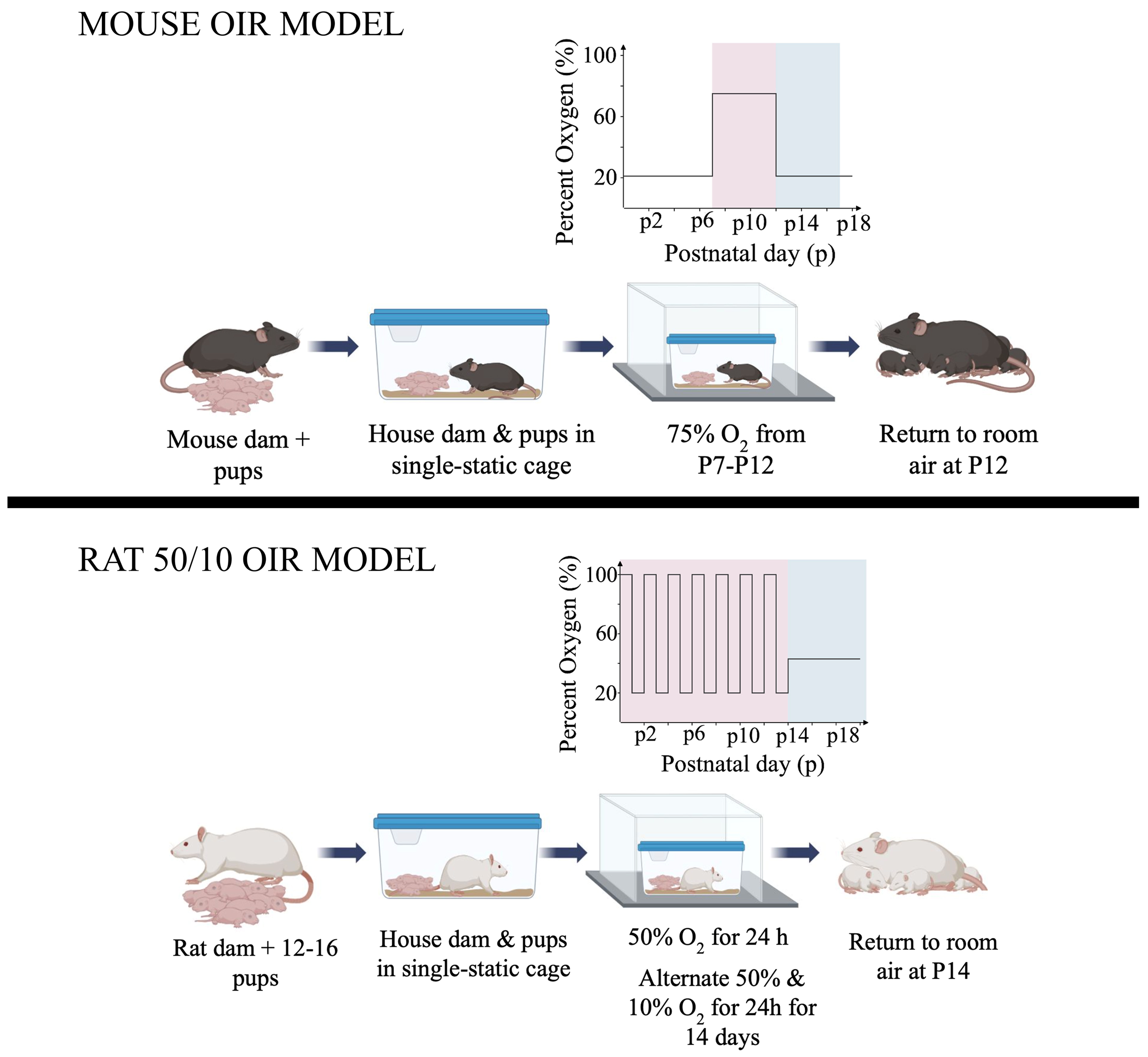
Studying molecular mechanisms of ROP in human preterm infants is not safe; as such, animal models are used to understand pathophysiology and investigate potential therapeutic targets. Oxygen-induced retinopathy (OIR) models have been used to study ROP pathophysiology [5–9], with the mouse and rat 50/10 OIR models being commonly used [9,10]. Mouse OIR recreates the vaso-obliteration of phase I and the vaso-proliferation of Phase II [3,10]. The rat 50/10 OIR model describes the refined hypothesis [4] of delayed physiological retinal vascular development, compromised vascularity (phase I), and vaso-proliferation as IVNV (phase II) [9]. However, model variability from inter-litter differences, consistency of oxygen delivery across experiments, incomplete removal of the hyaloid and vitreous affecting immunostaining, and loss of avascular peripheral retina during dissection in the rat model affect results and interpretation. While factors such as inter-litter variability are difficult to avoid and can only be controlled by including both experimental and control pups within each litter, other confounders can be minimized through reproducible experimental protocols. Here, we outline standardized protocols for (i) induction of mouse and rat OIR using a controlled OxyCycler system, (ii) retinal dissection and flat-mount preparation to ensure consistent staining, (iii) image acquisition capturing the entire retinal vasculature, and (iv) quantitative analysis of total retinal area, avascular area, and IVNV.
Materials and reagents
Biological materials
1. Mice: Timed-pregnant C57BL/6J (Jackson Laboratories, strain number: 000664) or specified transgenic model(s)
2. Rats: Timed-pregnant Sprague Dawley dams (Charles Rivers, strain number: 001)
Reagents
1. Paraformaldehyde (PFA), 4% (Electron Microscopy Sciences, catalog number: 1573520S1L)
2. Phosphate-buffered saline (PBS), 10× (Thermo Fisher Scientific, catalog number: AAJ75889)
3. Triton X-100 stock solution 20% v/v (Sigma-Aldrich, catalog number: T9284)
4. Normal horse serum (Thermo Fisher Scientific/Invitrogen, catalog number: 31874)
5. Isolectin GS-IB4 from Griffonia simplicifolia, Alexa Fluor 488 conjugate (ThermoFisher Scientific, catalog number: I21411)
6. 75% ethanol (Gold Shield Distributors, catalog number: 412811)
7. Fluoromount-G mounting medium (Invitrogen, catalog number: 50-187-88)
8. Triamcinolone acetonide (400 mg per 10 mL) (Teva Pharmaceuticals, catalog number: NDC 0703-0245-01)
Solutions
1. 1× PBS (see Recipes)
2. Blocking buffer (see Recipes)
3. Lectin staining solution (see Recipes)
Recipes
1. 1× PBS
Dilute 10× PBS to 1× PBS with distilled water. Standard room temperature storage.
2. Blocking buffer
1× PBS
10% normal horse serum
0.5% Triton X-100
We recommend preparing fresh blocking buffer; however, we found that storing at 4 °C for up to 1 week will not affect tissue quality or staining. Warm blocking buffer to room temperature and mix before use.
3. Lectin staining solution
Dilute isolectin GS-IB4, Alexa Fluor 488 in blocking buffer, 1:100. Prepare fresh (recommended) and protect from light.
Laboratory supplies
1. 96-well plates (Genesee Scientific, catalog number: 25-109) (optional)
2. 1.5 mL microcentrifuge tubes (CELLTREAT, catalog number: 229442)
3. Kimwipes (Fisher Scientific, catalog number: 06-666)
4. Transfer pipettes (Fisher Scientific, catalog number: 13-711)
5. Superfrost Plus microscope slides (Fisher Scientific, catalog number: 1255015)
6. Coverslips/micro cover glass No. 1.5 (VWR, catalog number: 48393-151)
7. Coverslip weights (Mettler Toledo, catalog number: 01-912-181)
8. Gloves (Fisher Scientific, catalog number: 19149863)
9. FalconTM standard tissue culture dishes 21.29 cm2 (Corning, catalog number: 08-772B)
Equipment
1. OxyCycler (Biospherix, model: A-84XOV) with OxyCycler software
2. Medical-grade oxygen tank, size K (Linde, catalog number: OX M-K) (or equivalent)
3. Medical-grade nitrogen tank, size K (Linde, catalog number: NI M-K) (or equivalent)
4. Portable Combustion Analyzer (Bacharach, model: 0024-8511-Fyrite InTech)
5. Stereomicroscope (Leica, model: EZ4 W)
6. Fluorescence microscope for tiled imaging and stitching (Keyence, model: BZ-X800)
7. Dumont #5 blunt straight tip forceps (FST, catalog number: 11251-30)
8. Fine grasping forceps (FST, catalog number: 11090-10)
9. Iris forceps: Bonn iris suture forceps (Titan Medical, catalog number: TMF612.50)
10. Curved tip forceps (Fisher Scientific, catalog number: 16-100-122)
11. Micro scissors: Noyes micro scissors (WPI, catalog number: 503306)
12. Platform Rocker (Corning, product number: 6702)
13. 30 G insulin needles (BD, SKU 305106)
Software and datasets
1. Fiji (ImageJ) (version: 1.54p)
2. Keyence BZ-X800 imaging software (version: 01.03.00.01)
3. Keyence BZ-X800 Analyzer software (version: 1.1.2.4)
4. Microsoft Excel (version: Microsoft Office LTSC Professional Plus 2021)
Procedure
登录/注册后免费查看全文
文章信息
稿件历史记录
提交日期: Mar 25, 2026
接收日期: Jun 21, 2026
在线发布日期: Jul 7, 2026
出版日期: Aug 5, 2026
版权信息
© 2026 The Author(s); This is an open access article under the CC BY license (https://creativecommons.org/licenses/by/4.0/).
如何引用
Tankersley, M. P., Beri, S., Ramshekar, A., Asare-Bediako, B., Shah, N. S., Karmoker, J. R., Huang, H. and Hartnett, M. E. (2026). Mouse and Rat Oxygen-Induced Retinopathy Models to Study Vascular Features Seen in Retinopathy of Prematurity. Bio-protocol 16(15): e5771. DOI: 10.21769/BioProtoc.5771.

 Download Citation in RIS Format
分类
神经科学 > 感觉和运动系统 > 视网膜
医学 > 眼科
神经科学 > 基础技术 > 组织解剖
您对这篇实验方案有问题吗?
在此处发布您的问题,我们将邀请本文作者来回答。同时,我们会将您的问题发布到Bio-protocol Exchange,以便寻求社区成员的帮助。
 发布问题
发布问题


