


In Vivo and In Vitro SUMOylation Assays in Arabidopsis
拟南芥体内与体外SUMO化检测方法

发布: 2026年08月05日第16卷第15期 DOI: 10.21769/BioProtoc.5769 浏览次数: 193
评审: Tasleem JavaidAnonymous reviewer(s)
 PDF
PDF  Q&A
Q&A
参见作者原研究论文
The authors used this protocol in:
Feb 2026
Advertisement






Abstract
Small ubiquitin-like modification (SUMOylation) is a crucial post-translational modification that modulates protein stability, localization, and interaction dynamics. Despite the identification of thousands of putative small ubiquitin-like modifier (SUMO) substrates, functional validation remains challenging due to the low abundance and highly dynamic nature of SUMOylated proteins. Here, we present a protocol for detecting protein SUMOylation, integrating bioinformatic site prediction, and rapid substrate screening via in vivo tobacco transient expression and in vitro E. coli assay, followed by precise validation using transgenic Arabidopsis lines. However, detection of low-abundance SUMOylated proteins may require coupling with mass spectrometry, and the in vitro system does not fully recapitulate the complex regulatory network in vivo. This workflow provides a useful tool for studying SUMOylation in plants.
Key features
• Integrates bioinformatic prediction, in vitro validation, and in vivo confirmation for SUMOylation analysis.
• E. coli co-expression system enables rapid SUMOylation detection without protein purification.
• Arabidopsis transgenic line system confirms SUMOylation under physiological conditions.
• The protocol is applicable to most Arabidopsis proteins.
Keywords: SUMOylation (SUMO化) Post-translational modification (翻译后修饰)
In vitro SUMOylation assay (体外SUMO化检测)
In vivo SUMOylation assay (体内SUMO化检测)
Arabidopsis (拟南芥)
Post-translational modification (翻译后修饰)
In vitro SUMOylation assay (体外SUMO化检测)
In vivo SUMOylation assay (体内SUMO化检测)
Arabidopsis (拟南芥)
Graphical overview
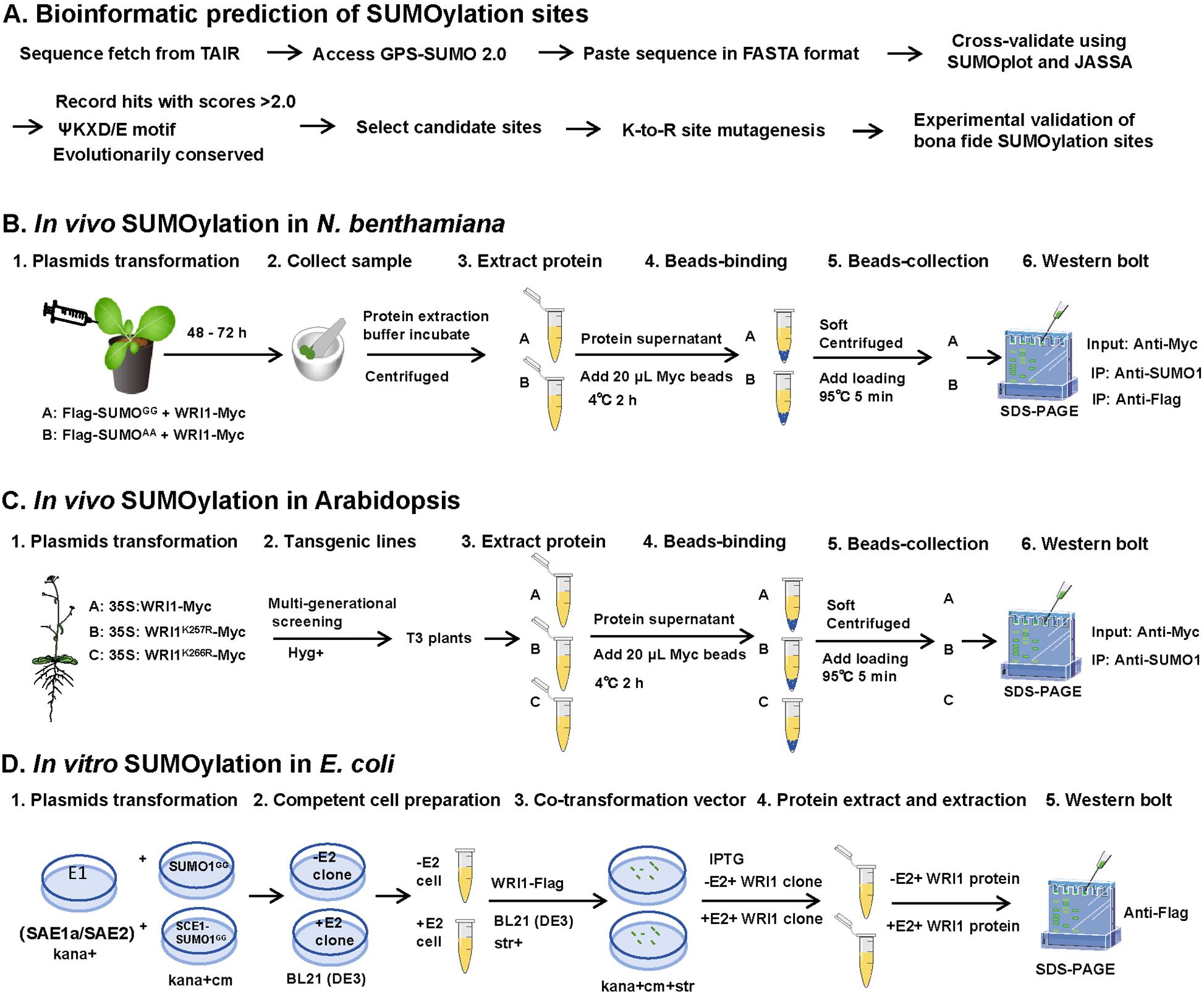
In vivo and in vitro SUMOylation. (A) Bioinformatic prediction of SUMOylation sites (dry-lab; expected time: 1–2 days). The target protein sequence is retrieved from TAIR and analyzed using GPS-SUMO 2.0, SUMOplot, and JASSA. GPS-SUMO 2.0 is suitable for large-scale prediction with customizable thresholds; SUMOplot focuses on the canonical ΨKXD/E motif; JASSA provides additional features such as secondary structure analysis and database hit searching. (B) In vivo SUMOylation assay in N. benthamiana (wet-lab; expected time: 3–4 days). Plasmids encoding Flag-SUMOGG or Flag-SUMOAA together with WRI1-Myc were transformed into N. benthamiana leaves. (C) In vivo SUMOylation assay in Arabidopsis (wet-lab; expected time: 9–10 months). Transgenic lines of 35S:WRI1-Myc, 35S:WRI1K257R-Myc, or 35S:WRI1K266R-Myc were generated through multi-generational screening. (D) In vitro SUMOylation in E. coli (wet-lab; expected time: 15–20 days). The competent cells of BL21 (DE3) containing pET28a-AtSAE1a-His-SAE2 + pACYCDuet-AtSUMO1GG (-E2) or pET28a-AtSAE1a-His-SAE2 + pACYCDuet-1-His-AtSCE1-Myc-AtSUMO1GG (+E2), respectively. The pCDFDuet-WRI1-Flag, WRI1K266R-Flag, WRI1K257R-Flag, and WRI12KR-Flag were transformed into -E2 and +E2 competent cells.
Background
Small ubiquitin-like modification (SUMOylation) is a reversible post-translational modification that plays essential roles in plant development, stress responses, and metabolic regulation [1]. The modification process involves a cascade of enzymes: small ubiquitin-like modifier (SUMO) activating enzyme E1, SUMO conjugating enzyme E2, and, usually, a SUMO ligase E3, which together conjugate SUMO molecules to lysine residues of target proteins [2]. Despite the identification of thousands of potential SUMO substrates through proteomic screens [3,4], functional validation of specific SUMOylation events remains technically challenging due to the low abundance of SUMOylated proteins and the presence of active SUMO proteases that rapidly remove SUMO conjugates.
Several methods have been developed to detect protein SUMOylation in plants. Qu et al. (2020) comprehensively described classic approaches, including in vitro assays using purified recombinant proteins and in vivo assays using tobacco transient expression, Arabidopsis protoplasts, and transgenic plants [5]. Similarly, a protocol for detecting SUMOylated phytochromes in plants was established to address the specific challenges of monitoring phytochrome SUMOylation [6]. Huang et al. (2022) established an efficient in vitro SUMOylation detection system in E. coli [7]. Lai et al. (2023) further expanded this system for high-throughput substrate screening, validating SUMOylation of 95% of candidate proteins from a maize cDNA library [8]
For large-scale identification of SUMO substrates, Miller et al. (2010) generated transgenic Arabidopsis lines expressing a His-tagged SUMO1 mutant H89R and identified 357 SUMOylated proteins by affinity purification coupled with mass spectrometry [9]. To improve detection efficiency, Hendriks et al. (2014) introduced a His-tagged SUMO2 mutant (KO-Q87R) and developed a two-step IMAC enrichment strategy, identifying over 4,300 SUMOylation sites and 1,600 target proteins [10]. A key technical breakthrough came with the discovery of the α-lytic protease WaLP from Lysobacter enzymogenesis, which specifically cleaves SUMO-modified peptides to generate KGG-containing peptides, enabling enrichment with anti-KGG antibodies [11]. Using this approach, Lumpkin et al. (2017) identified 1,209 endogenous SUMOylation sites [12]. Recently, Sang et al. (2024) employed a lysine-null SUMO1 in the sumo1 sumo2 mutant background, combined with a two-step lysine-null SUMO enrichment method, to identify 2,235 SUMOylation sites across 1,300 substrates [13]. Despite the identification of numerous SUMOylated proteins, in vivo experimental validation of these substrates remains challenging.
Building upon these established methods, we present an integrated protocol that combines bioinformatic site prediction, the E. coli co-expression system for in vitro validation, and both tobacco transient expression and transgenic plants for in vivo confirmation. Using the master regulator of seed oil synthesis WRINKLED1 (WRI1) as a case study, we provide detailed step-by-step instructions from SUMOylation site prediction to functional validation. The protocol is applicable to most target proteins in Arabidopsis and can be adapted for use in other plant species.
Materials and reagents
Biological materials
1. Escherichia coli DH5α (store at -80 °C)
2. Escherichia coli BL21 (DE3) (store at -80 °C)
3. Agrobacterium tumefaciens GV3101 (store at -80 °C)
4. Arabidopsis thaliana Columbia-0 (Col-0) seeds (store at -20 °C)
5. Nicotiana benthamiana seeds (store at -20 °C)
Reagents
1. Tris (Sigma-Aldrich, catalog number: T1503)
2. SDS (Sigma-Aldrich, catalog number: 436143)
3. NaCl (Sigma-Aldrich, catalog number: S3014-500G)
4. EDTA (Sigma-Aldrich, catalog number: E9884)
5. Triton X-100 (Sangon Biotech, catalog number: 73049-73-7)
6. Na2HPO4·12H2O (Sigma-Aldrich, catalog number: 71649)
7. KH2PO4 (Sigma-Aldrich, catalog number: P5655)
8. KCl (Sigma-Aldrich, catalog number: P9541)
9. Glycine (MACKLIN, catalog number: 50-01-1)
10. Coomassie Brilliant Blue R-250 (Sigma-Aldrich, catalog number: B1047)
11. Tween-20 (Biosharp, catalog number: BS100-500)
12. MES (Sangon Biotech, catalog number: A610214)
13. Acetosyringone (AS) (Sigma-Aldrich, catalog number: D134406)
14. MgCl2·6H2O (Sigma-Aldrich, catalog number: M2670)
15. Kanamycin sulfate (Sangon Biotech, catalog number: A506636-0100); 50 μg/mL
16. Chloramphenicol (Sangon Biotech, catalog number: A600118-0050); 50 μg/mL
17. Streptomycin sulfate (Sangon Biotech, catalog number: A610494); 50 μg/mL
18. Rifampicin (Sigma-Aldrich, catalog number: R3510); 50 μg/mL
19. Methanol (Thermo Fisher, catalog number: A412-4)
20. Glacial acetic acid (Thermo Fisher, catalog number: A38C212)
21. Protease inhibitor cocktail (Roche, catalog number: 04693159001)
22. Isopropyl-β-D-thiogalactopyranoside (IPTG) (Aladdin, catalog number: I108498)
23. BSA (Sangon Biotech, catalog number: A500023)
24. Coomassie Brilliant Blue R-250 (Sigma-Aldrich, catalog number: B1047)
25. Ponceau S staining solution (Affinibody, catalog number: AIWB-013)
26. ColorMixed Protein Marker 180 (10–180 kDa) (Abclone, catalog number: RM19001)
27. Anti-Myc agarose beads (Abmart, catalog number: M20012M)
28. Anti-FLAG agarose beads (Abmart, catalog number: M20008L)
29. Anti-Myc antibody (Abmart, catalog number: M20002L, 1:5,000)
30. Anti-FLAG antibody (Abmart, catalog number: M20008L, 1:5,000)
31. Anti-SUMO1 antibody (Abcam, catalog number: ab5316, 1:5,000)
32. Anti-actin antibody (Abmart, catalog number: M20009L, 1:5,000)
33. Goat anti-mouse IgG-HRP (Abmart, catalog number: M21001, 1:5,000–1:10,000)
34. Goat anti-rabbit IgG-HRP (Abmart, catalog number: M21002,1:5,000–1:10,000)
35. Competent Cell Preparation kit (Sangon Biotech, catalog number: B529305)
36. One-step PAGE Gel Preparation kit 10% (Affinibody, catalog number: NSF100)
37. BCA Protein Assay kit (Sangon Biotech, catalog number: C503031-1000)
38. High-sensitivity ECL chemiluminescence reagent (Affinibody, catalog number: AIWB-006)
39. Non-fat milk (Sangon Biotech, catalog number: A600669-0250)
Solutions
1. LB liquid medium (see Recipes)
2. Plant protein extract buffer (see Recipes)
3. 10× SDS-PAGE running buffer (see Recipes)
4. 10× PBS buffer (see Recipes)
5. 10× TBS buffer (see Recipes)
6. 10× Transfer buffer (see Recipes)
7. Coomassie blue staining solution (R-250 buffer) (see Recipes)
8. Destaining buffer (see Recipes)
9. 5% non-fat milk (10 mL)
10. 3% BSA (10 mL)
Recipes
1. LB liquid medium (1 L)
Tryptone (10 g)
Yeast extract (5 g)
NaCl (10 g)
LB solid medium: same as LB liquid medium with the addition of 15 g of agar; autoclave at 121 °C for 20 min.
Store at room temperature for up to 6 months. After addition of antibiotics, store at 4 °C and use within 2 weeks.
2. Plant protein extract buffer (1 L)
50 mM Tris-HCl pH 7.4 (7.88 g)
150 mM NaCl (8.77 g)
1 mM EDTA pH 8.0 (0.37 g)
0.5% (v/v) Triton X-100 (5 mL)
1× protease inhibitor cocktail (100 µL)
Store at -20 °C for 6 months. Add protease inhibitor cocktail fresh before use; after adding inhibitor, use within 24 h.
3. 10× SDS-PAGE running buffer (1 L)
250 mM Tris base (30 g)
1.92 M glycine (144 g)
1% (w/v) SDS (10 g)
Prepare 1× SDS-PAGE running buffer fresh by diluting 10× SDS-PAGE buffer 1:10 with ddH2O.
Store at room temperature for up to 6 months.
4. 10× PBS buffer (1 L)
100 mM Na2HPO4·12H2O (35.8 g)
18 mM KH2PO4 (2.4 g)
1.37 M NaCl (80 g)
27 mM KCl (2 g)
Prepare 1× PBS buffer fresh by diluting 10× PBS buffer 1:10 with ddH2O. Add 1× protease inhibitor cocktail to the 1× PBS buffer before use.
Store at room temperature for up to 6 months.
5. 10× TBS buffer (1 L)
200 mM Tris-HCl pH 8.0 (3.03 g)
1.5 M NaCl (87.66 g)
Prepare 1× TBST buffer fresh by diluting 10× TBS buffer 1:10 with ddH2O and add 1 mL of Tween-20.
Store at room temperature for up to 6 months.
6. 10× transfer buffer (1 L)
250 mM Tris (30.03 g)
1.92 M glycine (144 g)
1× Transfer buffer: Add 80 mL of 10× transfer buffer, 200 mL of methanol, and 720 mL of ddH2O to a total volume of 1 L. Prepare the 1× transfer buffer fresh on the day before use. Store 10× transfer buffer at room temperature for up to 6 months.
7. Coomassie blue staining solution (R-250 buffer) (1 L)
0.1% (w/v) Coomassie Brilliant Blue R-250 (1 g)
45% (v/v) methanol (450 mL)
10% (v/v) glacial acetic acid (100 mL)
45% (v/v) ddH2O (450 mL)
Store at room temperature in a dark bottle.
8. Destaining buffer (1 L)
10% (v/v) methanol (100 mL)
10% (v/v) glacial acetic acid (100 mL)
ddH2O (800 mL)
Store at room temperature. Can be reused until saturated with dye.
9. 5% non-fat milk (10 mL)
Non-fat milk (0.5 g)
1× TBST buffer (prepared fresh) (10 mL)
Prepare fresh and store at 4 °C for up to 1 day.
10. 3% BSA (10 mL)
3% BSA (0.3 g)
1× TBST buffer (prepared fresh) (10 mL)
Prepare fresh and store at 4 °C for up to 1 day.
Laboratory supplies
1. Microcentrifuge tubes, 1.5 mL (Pierce, catalog number: 69715)
2. Pipette tips 10, 200, 1000 μL (Axygen, catalog numbers: T-300, T-200-Y, T-1000-B)
3. Centrifuge tubes 15, 50 mL (Corning, catalog number: CLS430828-100EA)
4. PVDF membrane 0.45 μm (Merck Millipore, catalog number: IPVH00010)
5. Western blot filter papers (Bio-Rad, catalog number: 1703965)
6. Western blot sponge pads (Bio-Rad, catalog number: 1703932)
7. Syringe, 1 mL, needle-free (Welch Materials, catalog number: 00824-11121)
8. Tissue grinder or mortar and pestle (Aladdin, catalog number: 1245-160mm-1EA)
9. Liquid nitrogen (self-prepared)
Equipment
1. Ultrapure water system (Sartorius, model: Arium Pro Ultrapure Water System)
2. End-over-end rotator (Elmi, model: ROTAMIX RM1)
3. High-pressure cell disruptor (JNBIO, model: JN-10C)
4. Refrigerated centrifuge (Eppendorf, model: 5424R)
5. SDS-PAGE electrophoresis system (Bio-Rad, model: Mini-PROTEAN® Tetra)
6. Transfer system (Bio-Rad, model: Mini Trans-Blot®)
7. e-Blot for chemiluminescence imaging (Touch IMAGETM, Version 2.1)
Software and datasets
1. GPS-SUMO2.0 [1] (http://sumosp.biocuckoo.org/)
2. SUMOplot1.0 (http://www.abgent.com/sumoplot)
3. JASSA1.0 (http://www.jassa.fr/index.php?jassa)
4. GraphPad Prism9.0 (GraphPad Software, Version 9.0)
5. ImageJ 1.54 (https://imagej.nih.gov/ij/)
Procedure
登录/注册后免费查看全文
文章信息
稿件历史记录
提交日期: Apr 14, 2026
接收日期: Jun 17, 2026
在线发布日期: Jul 3, 2026
出版日期: Aug 5, 2026
版权信息
© 2026 The Author(s); This is an open access article under the CC BY-NC license (https://creativecommons.org/licenses/by-nc/4.0/).
如何引用
Liu, X., Tang, S., Guo, X., Fan, C. and Hu, Z. (2026). In Vivo and In Vitro SUMOylation Assays in Arabidopsis. Bio-protocol 16(15): e5769. DOI: 10.21769/BioProtoc.5769.

 Download Citation in RIS Format
分类
植物科学 > 植物生物化学 > 蛋白质 > 修饰
生物化学 > 蛋白质 > 修饰 > SUMO化
您对这篇实验方案有问题吗?
在此处发布您的问题,我们将邀请本文作者来回答。同时,我们会将您的问题发布到Bio-protocol Exchange,以便寻求社区成员的帮助。
 发布问题
发布问题


