


Construction and Functional Evaluation of Cyclic Peptide-Based CAR T Cells in Tumor Models
基于环肽的 CAR-T 细胞构建及其在肿瘤模型中的功能评价

发布: 2026年07月05日第16卷第13期 DOI: 10.21769/BioProtoc.5715 浏览次数: 296
评审: Navnita DuttaRan ChenMichael Enos
 PDF
PDF  Q&A
Q&A
参见作者原研究论文
The authors used this protocol in:
Jan 2026
Advertisement







Abstract
Cyclic peptides are emerging as a promising class of recognition modules for chimeric antigen receptor (CAR) engineering. Compared with single-chain variable fragment (scFv)-based CARs, disulfide-directed multicyclic peptides (DDMPs) represent a novel alternative, offering a markedly smaller molecular size (<5 kDa), enhanced structural stability through disulfide-directed cyclization, and broad tolerance to sequence diversification that supports systematic affinity and specificity optimization. DDMP-based CAR T cells leverage these properties to mediate antigen-dependent cytotoxicity while exhibiting an attenuated cytokine secretion profile, supporting the development of potentially safer immunotherapies for solid tumors. Here, we present a comprehensive workflow spanning CAR construct design and generation through in vitro and in vivo functional evaluation. While DDMPs are used as the exemplar recognition module, sections A and C–L of the protocol are directly applicable to any CAR format, including scFv- and nanobody-based designs with minimal modifications, making the workflow accessible to the broader CAR T-cell research community. The protocol includes the generation of Jurkat NFAT reporter cell lines and luciferase-expressing tumor target lines, which are widely used in different assays. Together, these standardized readouts enable rigorous, objective comparison of CAR T-cell efficacy and safety across tumor models.
Key features
• DDMPs (<5 kDa) are compact, disulfide-cyclized antigen recognition modules that tolerate extensive sequence diversification, enabling affinity and specificity optimization beyond conventional scFv-based CARs.
• An integrated pipeline normalizes all functional comparisons to CAR-positive cell numbers, eliminating transduction efficiency as a confounding variable across construct designs.
• Complementary readouts cross-validate efficacy and specificity: NFAT activation, luminescence-based killing, flow cytometry–based cytolysis, and ELISA-based cytokine secretion.
• Xenograft imaging via the in vivo imaging system (IVIS) validates DDMP-CAR T-cell antitumor activity, extending cross-validation to preclinical tumor models.
Keywords: Chimeric antigen receptor (嵌合抗原受体) Autologous CAR T (自体 CAR-T 细胞)
Disulfide-directed multicyclic peptide (二硫键导向多环肽)
DDMP (DDMP)
NFAT reporter (NFAT 报告系统)
Cyclic peptide (环肽)
HER2 (HER2)
Xenograft (异种移植模型)
Preclinical CAR T evaluation (CAR-T 细胞临床前评价)
Autologous CAR T (自体 CAR-T 细胞)
Disulfide-directed multicyclic peptide (二硫键导向多环肽)
DDMP (DDMP)
NFAT reporter (NFAT 报告系统)
Cyclic peptide (环肽)
HER2 (HER2)
Xenograft (异种移植模型)
Preclinical CAR T evaluation (CAR-T 细胞临床前评价)
Graphical overview
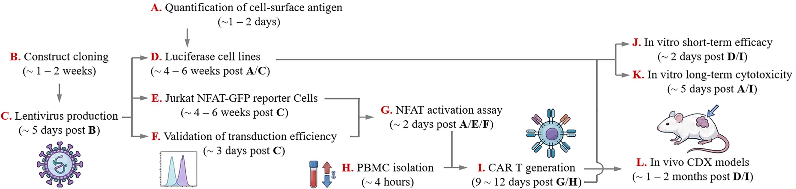
Experimental workflow. Schematic illustration of the experimental pipeline described in this protocol, from construct design to functional evaluation in vitro and in vivo. Arrows indicate the logical progression of the workflow. NFAT, nuclear factor of activated T cells; GFP, green fluorescent protein; PBMC, peripheral blood mononuclear cell; CAR, chimeric antigen receptor; CDX, cell line–derived xenograft; IVIS, in vivo imaging system. A detailed timeline of the experimental workflow is shown in Table 1.
Background
Standardized workflows for chimeric antigen receptor (CAR) T-cell manufacturing and functional testing are widely used to benchmark receptor designs across diverse tumor models and experimental readouts. Building on reported procedures for T-cell isolation/activation, viral gene transfer, and downstream in vitro/in vivo assays [1–6], this protocol provides a beginner-friendly, end-to-end pipeline for preclinical evaluation of autologous CAR T therapy. The workflow includes (i) quantitative profiling of target-antigen density on tumor cells, (ii) generation of reporter cell lines for functional readouts, and (iii) multi-assay validation spanning reporter activation, cytolysis/cytokine secretion, and xenograft efficacy. A key principle for comparing efficacy between different constructs is to normalize by the number of CAR-positive T cells, enabling functional comparisons that are independent of transduction efficiency.
This protocol is exemplified using disulfide-directed multicyclic peptides (DDMPs) as compact, engineerable antigen-recognition modules. DDMPs are disulfide-rich multicyclic peptides whose oxidative folding is guided by disulfide-directing motifs, such as biscysteine motifs CXC, CPPC, and CPXXC [7]. In conventional disulfide-rich peptides, a sequence containing 2n cysteine residues can theoretically form (2n)!/(2ⁿ n!) distinct isomers (e.g., 105 for n = 4), yielding complex mixtures. In contrast, incorporation of disulfide-directing motifs can drive highly efficient folding to a dominant product, which substantially simplifies isolation and characterization. Because DDMP folding is largely determined by these motifs, DDMP scaffolds also exhibit greater tolerance to extensive sequence manipulation, enabling highly diversified display libraries and systematic ligand discovery against a broad range of cell-surface receptors, including tumor-associated antigens and immune receptors [8–16]. While sections A and C–L are fully generalizable to any CAR format regardless of antigen-recognition module, section B (molecular cloning) includes DDMP-specific considerations, particularly sequence design principles that are specific to the DDMP CAR construct. Users employing single-chain variable fragment (scFv)- or nanobody-based CARs may adapt the cloning strategy accordingly. Thus, the integrated workflow described here is well suited for iterative optimization and head-to-head benchmarking of DDMP-based CAR designs, linking molecular construction to rigorous functional evaluation across complementary in vitro and in vivo tumor models [17,18].
Materials and reagents
Biological materials
1. HEK293T cells (Cell Bank of the Chinese Academy of Sciences, catalog number: GNHu17)
2. A549 cells (Cell Bank of the Chinese Academy of Sciences, catalog number: SCSP-503)
3. SK-OV-3 cells (Cell Bank of the Chinese Academy of Sciences, catalog number: SCSP-5214)
4. NCI-N87 cells (Procell, catalog number: CL-0169)
5. OE19 cells (Procell, catalog number: CL-0754)
Note: Users can use alternative HER2-positive tumor cell lines or any other cell line expressing the specific target antigen for their assays.
6. Jurkat E6–1 cells (Cell Bank of the Chinese Academy of Sciences, catalog number: SCSP-513)
7. Human peripheral blood (MileCell Bio; healthy donors; origin as supplied)
8. NCG mice, 6–8 weeks (GemPharmatech, catalog number: T001475)
Reagents
1. DMEM (Gibco, catalog number: 21063029)
2. RPMI 1640 (Gibco, catalog number: 11875093)
3. Click’s medium (Sigma, catalog number: C5572)
4. Opti-MEM (Gibco, catalog number: 31985-070)
5. FBS (TransGen Biotech, catalog number: FS401-02)
6. FBS (Gibco, catalog number: 10099141)
7. FBS (Sigma, catalog number: F8687)
Note: FBS was used as follows to balance performance and cost: Premium FBS (e.g., Gibco, Sigma) was used for all primary T cell and Jurkat cell cultures because these cells require low-endotoxin, high-consistency formulations; a single pre-screened lot was used for all related functional assays to ensure reproducibility. Economical domestic FBS (e.g., TransGen Biotech), was used for the routine maintenance of established tumor cell lines (e.g., HEK293T, A549, SK-OV-3, NCI-N87, and OE19), which are less sensitive to serum variations.
8. Penicillin/streptomycin (Gibco, catalog number: 15140122)
9. Lentiviral packaging plasmid: psPAX2 (Addgene, catalog number: 12260)
10. Lentiviral envelope plasmid: pMD2.G (Addgene, catalog number: 12259)
11. Lentiviral transfer plasmid backbone: pCDH (Addgene, catalog number: 72265/72266)
12. Lentiviral transfer plasmid for generating luciferase-expressing stable Luc-GFP cell lines: pCCLc-MNDU3-Luciferase-PGK-EGFP-WPRE (Addgene, catalog number: 89608)
13. Lentiviral transfer plasmid for generating NFAT-GFP reporter cell line: FLX1.8NFATGFPpd2HS4 (Addgene, catalog number: 169096)
14. PEI (Polysciences, catalog number: 24765) (linear polyethyleneimine MW 40,000; prepare as 1 μg/μL solution in sterile water, pH 7.0)
15. Ficoll (Cytiva, catalog number: 17544203) [1.077 g/mL at 20 °C, for human peripheral blood mononuclear cell (PBMC) isolation]
16. CryoStor CS10 (Stemcell, catalog number: 100-1061)
17. Dynabeads human T-Expander CD3/CD28 (Invitrogen, catalog number: 11141D)
18. Recombinant human IL-2 (Proteintech, catalog number: HZ-1015)
19. Retronectin (Takara, catalog number: T100A)
20. Polybrene (10 mg/mL) (Solarbio, catalog number: H8761-5), store at -20 °C and keep one tube as working solution at 4 °C
21. Violet 450 (Tonbo, catalog number: 13-0863)
22. PE anti-human HER2 (BioLegend, catalog number: 324406, clone 24D2)
23. FITC anti-human CD3 (4A Biotech, catalog number: FHF003-01-100, clone OKT3)
24. APC-Cy7 anti-human CD3 (BD, catalog number: 557832, clone SK7)
25. PE anti-V5 tag antibody (eBioscience, catalog number: 12-6796-42, clone TCM5)
26. PE anti-human TROP2 antibody (Invitrogen, catalog number: 12-6024-42, clone MR54)
27. Cell stimulation cocktail (Invitrogen, catalog number: 00-4970)
28. Counting beads (eBioscience, catalog number: 01-1234-42)
29. D-luciferin sodium salt (MedChemExpress, catalog number: HY-12591)
30. IVISbrite D-luciferin potassium salt (PerkinElmer, catalog number: 122799)
Note: D-luciferin sodium salt (MedChemExpress, catalog number: HY-12591) is used for in vitro bioluminescence assays on the plate reader (sections D and J). IVISbrite D-luciferin potassium salt (PerkinElmer, catalog number: 122799) is used for in vivo imaging system (IVIS) imaging (section L).
31. ELISA kits for IL-2 (Invitrogen, catalog number: 88-7025-88), TNF-α (Invitrogen, catalog number: 88-7346-77), IFN-γ (Invitrogen, catalog number: 88-7316-86), and GM-CSF (Invitrogen, catalog number: 88-8337-88) detection
32. Matrigel (Corning, catalog number: 356237)
33. PBS (Goonie Bio, catalog number: 251222)
34. Trypsin-EDTA (Gibco, catalog number: 25200072)
Solutions
1. 10% FBS in DMEM (see Recipes)
2. 10% FBS in RPMI 1640 (see Recipes)
3. 20% FBS in RPMI 1640 (see Recipes)
4. T-cell culture medium (see Recipes)
5. 10 mM D-luciferin (see Recipes)
6. 2% BSA (w/v) (see Recipes)
Recipes
1. 10% FBS in DMEM
89% DMEM + 10% FBS (TransGen) + 1% penicillin/streptomycin
2. 10% FBS in RPMI 1640
89% RPMI 1640 + 10% FBS (TransGen) + 1% penicillin/streptomycin
3. 20% FBS in RPMI 1640
79% RPMI 1640 + 20% FBS (Gibco) + 1% penicillin/streptomycin. This is used for culturing Jurkat cells.
4. T-cell culture medium
44.5% RPMI 1640 + 44.5% Click’s medium + 10% FBS (Sigma) + 1% penicillin/streptomycin
5. 10 mM D-luciferin
Dissolve 4.53 mg of D-luciferin in ddH2O to a final volume of 1.5 mL. Filter-sterilize through a 0.22 μm filter, aliquot 300 μL per tube, and store at -80 °C.
6. 2% BSA (w/v)
Dissolve 10 g of BSA in 1× PBS to a final volume of 500 mL. Filter-sterilize through a 0.22 μm filter. Store at 4 °C for up to 4 weeks. Discard if turbidity or precipitate is observed.
Laboratory supplies
1. 1.5 mL microcentrifuge tube (Axygen, catalog number: MCT-200-C)
2. 15 mL centrifuge tube (LabSelect, catalog number: CT-002-15A)
3. 50 mL conical tube (LabSelect, catalog number: CT-002-50A)
4. 5 mL tube for flow cytometry applications (Falcon, catalog number: 352235)
5. 300-mesh cell strainer (Solarbio, catalog number: YA0950)
6. Cryovials (ThermoFisher, catalog number: 377267)
7. Mr. Frosty freezing container (ThermoFisher, catalog number: 5100-0001)
8. 96-well cell culture plate (LabSelect, catalog number: 11510)
9. 96-well plate, white bottom (Biosharp, catalog number: BS-MP-96W-CL)
10. 12-well plate, non-TC treated (LabSelect, catalog number: 11220)
11. 24-well plate, non-TC treated (LabSelect, catalog number: 11320)
12. 24-well cell culture plates (LabSelect, catalog number: 11310)
13. 0.45 μm PES filter (Biosharp, catalog number: BS-PES-45)
14. 0.22 μm syringe filters (PES membrane; Millipore, catalog number: SLGP033RS)
Equipment
1. Class II biosafety cabinet (Haier, model: HR40-IIA2)
2. CO2 incubator (Haier, model: HCB-168)
3. 3D rotating mixer (Crystal, model: TR-02U)
4. Microcentrifuge (ThermoFisher, model: Pico 21)
5. High-speed centrifuge with a swinging-bucket rotor for 50 mL tubes (Eppendorf, model: 5910Ri)
6. Cell counter (DeNovix, model: CellDrop BF)
7. Flow cytometer (Beckman Coulter, model: CytoFLEX LX)
8. Cell sorter (Beckman Coulter, model: CytoFLEX SRT)
9. Microplate reader for bioluminescence detection (BioTek, model: Synergy Neo2)
10. In vivo imaging system (PerkinElmer, model: IVIS Spectrum)
Software and datasets
1. FlowJo (FlowJo, version 10.7)
2. Living Image (PerkinElmer, version 4.7.4)
Procedure
登录/注册后免费查看全文
文章信息
稿件历史记录
提交日期: Mar 23, 2026
接收日期: May 6, 2026
在线发布日期: May 21, 2026
出版日期: Jul 5, 2026
版权信息
© 2026 The Author(s); This is an open access article under the CC BY-NC license (https://creativecommons.org/licenses/by-nc/4.0/).
如何引用
Meng, X., Wu, Q. and Tsai, Y. (2026). Construction and Functional Evaluation of Cyclic Peptide-Based CAR T Cells in Tumor Models. Bio-protocol 16(13): e5715. DOI: 10.21769/BioProtoc.5715.

 Download Citation in RIS Format
分类
癌症生物学 > 肿瘤免疫学 > 癌症治疗 > 细胞移植治疗
细胞生物学 > 细胞工程 > 慢病毒递送
免疫学 > 免疫疗法 > CAR-T
您对这篇实验方案有问题吗?
在此处发布您的问题,我们将邀请本文作者来回答。同时,我们会将您的问题发布到Bio-protocol Exchange,以便寻求社区成员的帮助。
 发布问题
发布问题


