


Detection of Target Molecules Within One-Millimeter-Thick Mouse Brain Slices by Using Peroxidase-Fused Nanobodies and Fluorochromized Tyramide-Glucose Oxidase Reaction
利用过氧化物酶融合纳米抗体和荧光化酪酰胺-葡萄糖氧化酶反应检测 1 mm 厚小鼠脑切片中的靶分子

发布: 2026年06月05日第16卷第11期 DOI: 10.21769/BioProtoc.5713 浏览次数: 276
评审: Olga KopachJung-Wan MokAnya Bhavnani
 PDF
PDF  Q&A
Q&A
参见作者原研究论文
The authors used this protocol in:

Jun 2025
Advertisement







Abstract
Three-dimensional immunohistochemistry (3D-IHC) shows the organization of molecular assemblies in the context of tissue architecture. Deep and rapid antibody penetration into 3D tissues and highly sensitive detection are crucial for high-throughput analysis of 3D-IHC imaging. Here, we provide a detailed protocol for a nanobody (nAb)-based 3D-IHC technique, namely POD-nAb/FT-GO 3D-IHC, for high-speed and high-sensitivity detection of targets within 1-mm-thick mouse brain tissues. Peroxidase-fused nAb (POD-nAb) is a genetically encoded recombinant antibody, which consists of a camelid nAb and a variant of horseradish peroxidase, and fluorochromized tyramide-glucose oxidase (FT-GO) is a fluorescent tyramide signal amplification (TSA) system. POD-nAb/FT-GO 3D-IHC incorporates three main components: 1) tissue permeabilization, 2) POD-nAb binding, and 3) 3D-TSA reaction with FT-GO. POD-nAbs enhance signal penetration depth and allow for highly sensitive detection when combined with FT-GO signal amplification. By using the 3D-IHC protocol provided herein, we can visualize target molecules in mouse brain tissues of 1-mm thickness with drastic signal enhancement within three days. This protocol for POD-nAb/FT-GO 3D-IHC could facilitate structural and molecular interrogation of 3D tissues.
Key features
• A high-speed 3D-IHC technique that combines POD-nAbs and FT-GO signal amplification, offering faster results than conventional 3D-IHC using IgG antibodies.
• Visualization of somata and processes of neuronal and glial cells in millimeter-thick mouse brain tissues within three days.
• POD-nAb/FT-GO 3D-IHC offers 9.0- and 6.8-fold signal amplification compared with EGFP fluorescence and 3D-IHC using a synthetic fluorophore-conjugated anti-GFP nAb, respectively.
• POD-nAbs, camelid nanobodies fused with peroxidase, are immunoreagents with high specificity, selectivity, and reproducibility.
Keywords: FT-GO (FT-GO) Nanobody (纳米抗体)
Peroxidase (过氧化物酶)
POD-nAb (POD-nAb)
Three-dimensional immunohistochemistry (三维免疫组织化学)
Tyramide signal amplification (酪酰胺信号放大)
Nanobody (纳米抗体)
Peroxidase (过氧化物酶)
POD-nAb (POD-nAb)
Three-dimensional immunohistochemistry (三维免疫组织化学)
Tyramide signal amplification (酪酰胺信号放大)
Graphical overview
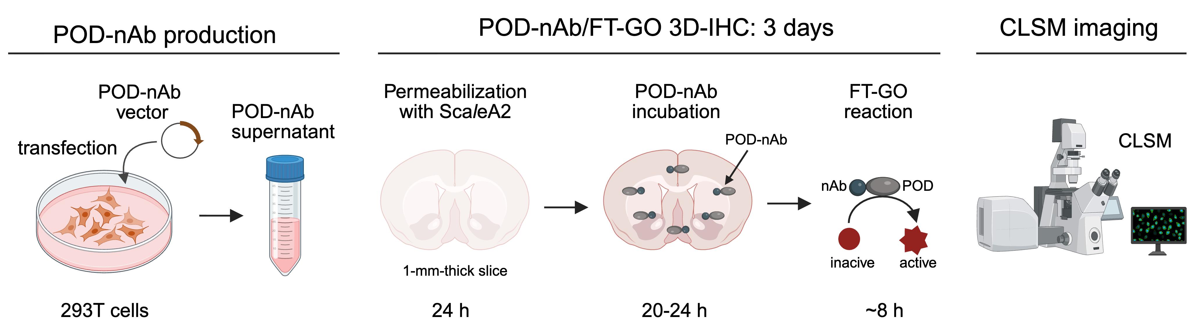
Overview of POD-nAb/FT-GO 3D-IHC in 1-mm-thick mouse brain slices. POD-nAbs are produced by transfection of 293T cells with POD-nAb vectors. The culture supernatant is used for 3D-immunolabeling. POD-nAb is a genetically encoded immunoreagent that consists of a camelid nAb and a variant of horseradish peroxidase. POD-nAb/FT-GO 3D-IHC incorporates three main components: (1) tissue permeabilization by incubation with ScaleA2 solution, (2) POD-nAb binding to their antigens, and (3) FT-GO reaction within large-scale tissues. Following the immunolabeling, brain slices are cleared with ScaleS4 solution and imaged by confocal laser scanning microscopy (CLSM). Somata and processes of neurons and glia in a millimeter-thick mouse brain slice are clearly and intensely labeled by the 3D-IHC method within three days. POD-nAb, peroxidase-fused nanobody; 3D-IHC, three-dimensional immunohistochemistry; FT-GO, fluorochromized tyramide-glucose oxidase.
Background
Three-dimensional immunohistochemistry (3D-IHC), the immunolabeling of 3D tissues, shows molecule–structure–function relationships in a biological system. 3D-IHC provides rich cellular and molecular information to permit reconstruction of microstructural details, unbiased sampling throughout a tissue, and a high data throughput. Recent advances in 3D-IHC techniques have allowed for the visualization of molecular distribution even within human embryos and organs [1–4].
Deep antibody (Ab) penetration in 3D tissues is crucial for staining homogeneity of 3D-IHC. Nanobodies (nAbs), recombinant minimal-antigen-binding fragments from single-chain Abs in camelids [5], should be suitable for 3D-IHC due to their much smaller size (12–15 kDa) than conventional immunoglobulin G (IgG) Abs (~150 kDa). Deep penetration of nAbs has been shown to occur into 3D biological tissues [6–9]. A major issue in the implementation of nAbs to 3D-IHC is signal strength, which imposes a limit on the throughput of 3D-IHC imaging. NAbs conjugated with one or two synthetic fluorophores are typically adopted for 3D-IHC without signal amplification [6–9].
Tyramide signal amplification (TSA) involves the catalytic activity of peroxidase (POD) to yield high-density labeling of targets [10,11], providing a possible solution to the relatively low sensitivity of 3D immunolabeling using nAbs. In our earlier study, we developed a straightforward and cost-effective fluorescent TSA system, fluorochromized tyramide-glucose oxidase (FT-GO) [12]. Unlike conventional TSA systems, FT-GO utilizes hydrogen peroxide (H2O2) produced by the oxidation of glucose by glucose oxidase to improve the operational stability of the TSA system [12].
Here, we describe a detailed protocol for nAb-based 3D immunolabeling, namely POD-nAb/FT-GO 3D-IHC, for high-speed and high-sensitivity detection of target molecules within 3D tissues. This protocol includes POD-nAb production, 3D-IHC staining with POD-nAbs, 3D-TSA reaction with FT-GO, and confocal laser scanning microscopy (CLSM) of immunolabeled 3D tissues. POD-nAbs (P-RAN-bodies [13]) are camelid nAbs fused with POD. FT-GO reaction within 3D tissues using peroxidase activity of POD-nAbs allows the visualization of target molecules with drastic signal enhancement. GFP POD-nAb/FT-GO 3D-IHC can offer ~10-fold signal amplification compared with EGFP fluorescence [14]. Using the 3D-IHC protocol provided herein, we can visualize somata and processes of neurons and glia in 1-mm-thick mouse brain tissues within three days.
Materials and reagents
Biological materials
1. pCAG-GFP1 POD-nAb-WPRE (1.0 μg/μL) (generated in-house [14], Addgene, #244973)
2. 293T cells (RIKEN BioResource Research Center, RCB2202)
3. AAV2/PHP.eB CAG-EGFP-WPRE (generated in-house [14])
Caution: Adeno-associated virus (AAV) is generally classified as a risk group 1 (RG1) virus. AAV vectors can be handled in Biosafety Level 1 (BSL-1) facilities in most cases. However, biosafety should be considered with respect to the precise nature of experiments being conducted.
4. C57BL/6J mice (8–16 weeks old) (Nihon SLC)
Reagents
1. Dulbecco’s modified Eagle’s medium (DMEM) (Thermo Fisher Scientific, catalog number: 11965-092)
2. Fetal bovine serum (FBS) (Sigma-Aldrich, catalog number: 173012)
3. L-glutamine (200 mM) (Thermo Fisher Scientific, catalog number: 25030-081)
4. MEM non-essential amino acid (100×) (Thermo Fisher Scientific, catalog number: 11140-050)
5. Penicillin-streptomycin (100×) (Thermo Fisher Scientific, catalog number: 15070-063)
6. Lipofectamine 3000 (Thermo Fisher Scientific, catalog number: L3000001)
7. Opti-MEM I reduced serum medium (Opti-MEM) (Thermo Fisher Scientific, catalog number: 31985062)
8. Thimerosal (Nacalai Tesque, catalog number: 21624-32)
9. Isoflurane (Pfizer)
10. Sodium chloride (NaCl) (Nacalai Tesque, catalog number: 31320-05)
11. Disodium hydrogen phosphate 12-water (Na2HPO4·12H2O) (Nacalai Tesque, catalog number: 31722-45)
12. Potassium chloride (KCl) (Nacalai Tesque, catalog number: 28514-75)
13. Potassium dihydrogenphosphate (KH2PO4) (Nacalai Tesque, catalog number: 28721-55)
14. Paraformaldehyde (PFA) (Merck Millipore, catalog number: 1.04005.1000)
15. 10 N sodium hydroxide (NaOH) solution (Nacalai Tesque, catalog number: 94611-45)
16. Sodium dihydrogen phosphate dihydrate (NaH2PO4·2H2O) (Nacalai Tesque, catalog number: 31718-15)
17. Pentobarbital sodium salt (Tokyo Chemical Industry, catalog number: P0776)
18. Agar (Nacalai Tesque, catalog number: 01028-85)
19. Sodium azide (NaN3) (Nacalai Tesque, catalog number: 31233-55)
20. Triton-X100 (Nacalai Tesque, catalog number: 35501-15)
21. Urea (FUJIFILM Wako Pure Chemical Industries, catalog number: 215-00616)
22. Glycerol (Nacalai Tesque, catalog number: 17018-25)
23. Hydrogen peroxide (H2O2) (31% aqueous solution) (Santoku Chemical Industries, catalog number: 18412-15)
24. CF tyramide (e.g., CF640R tyramide, Biotium, catalog number: 92175)
25. Dimethyl sulfoxide (DMSO) (FUJIFILM Wako Pure Chemical Industries, catalog number: 043-07216)
26. Glucose oxidase (Nacalai Tesque, catalog number: 16831-14)
27. Bovine serum albumin (BSA) (Nacalai Tesque, catalog number: 01863-77)
28. β-D-glucose (FUJIFILM Wako Pure Chemical Industries, catalog number: 049-31165)
29. D-sorbitol (Nacalai Tesque, catalog number: 06286-55)
30. Agarose (TaKaRa Bio, catalog number: 5003)
Solutions
1. 293T cell culture medium (see Recipes)
2. Lipofectamine 3000 solution (see Recipes)
3. Plasmid DNA with P3000 solution (see Recipes)
4. Plasmid DNA–lipid complex (see Recipes)
5. Thimerosal 10% (see Recipes)
6. Phosphate-buffered saline (PBS) (see Recipes)
7. AAV solution (see Recipes)
8. PFA 16% (see Recipes)
9. Phosphate buffer (PB) (pH 7.4) 0.2 M (see Recipes)
10. Fixative solution (see Recipes)
11. Sodium pentobarbital solution (see Recipes)
12. 4% agar in PBS (see Recipes)
13. PB (pH 7.4) 0.1 M (see Recipes)
14. Triton X-100 10% (w/v) (see Recipes)
15. ScaleA2 solution (see Recipes)
16. Endogenous peroxidase quenching solution (see Recipes)
17. Triton X-100 10% (v/v) (see Recipes)
18. PBS-X (see Recipes)
19. POD-nAb solution (see Recipes)
20. CF tyramide solution (see Recipes)
21. GO solution (see Recipes)
22. 2% BSA in 0.1 M PB (see Recipes)
23. FT-GO reaction mixture (see Recipes)
24. β-D-glucose solution (see Recipes)
25. ScaleS4 solution (see Recipes)
26. ScaleS4 D25(0) solution (see Recipes)
27. ScaleS4 gel (see Recipes)
Recipes
1. 293T cell culture medium
| Reagent | Final concentration | Volume |
|---|---|---|
| DMEM | 90% | 500 mL |
| FBS | 10% | 50 mL |
| L-glutamine | 2 mM | 5 mL |
| MEM non-essential amino acid (100×) | 1× | 5 mL |
| Penicillin-Streptomycin (100×) | 1× | 5 mL |
| Total | 565 mL |
Swirl the bottle to mix. The medium can be stored at 4 °C for 3 months.
2. Lipofectamine 3000 solution
| Reagent | Final concentration | Volume (for 1 well) |
|---|---|---|
| Lipofectamine 3000 Reagent | n/a | 7.5 μL |
| Opti-MEM | n/a | 125 μL |
| Total | 132.5 μL |
Mix well by vortexing for 2–3 s. The solution should be prepared before use. Recipes 2, 3, and 4 are part of the sequential steps for preparing transfection complexes.
3. Plasmid DNA with P3000 solution
| Reagent | Final concentration | Volume (for 1 well) |
|---|---|---|
| pCAG-GFP1 POD-nAb-WPRE (1.0 μg/μL) | 2.5 μg/tube | 2.5 μL |
| P3000 reagent | 2 μL/μg DNA | 5 μL |
| Opti-MEM | n/a | 125 μL |
| Total | 132.5 μL |
Dilute DNA in Opti-MEM and then add P3000 reagent. Mix well by pipetting. The solution should be prepared before use. Recipes 2, 3, and 4 are part of the sequential steps for preparing transfection complexes.
4. Plasmid DNA–lipid complex
| Reagent | Final concentration | Volume (for 1 well) |
|---|---|---|
| Lipofectamine 3000 solution | 50% | 125 μL |
| DNA with P3000 solution | 50% | 125 μL |
| Total | 250 μL |
Mix by tapping the tube. Incubate for 10–15 min at 20–25 °C. The solution should be prepared before use. Recipes 2, 3, and 4 are part of the sequential steps for preparing transfection complexes.
5. Thimerosal 10%
| Reagent | Final concentration | Volume (for 10 mL) |
|---|---|---|
| Thimerosal | 10% | 1 g |
| Double-distilled water (ddH2O) | n/a | up to 10 mL |
| Total | 10 mL |
Mix well by vortexing. The solution can be stored at 20–25 °C for 1 year.
Caution: Thimerosal is an alkylmercury compound. Avoid inhalation or contact with skin, eyes, and mucous membrane. Handle it inside a fume hood with appropriate protective gear.
Critical: NaN3 as a preservative should be avoided. NaN3 inactivates the enzymatic activity of POD.
6. PBS
| Reagent | Final concentration | Volume (for 1 L) |
|---|---|---|
| NaCl | 137 mM | 8 g |
| Na2HPO4·12H2O | 8.1 mM | 2.9 g |
| KCl | 2.7 mM | 200 mg |
| KH2PO4 | 1.5 mM | 200 mg |
| ddH2O | n/a | up to 1 L |
| Total | 1 L |
Mix well by stirring. The solution can be stored at 20–25 °C for 6 months.
7. AAV solution
| Reagent | Final concentration | Volume (for 100 μL) |
|---|---|---|
| AAV2/PHP.eB CAG-EGFP-WPRE | 5.0 × 10¹⁰ genome copies/mL | n/a |
| PBS | n/a | 100 μL |
| Total | 100 μL |
Mix well by pipetting. The solution should be prepared before use.
Caution: Biosafety should be considered with respect to the precise nature of experiments being conducted. AAV vectors can be handled in BSL-1 facilities in most cases.
Critical: The optimal concentration of the AAV-GFP vector should be determined for each experiment.
8. PFA 16%
| Reagent | Final concentration | Volume (for 1 L) |
|---|---|---|
| PFA | 16% | 160 g |
| 10 N NaOH solution | n/a | n/a |
| ddH2O | n/a | up to 1 L |
| Total | 1 L |
Add 160 g of PFA in ddH2O at 60–70 °C. Dissolve PFA by adding 10 N NaOH solution and stirring. Allow the solution to cool to 20–25 °C and bring the volume to 1 L with ddH2O. Filter the solution through filter paper and dispense to 10 mL each. The solution can be stored at -20 °C for 1 year.
Caution: PFA is toxic and teratogenic. Avoid inhalation or contact with skin, eyes, and mucous membranes. Handle it inside a fume hood with appropriate protective gear.
9. PB (pH 7.4) 0.2 M
| Reagent | Final concentration | Volume (for 1 L) |
|---|---|---|
| NaH2PO4·2H2O | 38 mM | 11.8 g |
| Na2HPO4·12H2O | 162 mM | 116 g |
| ddH2O | n/a | up to 1 L |
| Total | 1 L |
Mix well by stirring. The solution can be stored at 20–25 °C for 6 months.
10. Fixative solution
| Reagent | Final concentration | Volume (for 40 mL) |
|---|---|---|
| PFA 16% | 4% | 10 mL |
| PB (pH 7.4) 0.2 M | 0.1 M | 20 mL |
| ddH2O | n/a | up to 40 mL |
| Total | 40 mL |
Filter the solution through filter paper. Mix well by swirling. Use the solution within the same day.
Caution: PFA is toxic and teratogenic. Avoid inhalation or contact with skin, eyes, and mucous membranes. Handle it inside a fume hood with appropriate protective gear.
11. Sodium pentobarbital solution
| Reagent | Final concentration | Volume (for 20 mL) |
|---|---|---|
| Pentobarbital sodium salt | 50 mg/mL | 1 g |
| PBS | n/a | up to 20 mL |
| Total | 20 mL |
Mix well by stirring. The solution can be stored at 20–25 °C for 1 month.
12. 4% agar in PBS
| Reagent | Final concentration | Volume (for 50 mL) |
|---|---|---|
| Agar | 4% | 2 g |
| PBS | n/a | up to 50 mL |
| Total | 50 mL |
Mix well by stirring. Melt agar by heating in a microwave. The solution should be prepared before use.
13. PB (pH 7.4) 0.1 M
| Reagent | Final concentration | Volume (for 1 L) |
|---|---|---|
| PB (pH 7.4) 0.2 M | 0.2 M | 500 mL |
| ddH2O | n/a | up to 1 L |
| Total | 1 L |
Swirl the bottle to mix. The solution can be stored at 20–25 °C for 3 months.
14. Triton X-100 10% (w/v)
| Reagent | Final concentration | Volume (for 50 mL) |
|---|---|---|
| Triton X-100 | 10% (w/v) | 5 g |
| ddH2O | n/a | up to 50 mL |
| Total | 50 mL |
Mix well by stirring. The solution can be stored at 4 °C for 6 months.
15. ScaleA2 solution
| Reagent | Final concentration | Volume (for 100 mL) |
|---|---|---|
| Urea | 4 M | 24.02 g |
| Glycerol | 10% (w/v) | 10 g |
| Triton-X100 10% (w/v) | 0.1% (w/v) | 1 mL |
| ddH2O | n/a | up to 100 mL |
| Total | 100 mL |
Mix well by stirring. The solution can be stored at 4 °C for 1 month.
16. Endogenous peroxidase quenching solution
| Reagent | Final concentration | Volume (for 6.2 mL) |
|---|---|---|
| H2O2 31% | 1% | 200 μL |
| PBS | n/a | 6 mL |
| Total | 6.2 mL |
Mix well by swirling. The solution should be prepared before use.
Caution: H2O2 is corrosive and can cause local tissue damage. Avoid inhalation or contact with skin, eyes, and mucous membranes. Handle it inside a fume hood with appropriate protective gear.
17. Triton X-100 10% (v/v)
| Reagent | Final concentration | Volume (for 500 mL) |
|---|---|---|
| Triton X-100 | 10% (v/v) | 50 mL |
| ddH2O | n/a | up to 500 mL |
| Total | 500 mL |
Mix well by stirring. The solution can be stored at 4 °C for 6 months.
18. PBS-X
| Reagent | Final concentration | Volume (for 1 L) |
|---|---|---|
| Triton X-100 10% (v/v) | 0.3% (v/v) | 30 mL |
| PBS | n/a | up to 1 L |
| Total | 1 L |
Swirl the bottle to mix. The solution can be stored at 20–25 °C for 3 months.
19. POD-nAb solution
| Reagent | Final concentration | Volume (for 1,200 μL) |
|---|---|---|
| POD-nAb culture supernatant | 20% | 600 μL |
| Triton X-100 10% (v/v) | 0.3% (v/v) | 36 μL |
| Thimerosal 10% | 0.02% | 2.4 μL |
| PBS | n/a | up to 1,200 μL |
| Total | 1,200 μL |
Mix well by pipetting. The solution should be prepared before use.
Caution: Thimerosal is an alkylmercury compound. Avoid inhalation or contact with skin, eyes, and mucous membranes. Handle it inside a fume hood with appropriate protective gear.
Critical: The optimal concentration of POD-nAb culture supernatant should be determined for each experiment.
20. CF tyramide solution
| Reagent | Final concentration | Volume (for 1 mL) |
|---|---|---|
| CF tyramide | 2 mM | 2 μmol |
| DMSO | n/a | 1 mL |
| Total | 1 mL |
Mix well by pipetting. Dispense to 10 μL each. The solution can be stored at -80 °C for 1 year.
21. GO solution
| Reagent | Final concentration | Volume (for 1 mL) |
|---|---|---|
| Glucose oxidase | 1 mg/mL | 1 mg |
| PB (pH 7.4) 0.2 M | 0.1 M | 500 μL |
| ddH2O | n/a | up to 1 mL |
| Total | 1 mL |
Mix well by pipetting. Dispense to 50 μL each. The solution can be stored at -80 °C for 1 year.
22. 2% BSA in 0.1 M PB
| Reagent | Final concentration | Volume (for 50 mL) |
|---|---|---|
| BSA | 2% (w/v) | 1 g |
| PB (pH 7.4) 0.2 M | 0.1 M | 25 mL |
| ddH2O | n/a | up to 50 mL |
| Total | 50 mL |
Mix well by stirring. Sterilize the solution with a 0.22 μm filter. The solution can be stored at 4 °C for 3 months.
23. FT-GO reaction mixture
| Reagent | Final concentration | Volume (for 1 mL) |
|---|---|---|
| CF tyramide solution | 10 μM | 5 μL |
| GO solution | 3 μg/mL | 3 μL |
| 2% BSA in 0.1 M PB | n/a | 992 μL |
| Total | 1 mL |
Mix well by pipetting. The solution should be prepared before use.
24. β-D-glucose solution
| Reagent | Final concentration | Volume (for 1 mL) |
|---|---|---|
| β-D-glucose | 200 mg/mL | 200 mg |
| ddH2O | n/a | up to 1 mL |
| Total | 1 mL |
Mix well by pipetting. Dispense to 50 μL each. The solution can be stored at -80 °C for 1 year.
25. ScaleS4 solution
| Reagent | Final concentration | Volume (for 100 mL) |
|---|---|---|
| Urea | 4 M | 24.02 g |
| D-sorbitol | 40% (w/v) | 40 g |
| Glycerol | 10% (w/v) | 10 g |
| Triton-X100 10% (w/v) | 0.2% (w/v) | 2 mL |
| DMSO | 25% (v/v) | 25 mL |
| ddH2O | n/a | up to 100 mL |
| Total | 100 mL |
For a detailed procedure for preparation of this solution, refer to Miyawaki et al. [15]. The solution can be stored at 4 °C for 1 month.
26. ScaleS4 D25(0) solution
| Reagent | Final concentration | Volume (for 100 mL) |
|---|---|---|
| Urea | 4 M | 24.02 g |
| D-sorbitol | 40% (w/v) | 40 g |
| Glycerol | 10% (w/v) | 10 g |
| DMSO | 25% (v/v) | 25 mL |
| ddH2O | n/a | up to 100 mL |
| Total | 100 mL |
For a detailed procedure for preparation of this solution, refer to Miyawaki et al. [15]. The solution can be stored at 4 °C for 1 month.
27. ScaleS4 gel
| Reagent | Final concentration | Volume (for 100 mL) |
|---|---|---|
| Agarose | 1.5% (w/v) | 1.5 g |
| ScaleS4 D25(0) | n/a | up to 100 mL |
| Total | 100 mL |
For a detailed procedure for preparation of this solution, refer to Miyawaki et al. [15]. The solution can be stored at 4 °C for 1 year. ScaleS4 gel solidifies at 4 °C. Remelt the gel by heating it in a microwave before use.
Laboratory supplies
1. 50 mL conical tubes (Greiner Bio-One, catalog number: 227651)
2. Syringe filter (0.22 μm) (Merck Millipore, catalog number: SLGPR33RB)
3. Filter paper No. 2 (ADVANTEC, catalog number: 00021240)
4. 30-G insulin syringe (NIPRO, catalog number: 08-277)
5. 23-G regular bevel needle (Terumo Corporation, catalog number: NN-2332R)
6. 20 mL syringe (Terumo Corporation, catalog number: SS-20ESZ)
7. Surgical scissors (FRIGZ, catalog number: E221-288)
8. Tweezers (FRIGZ, catalog number: E380-546, E502-004)
9. 15 mL conical tubes (Greiner Bio-One, catalog number: 188271-N)
10. Cell culture multi-well plates, 6 well (6-well plate) (Greiner Bio-One, catalog number: 657160)
11. Razor blade (Feather Safety Razor, catalog number: FAS-10)
12. Superglue (Aron Alpha®) (Toagosei, catalog number: 201)
13. Micro spatula, stainless steel (AS ONE, catalog number: 9-891-03)
14. Safe-lock tubes, 2.0 mL (Eppendorf, catalog number: 0030120094)
15. Custom-made imaging chambers [16]; three-dimensional computer-aided design (3D CAD) data for the imaging chamber are provided in Furuta et al. [16]
16. Coverslips (24 × 40 mm) (Matsunami Glass, catalog number: C024401)
17. Kimwipe (NIPPON PAPER CRECIA, catalog number: S-200)
18. Slide glasses (26 × 76 mm) (Matsunami Glass, catalog number: S1225)
19. Metal weights (Hikari, catalog number: GZ25)
20. Glass petri dish (60 mm) (SANSYO, catalog number: 82-1683)
21. Blu-Tack® (Bostik, catalog number: CKBT-450000)
Equipment
1. Clean bench (Thermo Scientific, model: 1300 Series A2)
2. CO2 incubator (Thermo Fisher Scientific, model: Forma Steri-Cycle 370)
3. Centrifuge with rotors for 15 and 50 mL tubes (Tomy, model: MDX-310)
4. Fume hood (Dalton, model: Uni-optflow DFA10-AA15-AA10)
5. Rotary shaker (TAITEC, model: NR-20)
6. Vibratome (Dosaka EM, model: PRO7N)
7. Constant temperature incubator shaker (TAITEC, model: BR-23FP)
8. Confocal scanning microscope system (Leica Microsystems, model: TCS SP8)
9. 16× multi-immersion objective lens (Leica Microsystems, model: HC FLUOTAR 16×/0.60 IMM CORR VISIR)
Software and datasets
1. LAS-X (Leica Microsystems, v3.5.5.19976, February 2021)
2. Fiji (v2.16.0/1.54p, October, 2024)
Note: LAS-X software requires a license.
Procedure
登录/注册后免费查看全文
文章信息
稿件历史记录
提交日期: Mar 24, 2026
接收日期: Apr 27, 2026
在线发布日期: May 19, 2026
出版日期: Jun 5, 2026
版权信息
© 2026 The Author(s); This is an open access article under the CC BY license (https://creativecommons.org/licenses/by/4.0/).
如何引用
Yamauchi, K., Koike, M. and Hioki, H. (2026). Detection of Target Molecules Within One-Millimeter-Thick Mouse Brain Slices by Using Peroxidase-Fused Nanobodies and Fluorochromized Tyramide-Glucose Oxidase Reaction. Bio-protocol 16(11): e5713. DOI: 10.21769/BioProtoc.5713.

 Download Citation in RIS Format
分类
神经科学 > 神经解剖学和神经环路 > 免疫荧光
细胞生物学 > 细胞成像 > 固定组织成像
生物化学 > 蛋白质 > 免疫检测 > 免疫染色法
您对这篇实验方案有问题吗?
在此处发布您的问题,我们将邀请本文作者来回答。同时,我们会将您的问题发布到Bio-protocol Exchange,以便寻求社区成员的帮助。
 发布问题
发布问题


