


Versatile Dual Mounting Enables Larval Zebrafish Imaging Across Microscope Configurations
双向固定方法支持斑马鱼幼体在不同显微镜配置下成像

发布: 2026年06月05日第16卷第11期 DOI: 10.21769/BioProtoc.5709 浏览次数: 327
评审: Alberto RissoneIvonne SehringAnonymous reviewer(s)
 PDF
PDF  Q&A
Q&A
Advertisement








Abstract
Larval zebrafish are often mounted laterally to ensure consistent anatomical positioning and to standardize imaging of body axes across early development. However, this conventional approach often tethers sample orientation to a single microscope configuration and limits optical accessibility. We present a mounting protocol for larval zebrafish that enables optical access from both dorsal and ventral orientations while preserving lateral sample position. This approach uses common laboratory consumables to establish a mounting platform that eliminates any need to remount samples between the use of upright and inverted microscopes. By establishing a hydrophobic seal, mounted embryos can be inverted with ease to access the sample from either orientation. A seamless transition here facilitates reliable identification and longitudinal tracking of the same biological region of interest across microscope configurations. This protocol is broadly applicable to live imaging experiments requiring flexibility in imaging geometry, minimal sample handling, and high reproducibility.
Key features
• Dual-configuration mounting setup can be established from standard, low-cost consumables in any laboratory setting.
• Preserves lateral mounting in agarose for consistent anatomical positioning and experimental continuity.
• Enables seamless transition between upright and inverted microscopes without the need for specimen remounting.
Keywords: Zebrafish (斑马鱼) Larvae (幼体)
Mounting (样品固定)
Inverted (倒置显微镜)
Upright (正置显微镜)
Microscopy (显微成像)
Larvae (幼体)
Mounting (样品固定)
Inverted (倒置显微镜)
Upright (正置显微镜)
Microscopy (显微成像)
Graphical overview
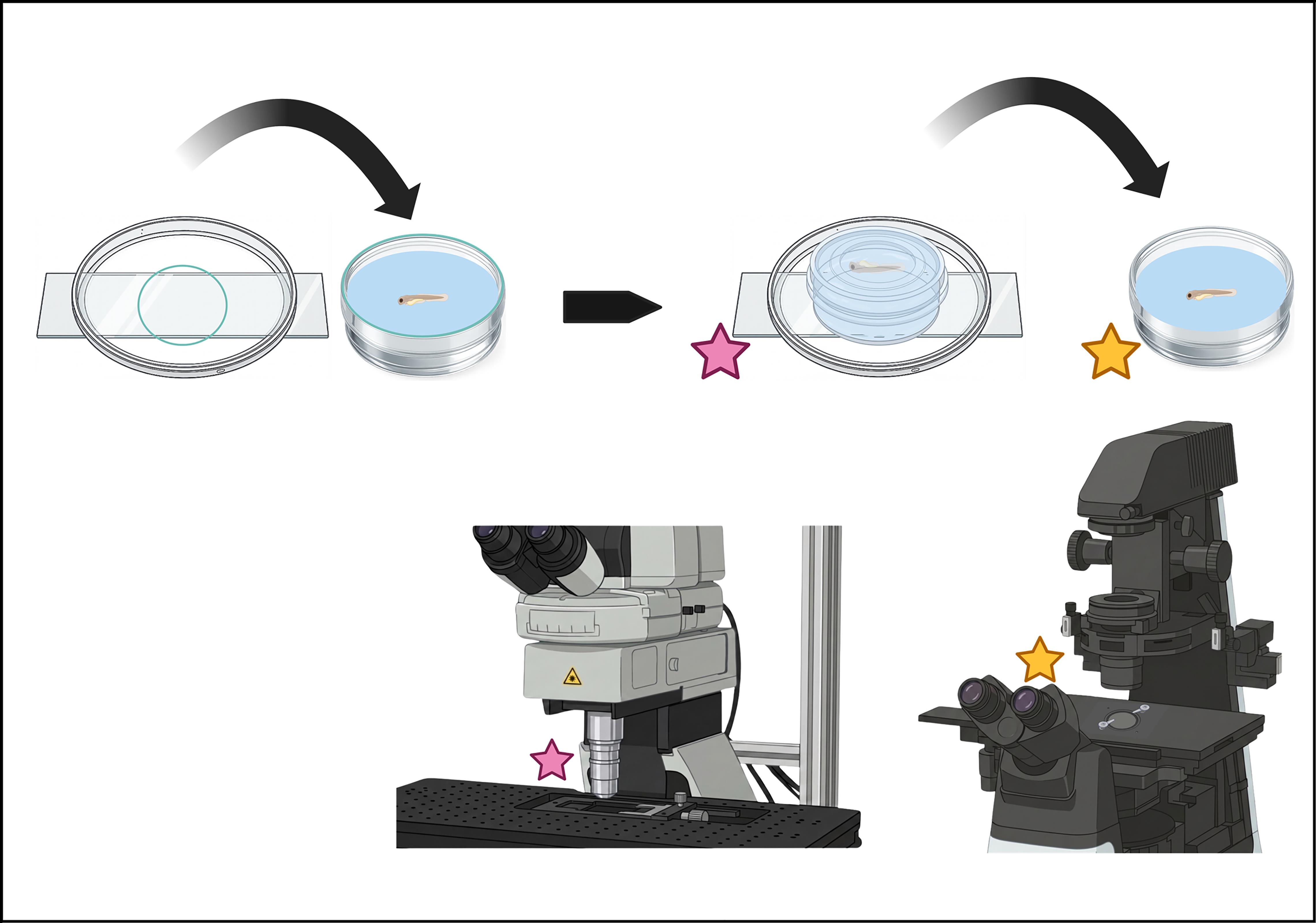
Dual-mounting configuration to transition between upright and inverted microscopes
Background
Zebrafish (Danio rerio) have become a widely popular vertebrate model organism given their early larval transparency, highly conserved genome with humans, external embryo fertilization, and rapid development compared to other vertebrates. These features position zebrafish as a well-suited model for in vivo observations across fields like developmental biology, neuroscience, genetics, and toxicology [1,2]. This has enabled their use as a robust predictive model for teratogenicity, with morphometric profiling of development offering insights into chemical-specific consequences and modes of action conserved across vertebrates [3]. More broadly, many modern experimental paradigms integrate different imaging modalities with genetic manipulations and behavioral assays to both influence and capture biological dynamics with larval zebrafish in real time [4]. However, fully exploiting these capabilities with non-invasive imaging techniques often depends on mounting orientation and optical accessibility.
These practical constraints become particularly important when selecting optical imaging modalities that differ in penetration depth, speed, and configuration. Techniques like calcium imaging, photochemical conversion of fluorophores via targeted stimulation, optogenetics, and photoinduced ablation benefit from the advantages of multiphoton microscopy, including deeper penetration than in linear techniques like confocal imaging [5]. A great example involves the increased engineering of in vivo indicators to exploit the nonlinear physics of multiphoton excitation. Recent advancements have leveraged rhodopsin and calcium-based sensors to establish genetically encoded calcium (GECI) and voltage (GEVI) indicators across wavelengths that maximize photon capture and signal-to-noise ratio in deep tissue [6,7]. On the other hand, the faster imaging speeds, greater ease of setup, and robust optical sectioning provided by linear microscopes, specifically confocal systems, are especially useful when imaging rapid biological dynamics where temporal resolution and precision in optical sectioning are required [8]. Combining both approaches can integrate the depth of multiphoton penetration while maintaining the rapid visualization and temporal resolution provided by confocal imaging, but the ease of use with larval embryos can be limited by opposing microscope objective orientations. Though varying modalities can be integrated into a single system, like the addition of a photostimulation laser to a spinning-disk confocal system, doing so often entails substantial cost and complexity.
Traditional lateral mounting for inverted imaging positions larval zebrafish against the bottom of a glass-bottom imaging dish, secured with a mound of agarose. This configuration allows for clear visibility of anterior–posterior axis development in zebrafish and helps to standardize anatomical positioning across samples. However, because the imaging dish is filled with liquid and embryos are fixed against the bottom of the dish, optimal accessibility is limited to imaging through the glass from below. Observation from above, as with top-down microscopes, is constrained by working distance limitations and obstruction from the mounting agar and anesthetic solution. This restricts flexibility across microscope geometries, as samples can only be imaged with the shortest optical distance from one side. Additionally, direct water immersion with the upright objective into the imaging dish risks damage to samples and mounting. The optical distance can be extended to target the region of interest across configurations without remounting, but this increases the limitations of multiphoton imaging in photobleaching, light scattering, and heat exposure [8]. Further, remounting samples can introduce variability in anatomical positioning, increase the required handling of anesthetized samples, and complicate the identification of the same biological region of interest for longitudinal assessment.
Here, we present a dual-configuration mounting model that enables easy transition across upright and inverted microscope geometries. This setup combines standard zebrafish mounting with a model constructed from common low-cost laboratory consumables to eliminate remounting across microscope configurations, supporting both upright and inverted imaging. Using a simple petroleum jelly assembly, we combine a 35 mm glass-bottom confocal imaging dish with a 60 mm Petri dish glued onto a microscope slide. This method successfully preserves the conventional lateral embryo positioning while enabling optical access from both upright and inverted perspectives to allow for rapid transition between imaging geometries. Ultimately, this configuration supports workflows that combine targeted interventions with long-term imaging and can be applied using any mounting orientation.
Materials and reagents
Biological materials
1. Tg(erbb3b:gal4)nkgsaizGFFD37A, Tg(UAS:eGFP)nkuasgfp1a, and Tg(olig2:dsRed2)vu19 zebrafish transgenic lines were used for mounting protocol validation [9,10].
Reagents
1. Instant Ocean salt (Instant Ocean, catalog number: 77780); store in an airtight container in a cool, dry place at room temperature (RT)
2. 3-Aminobenzoic acid ester (tricaine) (Pentair, catalog number: TRS1); store in an airtight container in a cool, dry place at RT
3. Tris (Tris[hydroxymethyl]aminomethane or Trimethamine) (Bio-Rad, catalog number: 1610716); store in an airtight container at RT
4. 1-Phenyl-2-thiourea (PTU) (Sigma, catalog number: P7629, CAS number: 103-85-5); store in an airtight container at RT, protected from light
5. Agarose, low gelling temperature (Sigma, catalog number: A94114); store in an airtight container at RT
Solutions
1. Egg water (see Recipes)
2. PTU stock solution (10×) (see Recipes)
3. PTU water (see Recipes)
4. Buffered tricaine methanesulfonate (MS-222) stock solution (see Recipes)
5. Anesthetic solution (see Recipes)
6. 0.8% low-gelling agarose (see Recipes)
Recipes
1. Egg water
| Reagent | Final concentration | Quantity or volume |
| Instant Ocean salt | 300 mg/L | 300 mg |
| Deionized H2O | – | 1 L |
2. PTU stock solution (10×)
| Reagent | Final concentration | Quantity or volume |
| PTU | 0.03% | 300 mg |
| Deionized H2O | – | 1 L |
3. PTU water
| Reagent | Final concentration | Quantity or volume |
| PTU 10× stock solution | 0.003% | 100 mL |
| Egg water | – | 900 mL |
4. MS-222 stock solution
| Reagent | Final concentration | Quantity or volume |
| Tricaine | 4 g/L | 4 g |
| Deionized H2O | – | up to 1 L |
| 1 M Tris (pH 9.0) | 21 mM | 21 mL |
5. Anesthetic solution
| Reagent | Final concentration | Quantity or volume |
| MS-222 stock solution | 4% | 400 μL |
| PTU water | 0.003% | 10 mL |
6. 0.8% low-gelling agarose
| Reagent | Final concentration | Quantity or volume |
| Low-gelling agarose | 0.8% | 8 mg |
| Egg water | – | 1 mL |
Laboratory supplies
1. Breeding tanks (Aquaneering, catalog number: ZHCT100)
2. Dumont Tweezers, pattern #5; 0.10 × 0.06 mm tip size (Roboz Surgical, catalog number: RS-5065)
3. 100 × 15 mm Petri dish (Falcon, catalog number: 351029)
4. 35 mm glass-bottom confocal dish, non-treated (Avantor, catalog number: 75856-746)
5. 60 mm Petri dish (Fisher, catalog number: FB0875713)
6. Disposable glass Pasteur pipettes (VWR, catalog number: 53283-916)
7. Pipette pump, 10 mL (Honbay, ASIN: B07P61YDXB)
8. Micro dissecting needle holder, 4 3/4’’ (Roboz Surgical, catalog number: RS-6061)
9. Insect pins 0.30 mm size 00 (Roboz Surgical, catalog number: RS-6081-30)
10. Glass microscope slides (Sail Brand, catalog number: 7105)
11. 50 mL borosilicate glass beaker (Maccx, catalog number: BKL050-012)
12. Borosilicate glass test tubes with autoclavable caps (ULAB, catalog number: UTT1009)
13. Petroleum jelly (Vaseline, catalog number: UNI34500)
14. Super glue (Krazy Glue, catalog number: KG58548R)
15. Wood applicator stick, 6” × 1/12” (Oxford Instruments, catalog number: ID 51-1625-0580)
16. Zeiss immersion W oil 2010 (Microscope World, catalog number: 444969-0000-000)
Equipment
1. Fisherbrand Basic 180 L Gravity Incubator (ThermoScientific)
2. ECLIPSE Ti2 (Nikon) equipped with a Dragonfly 200 high-speed confocal platform (Andor Oxford Instruments), a 40×/1.15 W APO LWD numerical aperture water immersion objective (Nikon), and a motorized stage (Applied Science Information)
3. Air-MP (Nikon), equipped with a 25×/1.10 W numerical aperture water immersion objective (Nikon), ProScan III motorized stage (Prior Scientific), Chameleon Vision II laser system (Coherent), and an HEC series Thermo-con cooling system (SMC)
Software and datasets
1. Fusion 2.6.0 (Andor Technology; 2026, license required)
2. Nikon Imaging Software, Elements Advanced Research 5.21.03 (Nikon; 2020, license required)
3. Imaris 11.0 (released 2025; license required)
Procedure
登录/注册后免费查看全文
文章信息
稿件历史记录
提交日期: Mar 17, 2026
接收日期: Apr 21, 2026
在线发布日期: May 14, 2026
出版日期: Jun 5, 2026
版权信息
© 2026 The Author(s); This is an open access article under the CC BY-NC license (https://creativecommons.org/licenses/by-nc/4.0/).
如何引用
Murphy, H. and Fontenas, L. (2026). Versatile Dual Mounting Enables Larval Zebrafish Imaging Across Microscope Configurations. Bio-protocol 16(11): e5709. DOI: 10.21769/BioProtoc.5709.

 Download Citation in RIS Format
分类
生物物理学 > 显微技术
细胞生物学 > 组织分析 > 组织成像
细胞生物学 > 细胞成像 > 活细胞成像
您对这篇实验方案有问题吗?
在此处发布您的问题,我们将邀请本文作者来回答。同时,我们会将您的问题发布到Bio-protocol Exchange,以便寻求社区成员的帮助。
 发布问题
发布问题


