


Measuring Electrophysiological Activity in Acute Brain Slices, Spheroids, and Organoids Using 3D High-Density Multielectrode Arrays
利用三维高密度多电极阵列检测急性脑片、脑球体和脑类器官的电生理活动

(*Contributed equally to this work, ΘContributed equally to this work, §Technical contact: ivan.verduci@3brain.com) 发布: 2026年06月05日第16卷第11期 DOI: 10.21769/BioProtoc.5708 浏览次数: 1088
评审: Elena A. OstrakhovitchXiaochen SunMario Valentino
 PDF
PDF  Q&A
Q&A
参见作者原研究论文
The authors used this protocol in:

Sep 2025
Advertisement








Abstract
Animal and human stem cell–derived three-dimensional models to study physio-pathological brain functioning are becoming a gold standard for in vitro electrophysiology, as they enable the recapitulation of complex network properties by accounting for spatial architectural features that better reflect in vivo conditions than simpler 2D models. Standard planar multielectrode arrays (MEAs), typically providing tens of recording electrodes, are commonly used to record activity from 2D neuronal cultures. However, when adapted for use with 3D models, planar 2D MEAs showed limited effectiveness. The main issues are limited specimen adhesion to the chip, a low number of sensing elements, inability to retrieve signals from within the tissue, and reduced perfusion and vitality of the tissue in contact with sensors. To overcome these limitations, a new generation of microchip-based 3D high-density MEAs (3D HD-MEA) has been developed and validated in recent years. This technological advancement has improved the sensing capabilities and the vitality of 3D models, providing a tool tailored to maximize their potential. Here, we present an optimized protocol for neural network activity recordings in 3D models (including acute slices, brain spheroids, and organoids) from various brain regions using 3D HD-MEAs. First, we summarize the critical steps for 1) obtaining viable acute slices from the mouse cerebellum, cortico-hippocampal circuit, and prefrontal cortex, 2) establishing efficient coupling of the slices with the chip, and 3) performing recordings and analyses. We then describe the main procedures required to obtain human and animal brain spheroids and neural organoids, as well as standardized routines to perform effective recordings and analyses. For each section, we highlight the crucial steps, identify tips for specific applications, and propose troubleshooting procedures. For example, the same type of preparation (e.g., acute slices) requires different adjustments when working with different brain areas. The specific information provided here is intended to assist researchers in their daily efforts to obtain efficient and reproducible functional recordings from 3D models by using the cutting-edge technique of 3D HD-MEA.
Key features
• Comprehensive all-in-one guide covering the complete workflow for acquiring electrophysiological data from brain slices, neural region-specific organoids, and brain spheroids.
• Intuitive, step-by-step protocol for brain slice preparation, enriched with practical tips and expert recommendations to ensure high-quality tissue viability.
• Detailed instructions for optimal use of 3D HD-MEA technology, including proper handling of the sample holder for recordings from brain slices, neural organoids, and spheroids.
• In-depth guidance on BrainWave6 software, providing clear procedures for data acquisition, signal detection, and advanced electrophysiological analysis across all sample types.
Keywords: 3D high-density multielectrode array (三维高密度多电极阵列) Acute slices (急性脑片)
Brain spheroids (脑球体)
Neural organoids (神经类器官)
Electrophysiological recordings (电生理记录)
Electrophysiological data analysis (电生理数据分析)
Acute electrophysiological measurements (急性电生理检测)
Acute slices (急性脑片)
Brain spheroids (脑球体)
Neural organoids (神经类器官)
Electrophysiological recordings (电生理记录)
Electrophysiological data analysis (电生理数据分析)
Acute electrophysiological measurements (急性电生理检测)
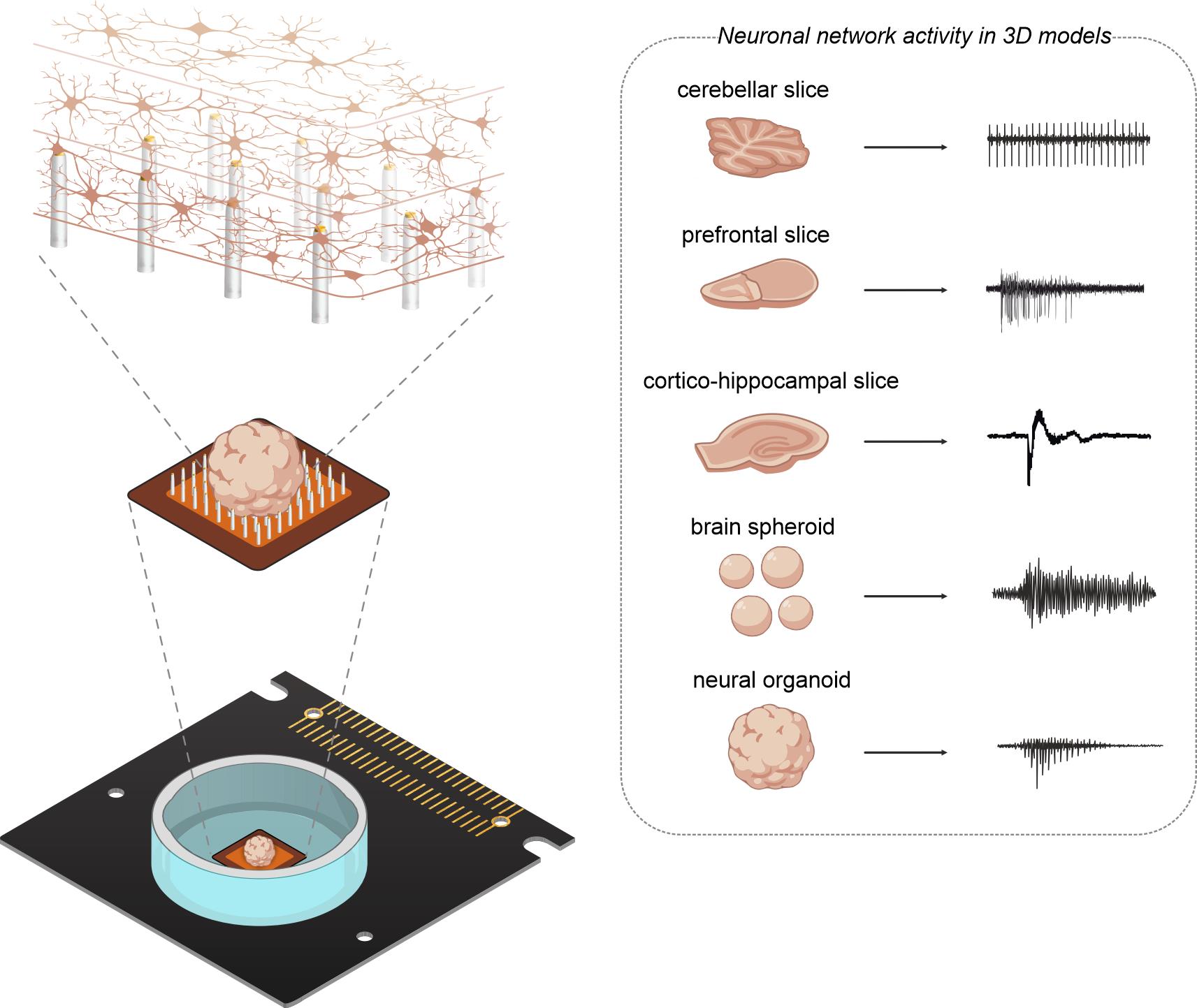
Graphical overview
Background
To date, 3D biological models with varying complexity are used to recapitulate the physiological features of animal and human nervous system circuitry, allowing for the study of physio-pathological brain activity [1–3]. Recent advances in stem cell technology have introduced the possibility of producing region-specific neural organoids from human-induced pluripotent stem cells (hiPSCs). Animal models offer fully mature, structurally accurate brain architectures, which are crucial for examining circuit functions and understanding how diseases affect the neuronal electrical activity [4,5]. In contrast, human-derived neural organoids and the simpler brain spheroids, which can be derived from human neural progenitors but lack the structural and regional organization typical of organoids, can replicate unique human features, thereby reducing challenges in translating research findings to humans [6–8]. Furthermore, as these models can be maintained in culture for extended periods (up to years), they provide opportunities for detailed neurodevelopmental studies. Additionally, neural organoids can generate diverse patterned cell types, such as cortical, thalamic, and spinal cord cells, and achieve greater functional maturation than 2D cultures [9,10], though not equivalent to adult human brains. This enables the study of complex, in vivo–like brain network formation [11–15].
Several techniques exist to study neuronal activity in 3D models, including patch-clamp recordings, calcium imaging, and multielectrode arrays (MEA). In particular, MEAs represent the ideal technique for characterizing the electrophysiological activity of neuronal networks in these models, enabling simultaneous non-invasive recordings from multiple locations within the network, maintaining single-cell resolution, and providing excellent spatial resolution with the recent introduction of microchip-based high-density MEAs (HD-MEAs) [16–18]. For these reasons, HD-MEA devices are already in use with several 3D models, such as acute slices [18–21] and organoids [2,22–24]. However, planar probes commonly used for electrophysiological recordings are insufficient to exploit the complex network architecture that provides the main advantage of using these models. For instance, the chip is in contact only with the surface of the sample, interfacing the electrodes solely with the outermost cell layers. As a result, it fails to capture the activity occurring within the deeper, denser, and more interconnected layers. Even more critical, signals may be dampened by the presence of a dead cell layer in acute slices or by scaffolding materials, such as Matrigel, that are required to embed organoids in some protocols [25–27]. Another important aspect concerns vitality, which is compromised when the tissue is pushed against the chip. This dramatically reduces the recorded surface supply of oxygen and nutrients required to sustain the sample's viability. To fully harness the potential of HD-MEA in combination with 3D tissues, a recent technological breakthrough is represented by the integration of 3D electrodes that can penetrate the tissue and record neuronal activity from inner layers. In addition, these new devices have been equipped with a microstructure at the base of the 3D electrodes that forms microchannels below the tissue, thereby enhancing viability and accelerating pharmacological testing [28].
This methodology is well-suited for detailed investigations of neural network dynamics within 3D biological models, opening new opportunities to understand the relationship between structure and function in preparations, such as brain slices and organoids, that display a significant circuit architecture. This makes the technique ideal for a range of applications across multiple areas of neuroscience, including neurodevelopment, neuropharmacology, and neuropathology. For example, the technique's sensitivity would allow the uncovering of subtle modifications in neuronal firing and network dynamics in disease models with altered neuronal excitability and anatomical connectivity. This aspect is particularly relevant for characterizing organoids' functional network connectivity and its modification during development and in pathological models. Moreover, another promising field of application is certainly the testing of neuroprotective or neurotoxic compounds, which will benefit from the high efficiency of activity recordings from 3D models that more closely recapitulate in vivo conditions and can be maintained in more physiological environments than 2D models. Overall, this methodology provides a versatile platform for researchers working with diverse 3D neural models to investigate physiological mechanisms and disease-related dysfunctions with high reproducibility.
Indeed, the introduction of this new generation of 3D HD-MEA devices addresses the limitations that previously hindered the effectiveness of recordings by using planar devices. However, their unique surface, made up of micropillars approximately 90 μm tall, arranged in a 64 × 64 matrix with an electrode pitch of 60 μm, combined with the complexity of 3D biological models, requires specific adjustments to the experimental preparation and the application of this technology, which are crucial to ensure high-quality recordings. This protocol offers a comprehensive overview of preparing, mounting, recording, and analyzing acute 3D HD-MEA experiments from various 3D models: cerebellar, prefrontal cortex, and cortico-hippocampal acute slices, brain spheroids, and cortical and spinal cord organoids. For clarity, the paper is organized into two independent sections, one for slices and the other for spheroids and organoids. The protocols reported here are intended to enhance researchers’ ability to record functional activity from different types of 3D biological samples.
Part I: Acute brain slices
Materials and reagents
Reagents
1. Sodium chloride (NaCl) (Sigma-Aldrich, CAS-number: 7647-14-5)
2. Potassium dihydrogen phosphate (KH2PO4) (Sigma-Aldrich, CAS-number: 7778-77-0)
3. Potassium chloride (KCl) (Sigma-Aldrich, CAS-number: 7447-40-7)
4. Magnesium sulfate heptahydrate (MgSO4·7H2O) (Sigma-Aldrich, CAS-number: 10034-99-8)
5. Calcium chloride dihydrate (CaCl2·2H2O) (Sigma-Aldrich, CAS-number: 10035-04-8)
6. Sodium phosphate (NaHPO4) (Sigma-Aldrich, CAS-number: 7558-79-4)
7. Sodium bicarbonate (NaHCO3) (Sigma-Aldrich, CAS-number: 144-55-8)
8. D-(+)-glucose (Sigma-Aldrich, CAS-number: 50-99-7)
Solutions
1. Krebs solution for cerebellar slices [29] (see Recipes)
2. Modified artificial cerebrospinal fluid (mACSF) for prefrontal cortex slices [30] (see Recipes)
3. Krebs high-potassium and high-calcium for cortico-hippocampal slices (see Recipes)
Recipes
1. Krebs solution for cerebellar slices
| Reagent | Concentration (mM) |
|---|---|
| NaCl | 120 |
| KH2PO4 | 1.18 |
| KCl | 2 |
| MgSO4·7H2O | 1.2 |
| CaCl2·2H2O | 2 |
| NaHCO3 | 26 |
| Glucose | 11 |
Adjust the solution to the desired volume with double-distilled water. pH of 7.4 is maintained when equilibrated with 95% O2, 5% CO2. Expected osmolarity: 300 ± 10 mOsm.
2. Modified ACSF solution for prefrontal cortex slices
| Reagent | Concentration (mM) |
|---|---|
| NaCl | 124 |
| KCl | 3.5 |
| NaHPO4* | 1 |
| CaCl2·2H2O | 1.25 |
| NaHCO3 | 26 |
| Glucose | 10 |
*After this step, add carbogen gas (95% O2, 5% CO2) to the solution for 30 min before adding calcium chloride to avoid salt precipitation.
Adjust the solution to the desired volume with double-distilled water. pH of 7.4 is maintained when equilibrated with 95% O2, 5% CO2. Expected osmolarity: 300 ± 10 mOsm.
3. Krebs high-potassium and high-calcium for cortico-hippocampal slices
| Reagent | Concentration (mM) |
|---|---|
| NaCl | 115.18 |
| KH2PO4 | 1.18 |
| KCl* | 6.82 |
| CaCl2·2H2O | 4 |
| NaHCO3 | 26 |
| Glucose | 11 |
*After this step, add carbogen gas (95% O2, 5% CO2) to the solution for 30 min before adding calcium chloride to avoid salt precipitation.
Adjust the solution to the desired volume with double-distilled water. pH of 7.4 is maintained when equilibrated with 95% O2, 5% CO2. Expected osmolarity: 300 ± 10 mOsm.
Laboratory supplies
1. Whatman® qualitative filter paper, Grade 1 (Sigma-Aldrich, catalog number: 1001-090)
2. Pasteur pipette 7 mL (Sigma-Aldrich, catalog number: BR747770)
3. Large weighing dishes (Sigma-Aldrich, catalog number: Z154881)
4. Small/medium weighing boats (Sigma-Aldrich, catalog number: W3001)
5. Extran® MA 02 (Sigma-Aldrich, catalog number: 1.07553)
6. Ethanol, pure (Sigma-Aldrich, catalog number: 32205-M)
7. Nitrile extra light gloves (VWR, Avantor, catalog number: 112-4195)
8. Plastic bag
9. Cyanoacrylate glue
10. Double-distilled water
Equipment
1. 500 mL Pyrex beaker
2. 250 mL Pyrex beaker
3. 50 mL beaker
4. Osmometer (Gonotec®, OsmomatTM 030) and calibration standard (Gonotec®, catalog number: 30.9.0500)
5. Vibroslicer (Leica BIOSYSTEMS, catalog number: VT1200S)
6. Vibroslicer chamber
7. Vibroslicer cutting plate
8. Glass Petri dish
9. Large scissors
10. Fine scissors
11. Fine tweezers
12. Semi-curved spatulas
13. Halved razor blade
14. Small flat-blade scalpel (No. 10)
15. Carbon steel scalpel (F.S.T., catalog number: 103115-12)
16. BioCAM (3Brain, DupleX)
17. 3D HD-MEA (3Brain, CorePlateTM 3D 1W 38-60-90)
18. Sample holder frame (3Brain, Sample Holder 2, Frame)
19. Sample holder insert (3Brain, Sample Holder 2, 1000 μN)
20. Sample holder silicone net (3Brain, Sample Holder 2, Silicone Net)
21. Sample holder handling tool (3Brain, Sample Holder 2, Handling tool)
22. Gas inlet/outlet tubes (Cole, Parmer, catalog numbers: 072-031-1, 072-063-1 C-FLEX)
23. Tubes (IsmatecTM, TYGON LMT, 55, ID 0.64 mm, WALL 0.90 mm)
24. Peristaltic pump (IsmatecTM, MS-4/12 Reglo Digital Pump)
Software and datasets
1. BrainWave6 (3Brain), requires a license
2. IC Capture (The Imaging Source, Version 2.4)
3. NEUROPulse (10.5281/zenodo.18196676)
Procedure
登录/注册后免费查看全文
文章信息
稿件历史记录
提交日期: Feb 17, 2026
接收日期: Apr 23, 2026
在线发布日期: May 14, 2026
出版日期: Jun 5, 2026
版权信息
© 2026 The Author(s); This is an open access article under the CC BY-NC license (https://creativecommons.org/licenses/by-nc/4.0/).
如何引用
Pali, E., Pellavio, G., Conforti, M., Sarkissian, A. A., Aliya, B., Sciacca, G., Wariyar, S. S., Mainardi, F., Tedesco, M., Verduci, I., Cadiao, G., Cervetto, C., Andersen, J., Birey, F., Maccione, A., D’Angelo, E. and Mapelli, L. (2026). Measuring Electrophysiological Activity in Acute Brain Slices, Spheroids, and Organoids Using 3D High-Density Multielectrode Arrays. Bio-protocol 16(11): e5708. DOI: 10.21769/BioProtoc.5708.

 Download Citation in RIS Format
分类
神经科学 > 基础技术
细胞生物学 > 组织分析 > 电生理学
细胞生物学
您对这篇实验方案有问题吗?
在此处发布您的问题,我们将邀请本文作者来回答。同时,我们会将您的问题发布到Bio-protocol Exchange,以便寻求社区成员的帮助。
 发布问题
发布问题


