


ROOT-ExM: Super-Resolution Imaging of Proteins in Arabidopsis Roots by Expansion Microscopy
ROOT-ExM:利用膨胀显微技术实现拟南芥根部蛋白质的超分辨率成像


(*contributed equally to this work) 发布: 2026年06月05日第16卷第11期 DOI: 10.21769/BioProtoc.5707 浏览次数: 555
评审: Shweta PanchalMalgorzata LichockaAnonymous reviewer(s)
 PDF
PDF  Q&A
Q&A
参见作者原研究论文
The authors used this protocol in:
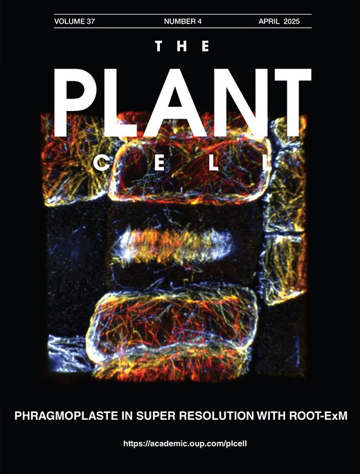
Apr 2025
Advertisement








Abstract
Conventional light microscopy is limited in resolution by the diffraction limit of light, restricting the visualization of the nanoscale organization of biomolecules. Expansion microscopy (ExM) has emerged as a powerful technique to overcome this barrier by physically expanding the specimen embedded in a swellable hydrogel without requiring specialized or high-cost imaging hardware. ExM is widely used in animal models, whereas its application to plant tissues has been challenging due to their multicellularity, in which each cell is encompassed by the rigid cell wall, which resists the expansion forces and prevents isotropic swelling. Here, we describe a robust and optimized ExM protocol specifically designed for Arabidopsis thaliana root tissues. This protocol details critical steps, including immunostaining, anchoring, gelation, denaturation, cell wall digestion, and expansion. Our method achieves an expansion factor of approximately 4.3×, enabling an effective lateral resolution of ~60 nm using a standard confocal microscope. We demonstrate the visualization of microtubules with preserved ultrastructure. This accessible protocol allows plant researchers to perform super-resolution imaging without specialized optical equipment, facilitating detailed structural analysis of plant cells.
Key features
• Expansion microscopy to break the diffraction barrier by increasing the physical distances between proteins while preserving relative spatial relationships and fluorescence signals.
• 4-fold expansion of Arabidopsis root tissues.
• 3D super-resolution imaging.
• Deep-tissue imaging thanks to optical clearing associated with expansion of hydrogel-embedded specimens.
Keywords: Expansion microscopy (膨胀显微技术) Super-resolution microscopy (超分辨率显微成像)
Fluorescence microscopy (荧光显微成像)
Immunostaining (免疫染色)
Tissue clearing (组织透明化)
Deep-tissue imaging (深层组织成像)
Plant biology (植物生物学)
Arabidopsis thaliana root (拟南芥根)
Super-resolution microscopy (超分辨率显微成像)
Fluorescence microscopy (荧光显微成像)
Immunostaining (免疫染色)
Tissue clearing (组织透明化)
Deep-tissue imaging (深层组织成像)
Plant biology (植物生物学)
Arabidopsis thaliana root (拟南芥根)
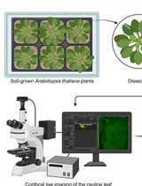
Graphical overview
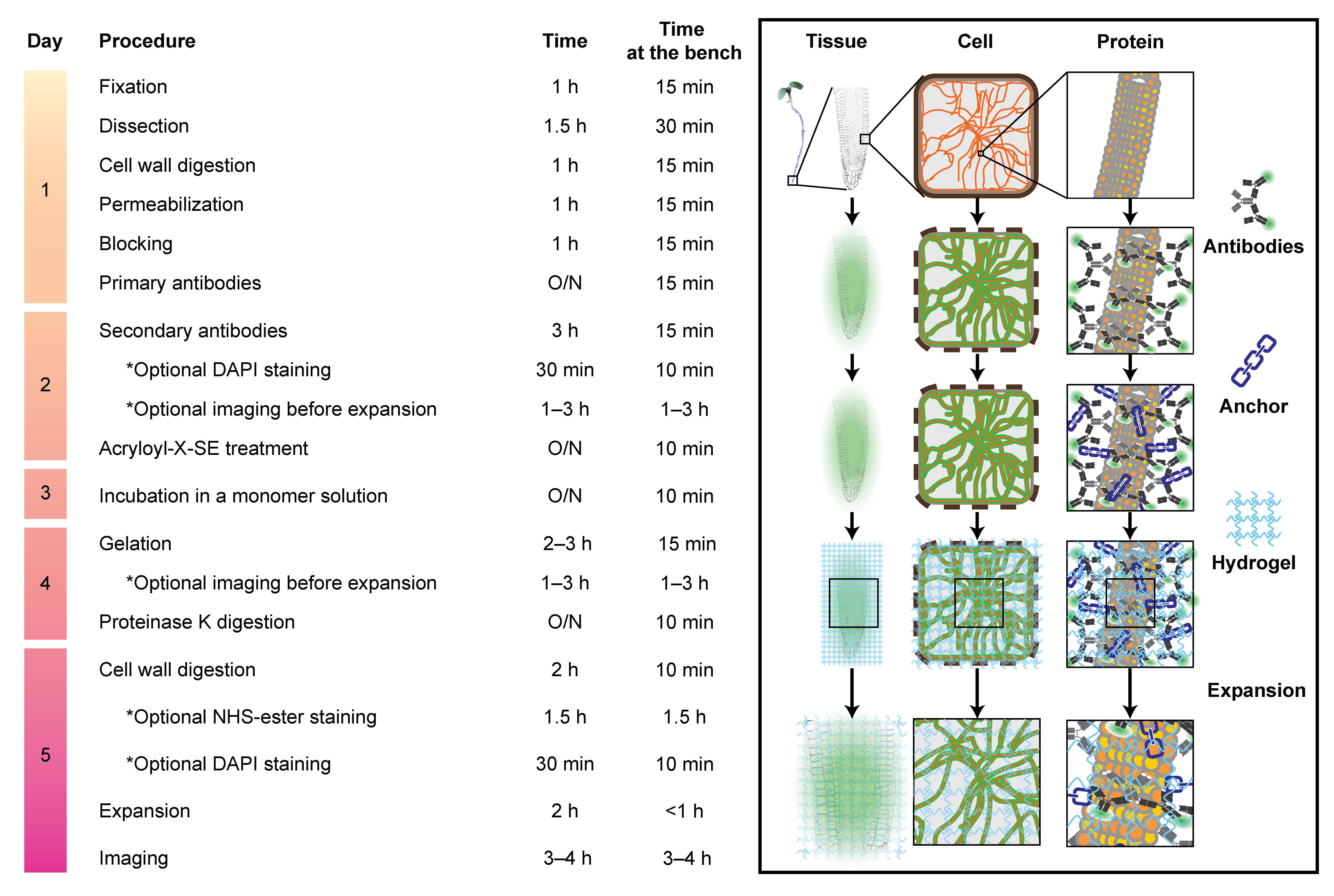
ROOT-ExM workflow and key steps. The required time indicated here includes associated procedures, such as washing.
Background
Determining molecular distribution within the cellular context is crucial for understanding biological functions. While fluorescence microscopy is a fundamental tool to fulfill this purpose in the life sciences, its resolution is, in principle, limited by the diffraction of the light. Super-resolution modalities—including stimulated emission depletion (STED) [1] and photoactivated localization microscopy (PALM) [2] or stochastic optical reconstruction microscopy (STORM) [3]—have revolutionized fluorescence microscopy by breaking this barrier, contributing to new discoveries in recent decades. However, these techniques often require expensive specialized optical setups and significant technical expertise, hindering their widespread adoption.
Imaging plant tissues presents additional unique challenges that render them optically non-ideal. Although Arabidopsis root tips are a classic model to study cell biology, physiology, and development, imaging the protein of interest within them at high resolution is difficult due to (i) cylindrical geometry and low adhesiveness of the root tissue, which often prevents close contact of the cell of interest with coverslips, (ii) refractive index mismatches caused by heterogeneous structures in plant cells, such as the cell wall, cytoplasm, and vacuole [4,5], and (iii) light scattering by the multi-layered organization of such cells. Particularly, routinely studied root epidermal cells in the meristematic zone are obscured by the lateral root cap cells. These factors collectively result in reduced signal-to-noise ratios and spatial resolution [6–9].
Expansion microscopy (ExM), a distinct paradigm for achieving super-resolution imaging first reported in 2015, offers a robust solution to these issues [10]. The principle of ExM is to physically expand the specimen and increase spatial distance between molecules. Thus, ExM enables super-resolution imaging even with conventional diffraction-limited microscopes. In ExM, specific biomolecules such as proteins are chemically modified with the so-called anchor. Subsequently, a swellable polyelectrolyte gel is synthesized throughout the biological sample, while the modified biomolecules can covalently bind to the hydrogel matrix. After gel polymerization, the embedded sample is treated by enzymatic digestion or heat- and detergent-induced denaturation, which is the so-called mechanical homogenization. This loosens adhesiveness derived from intra- and intermolecular interactions and creates appropriate uniformity in mechanical properties of the biological sample, which resists expansion, so that the resulting sample expands isotropically through dialysis in distilled water. As fluorophores are adequately preserved due to their stability to digestion or denaturation, the protein of interest can be localized at super-resolution after expansion. The expansion process also results in significant optical clearing and homogenizes the refractive index of the sample to match that of water. This makes ExM particularly advantageous for imaging in complex plant tissues.
Here, we present a step-by-step protocol for applying ExM to Arabidopsis root tips to achieve robust and reproducible super-resolution imaging of proteins of interest, which utilizes acryloyl-X, SE for anchoring, and sodium acrylate/acrylamide/N,N′-methylenebisacrylamide for the gel matrix. Our ROOT-ExM protocol is based on the pre-expansion staining protein-retention ExM (proExM) previously described in mammalian cells [11] and includes critical optimizations for plant tissues [12]. Most notably, we introduce a cell wall digestion step to overcome the mechanical rigidity imposed by the cell wall, allowing for uniform isotropic expansion. This protocol covers all steps from sample preparation and immunostaining to anchoring, gelation, mechanical homogenization, expansion, and image analysis. The resulting root tip undergoes an approximate 4.3-fold linear expansion and exhibits a water-matching refractive index, enabling super-resolution and deep-tissue imaging.
Materials and reagents
Biological materials
1. Arabidopsis thaliana ecotype Col-0: wild type
Note: Other ecotypes or transgenic lines expressing fluorescent proteins can also be used.
Reagents
1. Murashige and Skoog (MS) medium including vitamins powder (Duchefa Biochemie, catalog number: M0222.0050), storage temperature: 4 °C
2. Sucrose (Sigma-Aldrich, catalog number: 84100), storage temperature: room temperature (RT)
3. MES [2-(N-morpholino)ethanesulfonic acid] (Euromedex, catalog number: EU0033-A), storage temperature: RT
4. Plant agar (Duchefa Biochemie, catalog number: P1001.1000), storage temperature: RT
5. Potassium hydroxide (KOH) (Sigma-Aldrich, catalog number: P5958), storage temperature: RT
6. PIPES [piperazine-N,N′-bis(2-ethanesulfonic acid)] (Sigma-Aldrich, catalog number: P6757), storage temperature: RT
7. EGTA [ethylene glycol-bis(2-aminoethylether)-N,N,N′,N′-tetraacetic acid] (Sigma-Aldrich, catalog number: E4378), storage temperature: RT
8. Magnesium sulfate (MgSO4·7H2O) (Euromedex, catalog number: P027-A), storage temperature: RT
9. Paraformaldehyde solution, 16% (Ted Pella, catalog number: 18505), storage temperature: 4 °C
10. Driselase (Sigma-Aldrich, catalog number: D9515-5G), storage temperature: -20 °C
11. Macerozyme R-10 (Duchefa Biochemie, catalog number: M8002.0005), storage temperature: 4 °C
12. DMSO (dimethyl sulfoxide) (Sigma-Aldrich, catalog number: D8418), storage temperature: RT
13. DMSO, anhydrous (Invitrogen, catalog number: D12345), storage temperature: RT
14. IGEPAL CA-630 (Sigma-Aldrich, catalog number: I3021), storage temperature: RT
15. Bovine serum albumin (BSA) (Euromedex, catalog number: 1035-70-C), storage temperature: 4 °C
16. DAPI (4',6-diamidino-2-phenylindole) dihydrochloride (Sigma-Aldrich, catalog number: D8417-1MG), storage temperature: 4 °C
17. Acryloyl-X, SE [6-((acryloyl)amino)hexanoic acid, succinimidyl ester] (Thermo Fisher Scientific, catalog number: A20770), storage temperature: -20 °C
18. Sodium acrylate (Combi-Blocks, catalog number: QC-1489), storage temperature: 4 °C
19. Proteinase K, ≥30 units/mg (Sigma-Aldrich, catalog number: P2308-25MG), storage temperature: -20 °C
20. PBS (10×), pH 7.4 (Thermo Fisher Scientific, catalog number: 70011044), storage temperature: RT
21. 40% acrylamide solution (Sigma-Aldrich, catalog number: A4058-100ML), storage temperature: 4 °C
22. 2% N,N′-Methylenebisacrylamide solution (Sigma-Aldrich, catalog number: M1533-25ML), storage temperature: 4 °C
23. 4-Hydroxy-TEMPO (Sigma-Aldrich, catalog number: 176141-1G), storage temperature: 4 °C
24. TEMED (N,N,N′,N′-Tetramethylethylenediamine) (Sigma-Aldrich, catalog number: T7024-25ML), store at RT or 4 °C
25. Ammonium persulfate (APS) (Sigma-Aldrich, catalog number: A3678-25G), store at RT or 4 °C
26. Sodium chloride (NaCl) (Sigma-Aldrich, catalog number: S7653), storage temperature: RT
27. Tris base (Sigma-Aldrich, catalog number: 252859), storage temperature: RT
28. Ethylenediaminetetraacetic acid tetrasodium salt dihydrate (EDTA) (Sigma-Aldrich, catalog number: ED4SS), storage temperature: RT
29. Hydrochloric acid (HCl) (Sigma-Aldrich, catalog number: 258148), storage temperature: RT
30. Sodium hydroxide (NaOH) (Euromedex, catalog number: 2020), storage temperature: RT
31. Triton X-100 (Sigma-Aldrich, catalog number: T9284), storage temperature: RT
32. Guanidine hydrochloride (Sigma-Aldrich, catalog number: G3272), storage temperature: RT
33. Sodium bicarbonate (NaHCO3) (Sigma-Aldrich, catalog number: S6297), storage temperature: RT
34. 52.5% nitric acid (HNO3) (Prolabo, catalog number: 20420.291), storage temperature: RT
35. Ethanol (Fisher Scientific, catalog number: E-0650DF-17), storage temperature: RT
36. APTES [(3-Aminopropyl)triethoxysilane] (Sigma-Aldrich, catalog number: A3648), storage temperature: 4 °C
37. Mouse anti-α-tubulin monoclonal primary antibody (Sigma-Aldrich, catalog number: T5168) (1:500 dilution)
38. Donkey anti-mouse IgG secondary antibody conjugated with Alexa Fluor 488 (Abcam, catalog number: ab150105) (1:500 dilution), storage temperature: -20 °C (stock aliquots) or 4 °C for 4 weeks (thawed aliquots)
39. Goat anti-mouse IgG secondary antibody conjugated with Alexa Fluor 594 (Invitrogen, catalog number: A-11005) (1:500 dilution), storage temperature: -20 °C (stock aliquots) or 4 °C for 4 weeks (thawed aliquots)
40. NHS ester-ATTO647 (Sigma-Aldrich, catalog number: 07376), storage temperature: -20 °C
Note: Other fluorophore-conjugated NHS esters can also be used.
Solutions
1. 10 M KOH (see Recipes)
2. 1/2× MS solid media (see Recipes)
3. 4/3× microtubule stabilizing buffer (MTSB) (see Recipes)
4. MTSB (see Recipes)
5. Fixation solution (see Recipes)
6. Cell wall digestion solution I (see Recipes)
7. Permeabilization solution (see Recipes)
8. Blocking buffer (see Recipes)
9. DAPI solution (1,000×) (see Recipes)
10. PBS (see Recipes)
11. Acryloyl-X, SE stock solution (100×) (see Recipes)
12. 4-Hydroxy-TEMPO stock solution (50×) (see Recipes)
13. TEMED stock solution (50×) (see Recipes)
14. APS stock solution (50×) (see Recipes)
15. Sodium acrylate solution (see Recipes)
16. Monomer stock solution (see Recipes)
17. Gelation solution (see Recipes)
18. 1 M Tris (pH 8.0) (see Recipes)
19. 0.5 M EDTA (see Recipes)
20. Protein digestion buffer (see Recipes)
21. Proteinase K stock solution (100×) (see Recipes)
22. Protein digestion solution (see Recipes)
23. Cell wall digestion solution II (see Recipes)
24. NHS ester labeling buffer (see Recipes)
25. NHS ester stock solution (see Recipes)
26. NHS ester labeling solution (see Recipes)
Recipes
Caution: This protocol involves the use of chemicals classified as carcinogenic, mutagenic, and reprotoxic (CMR), such as paraformaldehyde and acrylamide. Therefore, appropriate personal protective equipment should be worn, and if necessary, all manipulations involving CMRs must be performed under a fume hood.
1. 10 M KOH
| Reagent | Final concentration | Amount |
|---|---|---|
| KOH | 10 M | 28.05 g |
| Ultrapure water | n/a | Make up to 50 mL |
Store in a plastic bottle/tube at RT
2. 1/2× MS solid media
| Reagent | Final concentration | Amount |
|---|---|---|
| MS powder | 1/2× | 2.2 g |
| Sucrose | 1% | 10 g |
| MES | 2.5 mM | 0.49 g |
| 10 M KOH | n/a | Adjust the pH to 5.8 |
| Plant agar | 0.8% | 8 g |
| Ultrapure water | n/a | Make up to 1 L |
Autoclave at 110 °C for 30 min and dispense 50 mL per 12 cm × 12 cm square Petri dish. Store the solidified agar media at 4 °C.
3. 4/3× MTSB
| Reagent | Final concentration | Amount |
|---|---|---|
| PIPES | 66.67 mM | 5.04 g |
| EGTA | 6.67 mM | 0.63 g |
| MgSO4·7H2O | 6.67 mM (MgSO4) | 0.41 g |
| KOH | n/a | Adjust the pH to 7 |
| Ultrapure water | n/a | Make up to 250 mL |
Autoclave at 110 °C for 30 min. Store at 4 °C.
4. MTSB
| Reagent | Final concentration | Amount |
|---|---|---|
| PIPES | 50 mM | 15.12 g |
| EGTA | 5 mM | 1.89 g |
| MgSO4·7H2O | 5 mM (MgSO4) | 1.23 g |
| KOH | n/a | Adjust the pH to 7 |
| Ultrapure water | n/a | Make up to 1 L |
Autoclave at 110 °C for 30 min. Store at 4 °C.
5. Fixation solution
| Reagent | Final concentration | Amount |
|---|---|---|
| 16% Paraformaldehyde solution | 4% (v/v) | 10 mL (1 ampoule) |
| 4/3× MTSB | 1× | 30 mL |
Note: This solution should be freshly prepared for each experiment. However, the rest can be stored at 4 °C for a week or at -20 °C for a month.
6. Cell wall digestion solution I
| Reagent | Final concentration | Amount per reaction |
|---|---|---|
| Driselase | 2% (w/v) | 4 mg |
| MTSB | 1× | 200 μL |
Centrifuge at 1,000× g for 1 min and use the supernatant.
Note: This solution should be freshly prepared for each experiment.
7. Permeabilization solution
| Reagent | Final concentration | Amount per reaction |
|---|---|---|
| DMSO | 10% (v/v) | 20 μL |
| IGEPAL CA-630 | 3% (v/v) | 6 μL |
| MTSB | 1× | 174 μL |
Note: This solution should be freshly prepared for each experiment.
8. Blocking buffer
| Reagent | Final concentration | Amount per reaction |
|---|---|---|
| BSA | 3% (w/v) | 6 mg |
| MTSB | 1× | 200 μL |
Note: This solution should be freshly prepared for each experiment.
9. DAPI solution (1,000×)
| Reagent | Final concentration | Amount |
|---|---|---|
| DAPI dihydrochloride | 2 mg/mL | 1 mg |
| Ultrapure water | n/a | 0.5 mL |
Prepare 10 μL aliquots in 0.5 mL tubes and store at -20 °C.
10. PBS
| Reagent | Final concentration | Amount |
|---|---|---|
| PBS (10×) | 1× | 5 mL |
| Sterilized ultrapure water | n/a | 45 mL |
Store at 4 °C.
11. Acryloyl-X, SE stock solution (100×)
| Reagent | Final concentration | Amount |
|---|---|---|
| Acryloyl-X, SE | 10 mg/mL | 5 mg |
| DMSO | n/a | 0.5 mL |
Prepare 10 μL aliquots in 0.5 mL tubes and store at -20 °C.
12. 4-Hydroxy-TEMPO stock solution (50×)
| Reagent | Final concentration | Amount |
|---|---|---|
| 4-Hydroxy-TEMPO | 0.5% (w/w) | 50 mg |
| Ultrapure water | n/a | 10 mL |
Prepare 50 μL aliquots in 0.5 mL tubes and store at -20 °C.
13. TEMED stock solution (50×)
| Reagent | Final concentration | Amount |
|---|---|---|
| TEMED | 10% (v/v) | 1 mL |
| Ultrapure water | n/a | 9 mL |
Prepare 50 μL aliquots in 0.5 mL tubes and store at -20 °C.
14. APS stock solution (50×)
| Reagent | Final concentration | Amount |
|---|---|---|
| APS | 10% (w/w) | 1 g |
| Ultrapure water | n/a | 10 mL |
Prepare 50 μL aliquots in 0.5 mL tubes and store at -20 °C.
15. Sodium acrylate solution
| Reagent | Final concentration | Amount |
|---|---|---|
| Sodium acrylate | 35.5% (w/w) | 2.2 g |
| Ultrapure water | n/a | 4 mL |
Critical: Check the purity by the color of the solution. It should be clear, slightly yellowish (Figure 1). If this is not the case, we recommend purchasing a new bottle of sodium acrylate.
Note: Make this immediately before preparing a monomer stock solution. The resulting volume will be approximately 4.5 mL.
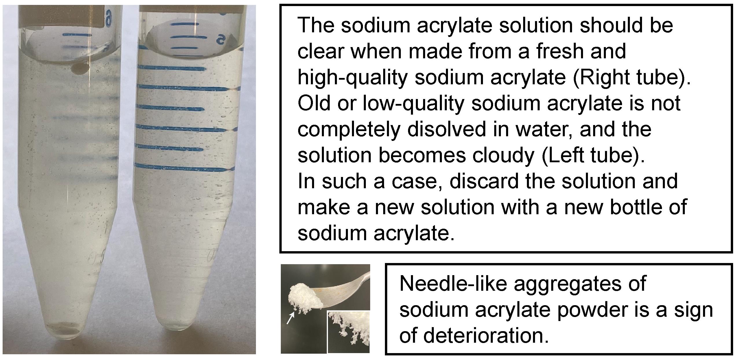
Figure 1. Quality check of sodium acrylate
16. Monomer stock solution
| Reagent | Final concentration | Amount |
|---|---|---|
| Sodium acrylate solution | 8.5% (8%) (w/w) | 4.5 mL |
| 40% Acrylamide solution | 2.1% (2%) (w/v) | 1 mL |
| 2% N,N′-Methylenebisacrylamide solution | 0.16% (0.15%) (w/v) | 1.5 mL |
| NaCl | 2.13 M (2 M) | 2.34 g |
| PBS (10×) | 1.06× (1×) | 2 mL |
| Ultrapure water | n/a | Make up to 18.8 mL |
Prepare 470 μL aliquots in 0.5 mL tubes and store at -20 °C.
Note: The concentration of each component will slightly decrease in a gelation solution with the addition of 4-Hydroxy-TEMPO, TEMED, and APS. These concentrations are shown in the brackets. One aliquot is generally used for two coverslips.
17. Gelation solution
| Reagent | Final concentration | Amount per reaction |
|---|---|---|
| Monomer solution | see above | 188 μL |
| 4-Hydroxy-TEMPO stock solution | 0.01% (w/w) | 4 μL |
| TEMED stock solution | 0.2% (v/v) | 4 μL |
| APS stock solution | 0.2% (w/w) | 4 μL |
Note: 4-Hydroxy-TEMPO, TEMED, and APS should be added sequentially in this order. Mix thoroughly after each addition. This solution should be prepared on ice immediately before the gelation step.
18. 1 M Tris (pH 8)
| Reagent | Final concentration | Amount |
|---|---|---|
| Tris base | 1 M | 12.11 g |
| HCl | n/a | Adjust the pH to 8 |
| Ultrapure water | n/a | Make up to 100 mL |
Store at RT.
19. 0.5 M EDTA
| Reagent | Final concentration | Amount |
|---|---|---|
| EDTA·4Na·2H2O | 0.5 M (EDTA) | 20.81 g |
| HCl | n/a | Adjust the pH to 8 |
| Ultrapure water | n/a | Make up to 100 mL |
Store at RT.
20. Protein digestion buffer
| Reagent | Final concentration | Amount |
|---|---|---|
| 1 M Tris (pH 8) | 50 mM | 5000 μL |
| 0.5 M EDTA | 2.5 mM | 500 μL |
| Triton X-100 | 0.5% (v/v) | 500 μL |
| Guanidine hydrochloride | 0.8 M | 7.64 g |
| Ultrapure water | n/a | Make up to 100 mL |
Prepare 10 mL aliquots and store at -20 °C.
Note: After thawing, the protein digestion buffer can be stored at 4 °C.
21. Proteinase K stock solution (100×)
| Reagent | Final concentration | Amount |
|---|---|---|
| Proteinase K, ≥30 units/mg | ≥800 units/ml | 25 mg |
| Ultrapure water | n/a | 937.5 μL |
Prepare 10 μL aliquots in 0.5 mL tubes and store at -20 °C.
22. Protein digestion solution
| Reagent | Final concentration | Amount per reaction |
|---|---|---|
| Proteinase K stock solution (100×) | ≥8 units/ml | 5 μL |
| Protein digestion buffer | n/a | 495 μL |
Note: This solution should be freshly prepared for each experiment.
23. Cell wall digestion solution II
| Reagent | Final concentration | Amount per reaction |
|---|---|---|
| Driselase | 2% | 10 mg |
| Macerozyme R-10 | 0.5% | 2.5 mg |
| PBS | 1× | 500 μL |
Centrifuge at 1,000× g for 1 min and use the supernatant.
Note: This solution should be freshly prepared for each experiment.
24. NHS ester labeling buffer
| Reagent | Final concentration | Amount per reaction |
|---|---|---|
| NaHCO3 | 100 mM | 0.42 g |
| HCl (0.1 or 1 M) | n/a | Adjust the pH to 8 |
| Ultrapure water | n/a | 50 mL |
Store at -20 °C.
Note: CO2 loss during storage leads to a gradual rise in pH. Keep the bottle tightly sealed with minimal headspace. It is highly recommended to verify the pH immediately before use, especially after prolonged storage.
25. NHS ester stock solution (100×)
| Reagent | Final concentration | Amount |
|---|---|---|
| NHS ester dye | 2 mg/mL | 1 mg |
| DMSO, anhydrous | n/a | 500 μL |
Prepare 10 μL aliquots in 0.5 mL tubes and store at -20 °C. Thawed aliquots can be stored at 4 °C for 4 weeks.
26. NHS ester labeling solution
| Reagent | Final concentration | Amount per reaction |
|---|---|---|
| NHS ester labeling buffer | n/a | 990 μL |
| NHS ester stock solution (100×) | 20 μg/mL | 10 μL |
Note: This solution should be freshly prepared for each experiment.
Laboratory supplies
1. Corning® BioCoat® Poly-D-Lysine 12 mm #1 German glass coverslip (Corning, catalog number: 354086)
Note: Homemade Poly-D-Lysine-coated round coverslips (ø 12 or 18 mm) can also be used.
2. Hydrophobic barrier pen (Vector Laboratories, catalog number: H-4000)
3. Square Petri dish, 120 × 120 × 17 mm (Greiner Bio-One, catalog number: 688102)
4. Round Petri dish, 100 × 20 mm (Greiner Bio-One, catalog number: 664102)
5. 6-well cell culture plate (Thermo Fisher Scientific, catalog number: 140675)
6. Tweezer (Hammacher, catalog number: HWC110-10)
7. Tweezer style 7 (Dumont, catalog number: 0304-7-PO)
8. Grease (Dow Corning Toray, catalog number: Dow Corning® High Vacuum Grease)
9. Glass microscope slide (Epredia, catalog number: AG00000112E04CML21
10. 18 × 18 mm coverslip (Knittel, catalog number: VD11818Y1A.01)
11. Razor blade (Accutec, catalog number: AGBL-7032-0000)
12. Parafilm (Amcor, catalog number: PM-996)
13. Aluminum foil (Alujet, catalog number: AP326)
Equipment
1. Stereomicroscope (Leica, model: MZ16F)
2. Confocal laser-scanning microscope (Carl Zeiss, model: LSM 880)
3. Dry objective lens, 10× (Carl Zeiss, model: Objective Plan-Apochromat 10×/0.45 M27)
4. Dry objective lens, 20× (Carl Zeiss, model: Objective Plan-Apochromat 20×/0.8 M27
5. Water-immersion objective lens, 40× (Carl Zeiss, model: Objective W Plan-Apochromat 40×/1.0 DIC M27)
Software and datasets
1. ZEN black 2.3 SP1 FP3 software (Carl Zeiss, version 14.0)
2. Fiji/ImageJ (version 2.16.0/1.54p) (https://imagej.net/software/fiji/)
3. All computational analyses were performed using Python 3.10.19 with SimpleITK-SimpleElastix (v2.0.0rc2.dev910) on Windows 10/11 (64-bit). The exact computational environment is archived in a public repository to ensure reproducibility (https://src.koda.cnrs.fr/bic-tai/expansion_analysis_protocol).
Procedure
登录/注册后免费查看全文
文章信息
稿件历史记录
提交日期: Mar 10, 2026
接收日期: Apr 27, 2026
在线发布日期: May 13, 2026
出版日期: Jun 5, 2026
版权信息
© 2026 The Author(s); This is an open access article under the CC BY-NC license (https://creativecommons.org/licenses/by-nc/4.0/).
如何引用
Shimizu, Y., Maucort, G., Fernández-Monreal, M., Dumas, F., Bayer, E., Boutté, Y. and Grison, M. (2026). ROOT-ExM: Super-Resolution Imaging of Proteins in Arabidopsis Roots by Expansion Microscopy. Bio-protocol 16(11): e5707. DOI: 10.21769/BioProtoc.5707.

 Download Citation in RIS Format
分类
植物科学 > 植物细胞生物学 > 细胞成像
细胞生物学 > 细胞成像 > 超分辨率成像
细胞生物学 > 组织分析 > 组织成像
您对这篇实验方案有问题吗?
在此处发布您的问题,我们将邀请本文作者来回答。同时,我们会将您的问题发布到Bio-protocol Exchange,以便寻求社区成员的帮助。
 发布问题
发布问题


