


Oligo(dT) Fluorescence In Situ Hybridization to Visualize the Poly(A) mRNAs in the Internal Tissues of Drosophila
利用 Oligo(dT) 荧光原位杂交观察果蝇内部组织中的 poly(A) mRNA


发布: 2026年06月05日第16卷第11期 DOI: 10.21769/BioProtoc.5702 浏览次数: 349
评审: Vibha DwivediIvonne SehringAnonymous reviewer(s)
 PDF
PDF  Q&A
Q&A
参见作者原研究论文
The authors used this protocol in:

Mar 2019
Advertisement








Abstract
Fluorescence in situ hybridization (FISH) is a cytological method used to visualize specific oligonucleotide sequences within the cell. This method relies on the specific binding of a fluorescence-tagged probe, a short stretch of single-stranded polynucleotide, to its complementary sequence in the DNA or RNA, forming stable double-stranded hybrids. Fluorochromes, such as fluorescein, Alexa Fluor, cyanine dyes, or rhodamine, are attached to these probes to help in detecting their presence within the cell. Based on sequence complementarity, FISH allows for the visualization of the DNA or RNA with which they have hybridized. The distribution of these fluorochrome-tagged probes can be observed under a fluorescence or confocal microscope. The oligo(dT) FISH technique specifically utilizes a fluorochrome-tagged stretch of 40–50 thymidine (T) oligonucleotides that binds to the poly(A) tails of mature mRNAs within the cell. Newly transcribing pre-mRNAs and certain non-coding RNAs may not have poly(A) tails and therefore cannot be detected by this method. This step-by-step protocol outlines the oligo(dT) FISH technique for visualizing the cellular distribution of polyadenylated mRNAs in the tissues of Drosophila and other related model organisms.
Key features
• Oligo (dT) fluorescence in situ hybridization is a robust cytological technique to visualize polyadenylated RNAs in the cell.
• This oligo(dT) FISH protocol is applicable for a wide range of animals and cell culture model systems.
• This protocol is particularly useful to visualize mRNAs in thin tissues with a few layers of cells.
Keywords: Probe (探针) FISH (荧光原位杂交)
Cytology (细胞学)
RNA (RNA)
Drosophila (果蝇)
Microscopy (显微成像)
FISH (荧光原位杂交)
Cytology (细胞学)
RNA (RNA)
Drosophila (果蝇)
Microscopy (显微成像)
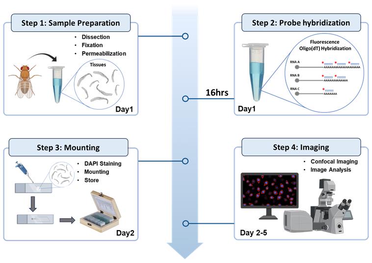
Graphical overview
Background
Fluorescence in situ hybridization (FISH) is a powerful technique for detecting specific nucleic acid sequences within fixed cells and tissues [1-3]. This technique has been widely used to visualize specific sequences in DNA (DNA-FISH) or RNA (RNA-FISH) molecules within the cell [4–7]. Traditional RNA-FISH uses gene-specific probes to localize individual transcripts. In contrast, the oligo(dT) FISH strategy employs a fluorescently labeled stretch of 40–50 thymidine (T) nucleotides that specifically hybridizes to the poly(A) tails of mature mRNAs [8,9]. Unlike gene-specific RNA in situ hybridization, which targets a specific transcript [10], this method allows for the visualization of the entire pool of poly(A) mRNAs [9]. This makes oligo(dT) FISH an essential tool for studying mRNA abundance and subcellular localization on a global scale. While earlier studies in yeast and mammalian systems have successfully utilized oligo(dT) probes to examine nuclear-cytoplasmic mRNA distribution, mRNA export defects, and stress-induced RNA retention, there is a notable absence of comprehensive, standardized oligo(dT) FISH protocols specifically optimized for Drosophila tissues [11–14]. This protocol can be widely applied to investigate mRNA dynamics during development, disease, and stress responses in Drosophila, C. elegans, or mammalian cell cultures. Furthermore, its compatibility with co-immunostaining makes it a versatile dual-track system for observing how specific proteins interact with the global mRNA pool under various cellular conditions.
Materials and reagents
Biological materials
Set the desired cross in vials containing standard cornmeal food and collect the specific stage of Drosophila by anesthetizing them using ether in an etherizer. In this study, we used actively wandering third-instar larvae of Nup107GFP/Nup107GFP (Bloomington Stock No. 35514) genotype, which enables visualization of the nuclear membrane in the larval salivary gland.
Reagents
1. Ether (SRL, catalog number: 25049)
2. NaCl (Himedia, catalog number: MB023)
3. KCl (Himedia, catalog number: TC010)
4. Na2HPO4 (Himedia, catalog number: GRM168)
5. KH2PO4 (Himedia, catalog number: GRM1188)
6. Sodium citrate (Himedia, catalog number: TC526U)
7. NP-40 (Thermo, catalog number: 85124)
8. Triton X-100 (Himedia, catalog number: MB031)
9. 4′,6-diamidino-2-phenylindole (DAPI) (Thermo, catalog number: 62247)
10. Paraformaldehyde (PFA) (Himedia, catalog number: RM3660)
Note: PFA is a polymer with a wide range of monomeric and oligomeric units of formaldehyde. It dissolves in water and depolymerizes into a monomeric form of formaldehyde. Formaldehyde can crosslink cellular constituents, particularly proteins with lysine residues. Formaldehyde is a toxic chemical; therefore, always wear a mask and gloves during handling.
11. Diethyl pyrocarbonate (DEPC) (Himedia, catalog number: MB076)
Note: DEPC irreversibly inactivates RNase contamination that may be present in dH2O. It is carcinogenic and, therefore, needs to be handled carefully.
12. Yeast tRNA (Thermo, catalog number: AM7119)
Note: Yeast tRNA is used as a nonspecific blocking agent, protecting both the probe and cellular RNA.
13. Dextran sulfate (Merck, catalog number: 265152-M)
Note: Dextran sulfate is a highly hydrated polysaccharide that acts as an effective volume exclusion agent. It affects the probe concentration by preventing its solubilization in water and thus gives higher hybridization rates in aqueous solutions.
14. RNase inhibitor (Thermo, catalog number: EO0382)
Note: RNase inhibitors directly bind with RNases in a non-competitive mode and thus block their activity. They have been widely used to stop the activity of RNases that are present within the cell. The presence of 1 U/μL RNase inhibitor can block most of the RNase activity during FISH.
15. Formamide (SIGMA, catalog number: 15706)
Note: Formamide interferes with hydrogen bonding in double-stranded polynucleotides and thus destabilizes double-strand formation. The presence of formamide in hybridization buffer, therefore, reduces the melting and annealing temperature, which keeps the tissue morphology relatively intact due to low incubation temperature. The working concentration of formamide in hybridization buffer varies from 10% to 50%, depending upon the probe length and its GC content.
16. 4′,6-diamidino-2-phenylindole (DAPI) (Thermo, catalog number: 62247)
Note: DAPI is a fluorescent stain that binds to the AT-rich region of DNA, with excitation at 405 nm and emission spectrum between 420 and 480 nm (blue color). The fluorescence of DAPI is enhanced several times following its binding with DNA.
17. ProLongTM Gold antifade mountant (Thermo, catalog number: P10114)
Note: ProLongTM Gold antifade mountant is a commercially available glycerol-based mounting media supplemented with an antifading agent that protects the fluorescent signal intensity.
18. Riboprobe; probes used here for FISH are single-stranded, non-self-complementary short stretches of polynucleotides (20–1500 nucleotide long DNA or RNA) that have a complementary sequence to the target RNA. In the present study, we used RNase Free HPLC Purified 45 nucleotide long stretch of oligo(dT) probe tagged with Cy3 at the 5′ end. We customized the probe and procured it from Integrated DNA Technologies (IDT)
Solutions
1. 10× PBS (see Recipes)
2. 20× SSC (see Recipes)
3. 4% PFA (see Recipes)
4. 50% dextran sulfate (see Recipes)
5. 0.1% DEPC (see Recipes)
6. 0.3% PBST (see Recipes)
7. 100 mg/mL yeast tRNA (see Recipes)
8. 1 μg/mL DAPI (see Recipes)
9. Permeabilization buffer (see Recipes)
10. Hybridization buffer (see Recipes)
11. Wash buffer (see Recipes)
Recipes
1. 10× PBS (pH 7.4)
| Reagent | Concentration | Quantity |
|---|---|---|
| NaCl | 1.37 M | 80 g |
| KCl | 27 mM | 2 g |
| Na2HPO4 | 100 mM | 14.4 g |
| KH2PO4 | 18 mM | 2.45 g |
Take 800 mL of distilled water (dH2O) and dissolve all four components. Adjust the pH to 7.4 and make up the volume to 1 L with dH2O. Autoclave and store the 10× PBS at room temperature (RT). Dilute the 10× PBS with dH2O in a 1:9 ratio to make the 1× PBS as a working solution and store it at 4 °C.
2. 20× SSC (pH 7.0)
| Reagent | Concentration | Quantity |
|---|---|---|
| NaCl | 3 M | 175.3 g |
| Sodium citrate | 0.3 M | 77.4 g |
Take 800 mL of dH2O and dissolve both components. Adjust the pH to 7.0 with HCl and make up the volume to 1 L with dH2O. Autoclave and store at RT.
3. 4% PFA
Dissolve 4.0 g of PFA in 75 mL of 1× PBS at 60 °C with constant stirring for 10 min. Slowly add 5 N NaOH to the cloudy mixture until a clear solution is formed. Filter the solution with a Whatman filter paper and adjust the pH to 7.0 with HCl or NaOH. Make up the volume to 100 mL with 1× PBS and store in small aliquots at -20 °C. Defrost an aliquot and use it on the same day of the experiment. It is, however, preferred that small volumes of PFA solution be prepared fresh, as required. Prolonged storage of formaldehyde leads to its polymerization in the form of paraformaldehyde, thus reducing its fixative properties.
4. 50% dextran sulfate
Slowly dissolve 20 g of dextran sulfate powder in 30 mL of DEPC-treated dH2O and leave the tube overnight on the rotator. Make up the volume to 40 mL with dH2O and store at -20 °C in small aliquots. 50% dextran sulfate is very viscous, and special care should be taken during pipetting.
5. 0.1% DEPC
Mix 0.1 mL of DEPC in 100 mL of dH2O (0.1% DEPC) and autoclave after 4 h incubation at 37 °C. DEPC treatment is a very effective way to make RNase-free solutions; however, buffers like Tris or HEPES cannot be treated with DEPC as they absorb DEPC and make it unavailable for RNase. To prepare these buffers, DEPC-treated dH2O should be used to dissolve the RNase-free reagent powders.
6. 0.3% PBST
Triton X-100 is a non-ionic mild detergent that solubilizes proteins and is commonly used to permeabilize the cell membrane. Dissolve 0.03 mL of Triton X-100 in 10 mL of 1× PBS to make 0.3% PBST, which is used as a washing buffer in FISH.
7. 100 mg/mL yeast tRNA
Dissolve 25 mg of yeast tRNA powder in 250 μL of DEPC-treated dH2O and store at -20 °C.
8. 1 μg/mL DAPI
Dissolve 1 mg of DAPI in 1 mL of dH2O and store the stock (1 mg/mL) solution at -20 °C. Dilute the DAPI stock solution in 1× PBS (1:1000) to make the 1 μg/mL working DAPI solution. Store at 4 °C for several weeks.
9. Permeabilization buffer
| Reagent | Concentration | Quantity |
|---|---|---|
| Triton X-100 | 1% | 1 μL |
| NP-40 | 1% | 1 μL |
| 40 U/μL RNase inhibitor | 1 U/μL | 2.5 μL |
| 1× PBS | 95.5 μL |
Dissolve 1 μL of Triton X-100, 1 μL of NP-40, and 2.5 μL of 40 U/μL RNase-inhibitor in 95.5 μL of 1× PBS. Use fresh.
10. Hybridization buffer
| Reagent | Concentration | Quantity |
|---|---|---|
| Formamide | 25% | 25 μL |
| 20× SSC | 2× | 10 μL |
| 50% dextran sulfate | 10% | 20 μL |
| 100 mg/mL yeast tRNA | 1 mg/mL | 1 μL |
| 100 ng/mL Oligo(dT) riboprobe | 5 ng/mL | 5 μL |
| DEPC-treated dH2O | 39 μL |
Take 39 μL of DEPC-treated dH2O to dissolve 25 μL of formamide, 10 μL of 20× SSC pH 7.0, 20 μL of 50% dextran sulfate, 1 μL of 100 mg/mL yeast tRNA, and 5 μL of 100 ng/μL Cy3-labeled oligo(dT) riboprobe. Use fresh.
11. Wash buffer
| Reagent | Concentration | Quantity |
|---|---|---|
| Formamide | 25% | 25 μL |
| 20× SSC | 2× | 10 μL |
| DEPC-treated dH2O | 65 μL |
Dissolve 25 μL of formamide and 10 μL of 20× SSC in 65 μL of DEPC-treated dH2O. Use fresh.
Laboratory supplies
1. Glass slide (Blue Star microslide, 75 mm × 25 mm)
2. Fine forceps (Dumont Tweezer, catalog number: 72701-D)
3. Glass coverslips (Blue Star, 22 mm2, part no. CSI-130)
4. Transparent Nail polish (Shade-40, Lakme, Model No TWUY100)
5. Dissection needle (Syringes 31G, Dispovan, RDMS-01)
6. Round synthetic paintbrush (Round paintbrush Size 4)
7. Dark chamber (opaque plastic box)
8. Etherizer (Tarson, catalog numbers: 630020 and 500043)
Equipment
1. Stereomicroscope (Nikon, model: SMZ745)
2. Fluorescence microscope (Olympus, model: IX83)
3. Confocal microscope (Olympus, model: FV3000)
4. Incubators set at 25 °C and 42 °C (Orbitek, model: CINC450)
Software and datasets
1. Fiji/ImageJ software (NIH, USA) (Java 6, available at imagej.nih.gov/ij)
Procedure
A. Tissue pretreatment
1. Collect actively wandering, healthy third-instar larvae in a Petri dish.
2. Wash larvae with distilled water (dH2O) and dry them on tissue paper.
3. Dissect out the desired internal tissues of larvae in 1× PBS on a glass slide with the help of fine forceps or dissecting needles.
4. Fix the tissues by incubating them in freshly prepared 4% PFA at RT for 20 min in a microcentrifuge tube. Perform all FISH steps in the same microcentrifuge tube.
Note: Fix the tissue immediately after dissection to minimize any stress to the tissue. Ensure that the tissues are properly submerged in the fixing solution and not adhering to the container wall or floating on the surface.
5. Remove the fixative and wash the tissues with 200 μL of 0.3% PBST on a rotator for 10 min at RT. Repeat this washing step three times.
6. Incubate the tissue with permeabilization buffer for 20 min at 4 °C on a rotator to permeabilize the cell and nuclear membrane.
7. Rinse the tissue with 200 μL of 0.3% PBST three times for 10 min each.
8. Rinse the tissue further with 200 μL of 2× SSC twice for 10 min each.
B. Probe hybridization
1. For each sample of 5–6 larval tissue, prepare ~30 μL of hybridization solution. Thoroughly mix all components with a 200 μL pipette.
2. Incubate the tissue with hybridization solution in a 500 μL microcentrifuge tube at 42 °C for 12–16 h. Make sure that the cap of the tube is properly closed and sealed with parafilm during incubation to prevent drying.
Note: The composition of the hybridization buffer and incubation temperature may vary with probe length and its GC content. Smaller probes (~30 nt) need a relatively lower concentration of formamide (10%) and lower incubation temperature (35–37 °C), while longer probes (~500 nt) need a higher concentration of formamide (50%) and higher incubation temperature (60–65 °C).
3. Rinse the tissue at 42 °C with freshly prepared prewarmed wash buffer three times for 5 min each.
4. Rinse the tissue again at 42 °C with prewarmed 2× SSC, followed by 1× SSC, three times for 5 min each.
5. Rinse the tissue further with 1× PBS at RT three times for 5 min each.
Note: Immunostaining for a protein of interest can be performed to examine its interaction with poly(A) RNA in the cell. Immunostaining can be performed just after completion of FISH and before counterstaining with DAPI [2,7,15,16].
C. Counterstaining and microscopy
1. Counterstain the nuclei with DAPI (1 μg/mL) by incubating the tissue for 10 min at RT. Mount the tissue in antifade mounting media. Store the slides in a dark place at -20 °C until further examination.
2. Examine the fluorescence signals using appropriate excitation and emission filters under a fluorescence or confocal microscope.
Note: The excitation and emission spectra of the fluorochromes used in FISH should be sufficiently different for clear distinction of the two signals (without any bleeding effect).
Data analysis
In the example shown in Figure 1, we conducted RNA FISH with a Cy3-labeled oligo(dT) riboprobe in the salivary glands of a third-instar larva. The Nup107GFP protein is visualized at the nuclear membrane, clearly outlining the boundary between the nucleus and the cytoplasm. The image indicates that poly(A) mRNA is present in both the nucleus and the cytoplasm (Figure 1A–C). We measured fluorescence signal intensity using the Analyse > Measure tool in FIJI and found that the level of poly(A) mRNA in the cytoplasm was significantly higher than that in the nucleus (Figure 1D). Notably, the oligo(dT) FISH signal was nearly absent in the nucleolus, which is primarily composed of the non-polyadenylated ribosomal RNAs (rRNAs) and small nucleolar RNAs (snoRNAs).
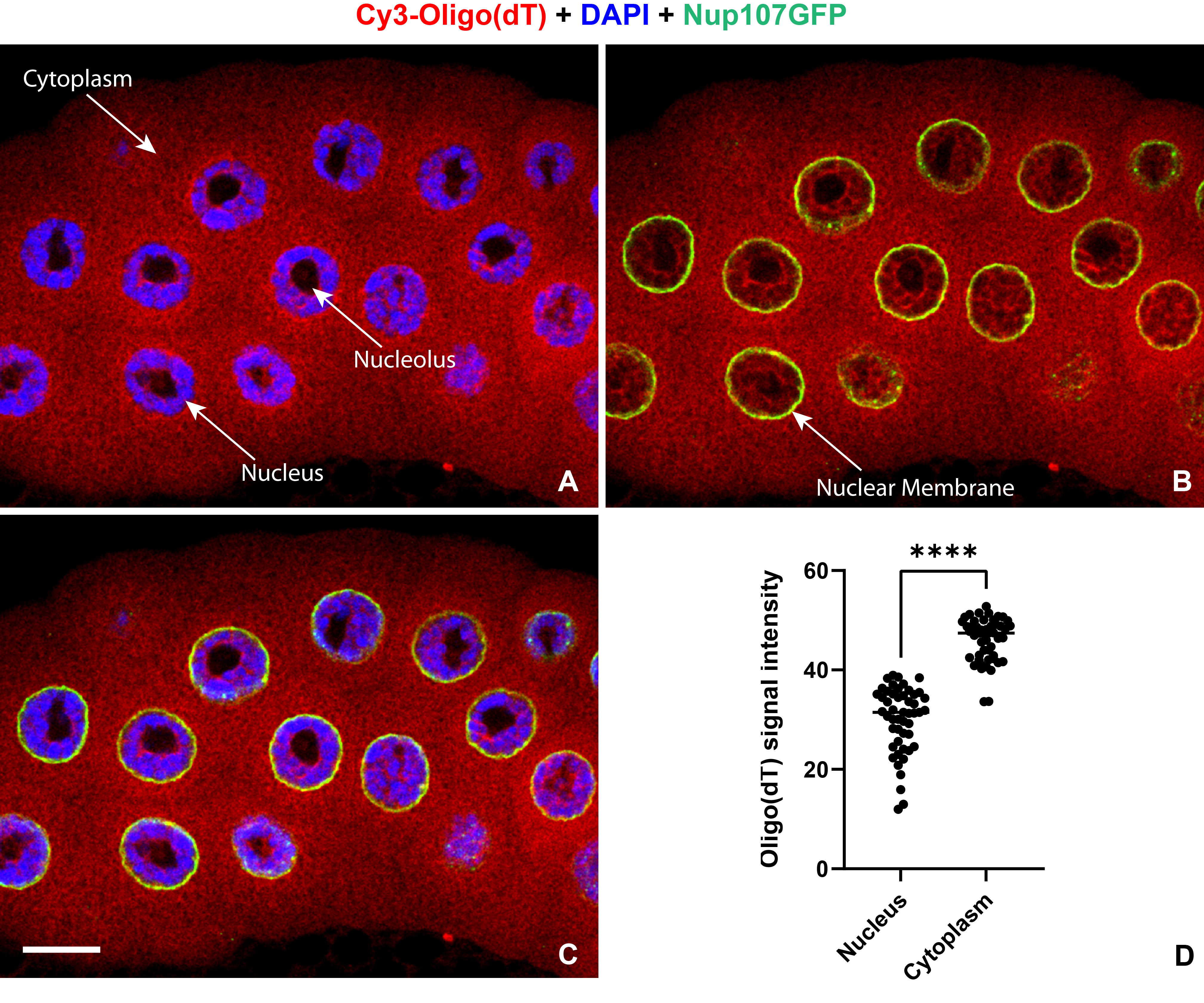
Figure 1. Oligo(dT) fluorescence in situ hybridization. (A–C) Confocal single-section image of Cy3-labeled oligo(dT) FISH showing the distribution of polyadenylated mRNAs (red) in the salivary gland of third-instar larva. The nuclear membrane is visualized by the GFP-tagged Nup107 protein (green). Nuclei are counterstained with DAPI (blue). Scale bars = 20 μm. (D) Oligo(dT) probe fluorescence intensity in the nucleus and cytoplasm of the salivary gland (n = 50). The difference in signal intensity was statistically significant (**** represents p < 0.001) tested by the unpaired t-test.
Validation of protocol
This protocol has been used and validated in the following research article:
Singh et al. [9]. The RNA helicase UPF1 associates with mRNAs co-transcriptionally and is required for the release of mRNAs from gene loci. eLife.
General notes and troubleshooting
General notes
1. Experimental controls for FISH: In order to ensure that the observed FISH signal is genuine and not a false-positive result, positive and negative experimental controls should also be processed in parallel. The positive control could be a widely expressed and well-studied gene with a known expression pattern, like tubulin or G3PDH. FISH with a probe against an mRNA that is expressed only in some specific cell types can also be used as a good control to confirm the specificity of the signal. Likewise, a negative control could be probed with the sense strand sequence of the same gene, which is expected to have no complementary sequence and, therefore, should not generate any hybridization signal. However, many genes express the antisense strand as well and, therefore, can also give a positive signal with the sense probe. Another negative control can be a parallel sample treated with RNase, which must not produce any FISH signal. However, great care is needed, as even a very small RNase contamination of the experimental sample can ruin the entire experiment.
2. RNases are the natural enemy of RNA molecules, and blocking their activity is a big challenge in FISH. They can be present anywhere, including reagents, solutions, instruments, and, most importantly, on our body surface. Therefore, special care is required in all experiments involving RNA. Some RNases are resistant to standard decontamination methods. Therefore, all the reagents should be kept in an RNase-free environment. The solutions and dH2O used in the experiment should be treated with DEPC. The working bench, pipettes, and plasticware should be wiped with 0.2 N NaOH and then with hot dH2O. All glassware should be treated with 0.5% SDS and baked (dry heating at 150 °C) after washing. The tubes and tips should be fresh and stored in properly sealed containers. It is important to wear disposable plastic or latex gloves to keep the sample safe from RNases present on our hands. If the glove has come into contact with RNase-contaminated lab equipment, such as pipettes, microscopes, or door handles, it should be immediately changed, as it would no longer be RNase-free.
3. Keep the probe stock at -80 °C and an aliquot of working concentration at -20 °C, as multiple freeze/thaw cycles can degrade the probe. The box containing the probe should be opaque to ensure a minimal possibility of photobleaching.
4. A few tissues, like the Drosophila embryo, have a thick cuticle layer. In order to perform FISH in an embryo, we need to perform dechorionation to ensure the proper penetration of the probe inside the embryo. Incubating the embryo with a 3% bleach solution for 90 s offers efficient dechorionation [17]. Likewise, tissues like adult brain need a higher concentration of detergent in washing buffer (0.5% PBST) to penetrate the probe. Tissues like Malpighian tubule, gut, fat body, etc. need an even smaller concentration of washing buffer (0.1% PBST).
5. Stopping the experiment at any step of the protocol can cause variability in the results. Therefore, it is suggested to complete the FISH protocol without any pause.
Troubleshooting
1. The sequence of the probe should be very specific and complementary to the RNA one wants to localize. The concentration of probe in hybridization buffer must be in an adequate range, as a very low or high concentration may lead either to no signal or to a nonspecific signal, respectively.
2. The concentration of Triton X-100 in the rinsing solution varies with tissue type, e.g., tissues with a thick outer membrane/sheath (larval salivary gland and brain) need a higher concentration (0.5%), while tissues with a thin outer membrane (larval imaginal disc, Malpighian tubules, fat body, etc.) need a low (0.1%) concentration of the detergent.
Acknowledgments
We thank the Science and Engineering Research Board, Grant/Award Number CRG/2022/002767 and the Department of Biotechnology, Ministry of Science and Technology, India, Grant/Award Number BT/RHD/35/02/2006 for funding support.
Competing interests
The authors declare no conflicts of interest.
References
- Gall, J. G. and Pardue, M. L. (1969). FORMATION AND DETECTION OF RNA-DNA HYBRID MOLECULES IN CYTOLOGICAL PREPARATIONS. Proc Natl Acad Sci USA. 63(2): 378–383. https://doi.org/10.1073/pnas.63.2.378
- Singh, A. K. and Lakhotia, S. C. (2015). Dynamics of hnRNPs and omega speckles in normal and heat shocked live cell nuclei of Drosophila melanogaster. Chromosoma. 124(3): 367–383. https://doi.org/10.1007/s00412-015-0506-0
- Tripathi, V., Fei, J., Ha, T. and Prasanth, K. V. (2014). RNA Fluorescence In Situ Hybridization in Cultured Mammalian Cells. Methods Mol Biol. 123–136. https://doi.org/10.1007/978-1-4939-1369-5_11
- Tam, R., Smith, K. P. and Lawrence, J. B. (2004). The 4q subtelomere harboring the FSHD locus is specifically anchored with peripheral heterochromatin unlike most human telomeres. J Cell Biol. 167(2): 269–279. https://doi.org/10.1083/jcb.200403128
- Johnson, C., Primorac, D., McKinstry, M., McNeil, J., Rowe, D. and Lawrence, J. B. (2000). Tracking Col1a1 RNA in Osteogenesis Imperfecta. J Cell Biol. 150(3): 417–432. https://doi.org/10.1083/jcb.150.3.417
- Singh, A. K. and Lakhotia, S. C. (2015). The hnRNP A1 homolog Hrb87F/Hrp36 is important for telomere maintenance in Drosophila melanogaster. Chromosoma. 125(3): 373–388. https://doi.org/10.1007/s00412-015-0540-y
- Singh, A. K. and Lakhotia, S. C. (2022). Cytological Approaches to Visualize Intracellular Dynamics of RNA-Binding Proteins at Active Genes in Drosophila. Methods Mol Biol. : 271–293. https://doi.org/10.1007/978-1-0716-2891-1_16
- Byron, M., Hall, L. L. and Lawrence, J. B. (2013). A Multifaceted FISH Approach to Study Endogenous RNAs and DNAs in Native Nuclear and Cell Structures. Curr Protoc Hum Genet. 76(1): ehg0415s76. https://doi.org/10.1002/0471142905.hg0415s76
- Singh, A. K., Choudhury, S. R., De, S., Zhang, J., Kissane, S., Dwivedi, V., Ramanathan, P., Petric, M., Orsini, L., Hebenstreit, D., et al. (2019). The RNA helicase UPF1 associates with mRNAs co-transcriptionally and is required for the release of mRNAs from gene loci. eLife. 8: e41444. https://doi.org/10.7554/elife.41444
- Aleman, J. R., Little, S. C. and Capelson, M. (2022). Using Single Molecule RNA FISH to Determine Nuclear Export and Transcription Phenotypes in Drosophila Tissues. Methods Mol Biol. 2502: 113–125. https://doi.org/10.1007/978-1-0716-2337-4_8
- Rivera, D. E., Lažetić, V., Troemel, E. R. and Luallen, R. J. (2022). RNA Fluorescence in situ Hybridization (FISH) to Visualize Microbial Colonization and Infection in Caenorhabditis elegans Intestines. J Visualized Exp. e3791/63980–v. https://doi.org/10.3791/63980-v
- Yoon, D. S., Pendergrass, D. L. and Lee, M. H. (2016). A simple and rapid method for combining fluorescent in situ RNA hybridization (FISH) and immunofluorescence in the C. elegans germline. MethodsX. 3: 378–385. https://doi.org/10.1016/j.mex.2016.05.001
- Jansova, D. (2015). Single Molecule RNA FISH in the Mammalian Oocyte. Bio Protoc. 5(23): e1666. https://doi.org/10.21769/bioprotoc.1666
- Wadsworth, G., Parikh, R. and Kim, H. (2018). Single-probe RNA FISH in Yeast. Bio Protoc. 8(11): e2868. https://doi.org/10.21769/bioprotoc.2868
- Onorati, M. C., Lazzaro, S., Mallik, M., Ingrassia, A. M. R., Carreca, A. P., Singh, A. K., Chaturvedi, D. P., Lakhotia, S. C. and Corona, D. F. V. (2011). The ISWI Chromatin Remodeler Organizes the hsrω ncRNA–Containing Omega Speckle Nuclear Compartments. PLos Genet. 7(5): e1002096. https://doi.org/10.1371/journal.pgen.1002096
- Choudhury, S. R., Singh, A. K., McLeod, T., Blanchette, M., Jang, B., Badenhorst, P., Kanhere, A. and Brogna, S. (2016). Exon junction complex proteins bind nascent transcripts independently of pre-mRNA splicing in Drosophila melanogaster. eLife. 5: e19881. https://doi.org/10.7554/elife.19881
- Legendre, F., Cody, N., Iampietro, C., Bergalet, J., Lefebvre, F.A., Moquin-Beaudry, G., Zhang, O., Wang, X., and Lécuyer, E. (2013). Whole Mount RNA Fluorescent in situ Hybridization of Drosophila Embryos. J Vis Exp. 50057. https://doi.org/10.3791/50057
文章信息
稿件历史记录
提交日期: Jan 17, 2026
接收日期: Apr 22, 2026
在线发布日期: May 13, 2026
出版日期: Jun 5, 2026
版权信息
© 2026 The Author(s); This is an open access article under the CC BY license (https://creativecommons.org/licenses/by/4.0/).
如何引用
Readers should cite both the Bio-protocol article and the original research article where this protocol was used:
- Kumar, A., Biswas, J. and Singh, A. K. (2026). Oligo(dT) Fluorescence In Situ Hybridization to Visualize the Poly(A) mRNAs in the Internal Tissues of Drosophila. Bio-protocol 16(11): e5702. DOI: 10.21769/BioProtoc.5702.
- Singh, A. K., Choudhury, S. R., De, S., Zhang, J., Kissane, S., Dwivedi, V., Ramanathan, P., Petric, M., Orsini, L., Hebenstreit, D., et al. (2019). The RNA helicase UPF1 associates with mRNAs co-transcriptionally and is required for the release of mRNAs from gene loci. eLife. 8: e41444. https://doi.org/10.7554/elife.41444

 Download Citation in RIS Format
分类
分子生物学 > RNA > RNA定位
细胞生物学 > 细胞染色 > 核酸
发育生物学 > 细胞生长和命运决定
您对这篇实验方案有问题吗?
在此处发布您的问题,我们将邀请本文作者来回答。同时,我们会将您的问题发布到Bio-protocol Exchange,以便寻求社区成员的帮助。
 发布问题
发布问题


