


Comprehensive Protocol for Handling Human Small Airway Epithelial Cells (HSAECs) to Establish Air–Liquid Interface (ALI) Cultures With TEER-Based Barrier Integrity Assessment
人小气道上皮细胞气液界面培养及基于 TEER 的屏障完整性评估操作方案

(*contributed equally to this work) 发布: 2026年06月05日第16卷第11期 DOI: 10.21769/BioProtoc.5699 浏览次数: 287
评审: Anonymous reviewer(s)
 PDF
PDF  Q&A
Q&A
Advertisement








Abstract
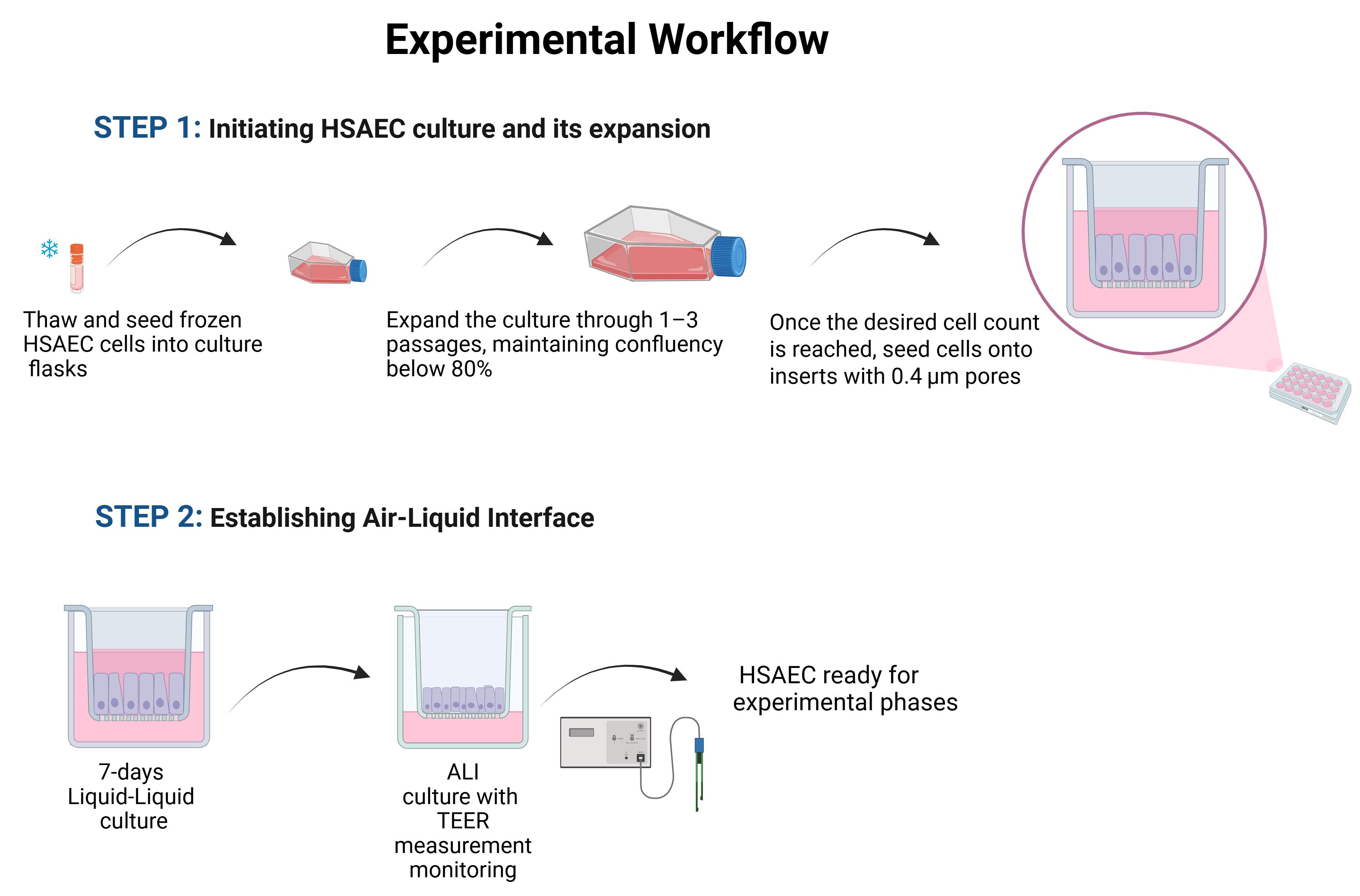
Understanding epithelial barrier function is essential for studying both its normal physiology and its role in disease, yet choosing an appropriate experimental model remains challenging. Animal models are commonly used but often suffer from interspecies differences that limit translational relevance. Human-derived cell lines offer a more suitable alternative, although establishing them often requires immortalisation strategies that involve overexpression of oncogenes, which can introduce phenotypic and functional changes. In contrast, primary cells, such as human small airway epithelial cells (HSAECs), provide a more physiologically accurate model. A critical aspect of replicating the native respiratory environment is maintaining continuous air exposure, which can be achieved through air–liquid interface (ALI) culture. This protocol provides a unified, step-by-step workflow for cultivating primary HSAECs under ALI conditions, covering the entire process from initial recovery after cryopreservation to the formation of a barrier-like layer. The protocol incorporates non-invasive methods such as transepithelial electrical resistance (TEER) measurements to monitor its integrity. While individual elements of this workflow have been described separately in different studies, a consolidated version encompassing the full workflow has not been widely available. This resource is intended for researchers with limited experience in airway epithelial culture and offers practical, clear guidance through each step of the process.
Key features
• Using primary HSAECs enables modelling the human respiratory barrier while avoiding limitations of immortalised or animal-derived cell lines.
• ALI culture technique allows continuous air exposure, closely resembling in vivo conditions for airway epithelial cells.
• TEER measurement offers a non-invasive, rapid method to assess epithelial barrier integrity without damaging the cultured cell layer.
• Protocol supports barrier function studies, including but not limited to respiratory infections, allergic responses, toxicology screening, microbiome interactions, and drug delivery investigation.
Keywords: Air–liquid interface (ALI) (气液界面培养) Primary cell culture (原代细胞培养)
Human small airway epithelial cells (HSAECs) (人小气道上皮细胞)
Transepithelial electrical resistance (TEER) (跨上皮电阻)
Basic protocol (基础实验方案)
Primary cell culture (原代细胞培养)
Human small airway epithelial cells (HSAECs) (人小气道上皮细胞)
Transepithelial electrical resistance (TEER) (跨上皮电阻)
Basic protocol (基础实验方案)
Graphical overview
Background
As the initial point of contact between external and internal environments, the epithelial barrier of the respiratory tract is essential for maintaining homeostasis. Humans inhale approximately 6 L of air per minute [1], with particle composition varying by location and reaching, for example, 1,300–1,900 particles per litre indoors [2]. Inhaled air may contain pollutants, viruses, bacteria, fungi, pollen, and other agents [3], which can significantly impact the integrity of the respiratory epithelial barrier and compromise health. Therefore, mapping the structure and understanding the function of the epithelial barrier are essential in studies of barrier physiology and pathophysiology, providing a foundation for exploring underlying mechanisms and evaluating therapeutic strategies. However, choosing an appropriate experimental system for such studies remains challenging. Animal models provide valuable biological insights but often exhibit interspecies differences that limit translational relevance. Immortalised human cell lines are easy to maintain, yet their establishment frequently involves genetic modifications that may alter phenotype and function. Therefore, primary human airway epithelial cells are increasingly used as an alternative model, as they retain many of the physiological characteristics necessary to reproduce the native epithelial behaviour in vitro.
Generally, primary cells are collected with the donor’s consent from specific body regions (e.g., nasal swab, bronchoscopy, intraoperative collection) and undergo minimal manipulation, thereby preserving their physiological relevance and making them suitable for studying native epithelial responses. One advantage of using primary cells is that they retain donor-specific biological diversity, allowing researchers to capture aspects of interindividual variability that are lost in immortalised systems. However, like most primary cultures, they are characterised by a limited lifespan and sensitivity to storage and handling conditions, which may influence their functional properties [4,5].
A characteristic feature of the respiratory epithelium is its continuous exposure to air, which shapes its structure, function, and responsiveness to external stimuli. When cultured under appropriate conditions, primary airway epithelial cells can form cohesive layers that reproduce essential traits, including barrier formation, allowing the examination of key aspects of epithelial physiology and pathology in a controlled setting. To support barrier development under in vitro conditions, culture systems that enable apical air contact have become fundamental tools in airway epithelial research. These approaches facilitate the formation of morphologically and functionally mature epithelial layers, providing a physiologically relevant environment for studying epithelial behaviour.
The structural integrity of such cultures can be evaluated using transepithelial electrical resistance (TEER), a non-invasive measurement of paracellular permeability [6]. A progressive increase and subsequent stabilisation of TEER values typically indicate maturation of the epithelial barrier, whereas reduced values reflect weakened tight-junction organisation. Fluctuations observed during differentiation may arise from biological changes in junctional architecture or cell morphology [7], but can also reflect the methodological differences, including electrode design, measurement conditions, calibration, or handling during assessment.
The protocol presented in this manuscript integrates these elements into a unified workflow for establishing primary cell culture and an airway-exposed model and assessing barrier integrity. While similar procedures have been reported independently across studies, consolidated, practical guidance spanning the entire process is less common. By outlining each stage in a clear, accessible manner, this protocol is intended to support researchers, particularly those with limited prior cell culture experience, in implementing reproducible, physiologically relevant airway epithelial models.
Materials and reagents
Biological materials
1. Primary small airway epithelial cells (HSAECs), normal, human, derived from the lung tissue of a healthy 16-year-old Hispanic/Latino male (ATCC, catalog number: PCS-301-010)
Reagents
1. Airway epithelial cell basal medium (ATCC, catalog number: PCS-300-030)
2. Bronchial Epithelial Growth kit (ATCC, catalog number: PCS-300-040)
3. Penicillin-streptomycin solution (Sigma-Aldrich, catalog number: P4333)
4. Dulbecco’s phosphate-buffered saline (D-PBS) (ATCC, catalog number: 30-2200)
5. Trypsin-EDTA for primary cells containing 0.05% trypsin and 0.02% EDTA (ATCC, catalog number: PCS-999-003)
6. Trypsin neutralizing solution (ATCC, catalog number: PCS-999-004)
7. Trypan Blue 0.4% (Invitrogen, catalog number: T10282)
8. Ethanol 96% (POCH, catalog number: 396420113)
9. Ultrapure sterile water (Sigma-Aldrich, catalog number: W3500)
Solutions
1. HSAEC culture medium (see Recipes)
2. 70% ethanol (see Recipes)
Recipes
1. HSAEC culture medium
Transfer the indicated volume of each Bronchial Epithelial Growth kit component directly to the airway epithelial cell basal medium according to the manufacturer’s procedure:
| Reagent | Final concentration | Quantity or volume |
|---|---|---|
| HLL supplement | HSA 500 μg/mL Linoleic acid 0.6 μM Lecithin 0.6 μg/mL | 1.25 mL |
| L-glutamine | 6 mM | 15 mL |
| Airway epithelial cell supplement | Epinephrine 1.0 μM Transferrin 5 μg/mL T3 10 nM Hydrocortisone 5 μg/mL, rh EGF 5 ng/mL rh insulin 0.1 μg/mL | 5.0 mL |
| Optional penicillin-streptomycin solution | Penicillin: 10 Units/mL Streptomycin: 10 μg/mL | 0.5 mL |
| Airway epithelial cell basal medium | n/a | 485 mL |
| Total | n/a | 506.75 mL |
Ensure the medium is formulated under sterile conditions using a biological safety cabinet. Due to light sensitivity, the complete growth medium must be kept in the dark at 2–8 °C and should not be frozen. Storage duration should not exceed 30 days. During the procedures, it is recommended to transfer the necessary volume of medium into a separate sterile container, e.g., a 50 mL centrifuge tube, and heat only the portion required.
2. Ethanol 70%
| Reagent | Final concentration | Quantity or volume |
|---|---|---|
| Ethanol 96% | 70% | 72.9 mL |
| Ultrapure sterile water | 27.1 mL | |
| Total | 70% | 100 mL |
Laboratory supplies
1. Tissue culture flasks, depending on the number of cells and culture stage: T25 (TPP, catalog number: 90026), T75 (TPP, catalog number: 90076), T150 (TPP, catalog number: 90151), and T300 (TPP, catalog number: 90301)
2. Dual-chamber cell counting slides (Bio-Rad, catalog number: 1450011)
3. Tissue culture test plate 24 well (TPP, catalog number: 92024)
4. ThinCerts-TC inserts 24 well, pore size 0.4 μm, transparent (Greiner Bio-One, catalog number: 662641)
5. Serological pipettes: 5 mL (GoogLab Scientific, catalog number: G33260011), 10 mL (GoogLab Scientific, catalog number: G33270011)
6. Pipetting tips: 2–100 μL (Biosphere Filter Tips, catalog number: 70.760.212), 1,000 μL (GenoPlast Biotech S.A., catalog number: GBFT100-R-NS)
7. Centrifuge tubes: 50 mL (GoogLab Scientific, catalog number: G66020522), 15 mL (GoogLab Scientific, catalog number: G66010522)
8. Eppendorf tubes 1.5 mL (Eppendorf, catalog number: EP0030120086)
Equipment
1. Cell culture incubator with controlled temperature (37 °C), CO2 (5%), oxygen (20%), and humidity (90%) (Memmert, model: ICO-150MED, catalog number: ICO-150MED)
2. Biosafety cabinet class II (Thermo Scientific, model: SAFE2020, catalog number: 51026637)
3. Inverted microscope (Olympus, model: CK2, catalog number: OLY-CK2)
4. Centrifuge (Eppendorf, model: 5804R, catalog number: 5805000010)
5. Automated cell counter (Bio-Rad, model: TC20, catalog number: 1450102)
6. Epithelial Volt-Ohm Meter Millicell (Millipore, model: ERS-2, catalog number: MERS00002)
7. Adjustable automatic pipettes: 2–20 μL (HTL Lab Solution, model: Discovery Comfort, catalog number: DV20), 20–200 μL (HTL Lab Solution, model: Discovery Comfort, catalog number DV200), 100–1,000 μL (HTL Lab Solution, model: Discovery Comfort, catalog number DV1000)
8. Automatic pipettor for serological pipettes (Finetech Research and Innovation Corporation, catalog number: WIZ-EP)
9. Water bath (BioSab, model: WB-4MS, catalog number: BS-010406-AAA)
10. Sterile tweezers (Sigma-Aldrich, catalog number 930229)
11. Plastic box, e.g., Tubby container with lid (Merck, catalog number: Z675946)
Software and datasets
1. GraphPad Prism (GraphPad Prism, Version 10), https://www.graphpad.com/scientific-software/prism/, commercial license required
2. Microsoft Excel (Microsoft Corporation, Microsoft 365 Version), https://www.microsoft.com/en-us/microsoft-365/excel, commercial license required
Note: The software listed above were used in this protocol; however, other tools capable of performing descriptive statistical analysis and data visualisation may also be suitable, e.g., LibreOffice Calc (The Document Foundation, Version 7.6), https://www.libreoffice.org/, free and open-source.
Procedure
登录/注册后免费查看全文
文章信息
稿件历史记录
提交日期: Oct 22, 2025
接收日期: Apr 14, 2026
在线发布日期: May 12, 2026
出版日期: Jun 5, 2026
版权信息
© 2026 The Author(s); This is an open access article under the CC BY license (https://creativecommons.org/licenses/by/4.0/).
如何引用
Jakubczyk, D., Pyclik, M., Kozakiewicz, D., Macała, J., Zabłocka, A. and Górska, S. (2026). Comprehensive Protocol for Handling Human Small Airway Epithelial Cells (HSAECs) to Establish Air–Liquid Interface (ALI) Cultures With TEER-Based Barrier Integrity Assessment. Bio-protocol 16(11): e5699. DOI: 10.21769/BioProtoc.5699.

 Download Citation in RIS Format
分类
细胞生物学 > 细胞分离和培养 > 细胞分化
细胞生物学 > 基于细胞的分析方法 > 电生理技术
生物工程 > 生物医学工程
您对这篇实验方案有问题吗?
在此处发布您的问题,我们将邀请本文作者来回答。同时,我们会将您的问题发布到Bio-protocol Exchange,以便寻求社区成员的帮助。
 发布问题
发布问题


