


Isolation and Biophysical Characterization of Extracellular Vesicles Released by Myocytes
肌细胞来源细胞外囊泡的分离与生物物理特性分析

发布: 2026年05月20日第16卷第10期 DOI: 10.21769/BioProtoc.5698 浏览次数: 544
评审: Anonymous reviewer(s)
 PDF
PDF  Q&A
Q&A
参见作者原研究论文
The authors used this protocol in:

Apr 2026
Advertisement







Abstract
Extracellular vesicles (EVs) are lipid bilayer–enclosed vesicles released by diverse cell types and found in various body fluids. Because their composition and cargo dynamically respond to physiological and environmental cues, EVs hold promise both as biomarkers and as carriers for therapeutic delivery. Skeletal muscle functions as an endocrine organ, secreting myokines and EVs that modulate a wide range of cellular processes. The murine C2C12 cell line is a widely used in vitro model for investigating muscle biology. Here, we describe a protocol for isolating EVs from differentiated C2C12 myocytes. The isolated EVs are characterized and validated using western blotting, transmission electron microscopy (TEM), and dynamic light scattering (DLS) analysis. This workflow provides a robust platform for studying the molecular composition and functional roles of muscle-derived EVs.
Key features
• Standardized protocol for isolating extracellular vesicles (EVs) from differentiated C2C12 myocytes.
• High-purity EV isolation achieved through sequential ultracentrifugation and size-exclusion chromatography.
• Generates EV samples compatible with diverse downstream applications, including western blotting, transmission electron microscopy (TEM), and dynamic light scattering (DLS) analysis.
• Enables comparative studies of muscle-derived EV composition and functional changes under both control and treatment conditions in C2C12 myocytes.
Keywords: Extracellular vesicles (EVs) (细胞外囊泡) Myocytes (肌细胞)
In vitro model (体外模型)
Skeletal muscle (骨骼肌)
Western blotting (蛋白质印迹)
Transmission electron microscopy (透射电子显微镜)
Dynamic light scattering (DLS) (动态光散射法)
Myocytes (肌细胞)
In vitro model (体外模型)
Skeletal muscle (骨骼肌)
Western blotting (蛋白质印迹)
Transmission electron microscopy (透射电子显微镜)
Dynamic light scattering (DLS) (动态光散射法)
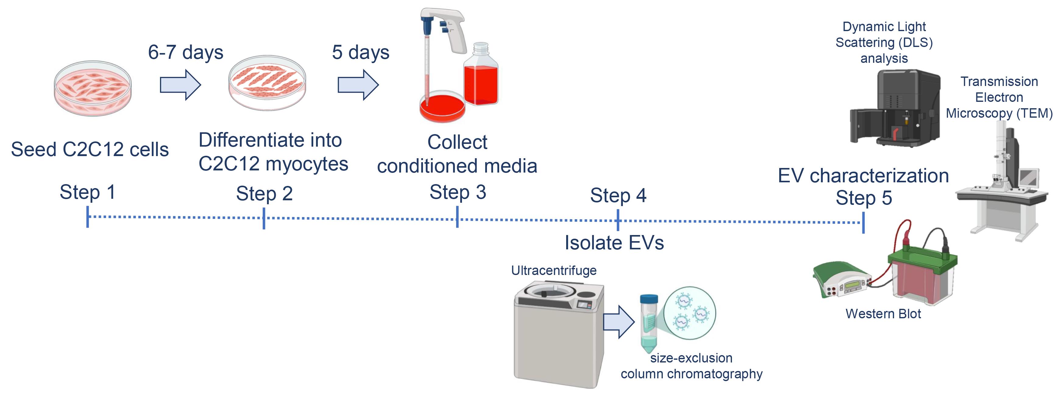
Graphical overview
Background
Extracellular vesicles (EVs) are nanosized, lipid bilayer–enclosed particles (30–150 nm) secreted by various cell types, including immune cells, neurons, adipocytes, and muscle cells [1]. They are present in body fluids such as plasma, urine, cerebrospinal fluid, breast milk, and saliva. EV composition and cargo change dynamically in response to cellular and physiological conditions, making them promising biomarkers and therapeutic tools in disease and regenerative medicine [2,3].
Skeletal muscle accounts for about 40%–50% of total body mass and plays a central role in movement, energy metabolism, and systemic homeostasis [4–8]. It secretes signaling molecules, including myokines and EVs, which regulate lipid and glucose metabolism, myogenesis, lipogenesis, angiogenesis, and inflammation [9,10]. The murine C2C12 skeletal muscle cell line, established by Yaffe and Saxel [11], is a well-characterized in vitro model for studying muscle biology and the muscle secretome [12]. Upon differentiation, C2C12 myoblasts form multinucleated myotubes resembling mature skeletal muscle fibers, providing a robust system to investigate EV biogenesis and function [12].
Isolation of EVs from intact muscle or explant cultures is technically challenging due to mixed cell populations and the lack of markers to determine vesicle origin. To address this, we developed a standardized protocol to isolate EVs from C2C12 myocytes. Conditioned media are collected and subjected to sequential ultracentrifugation followed by size-exclusion chromatography [13,14]. The resulting myocyte-derived EVs can be validated using western blotting, transmission electron microscopy (TEM), and dynamic light scattering (DLS) analysis. This protocol enables the isolation and characterization of muscle-derived EVs with high purity, reducing contamination and avoiding misinterpretation that can arise from isolating EVs directly from heterogeneous muscle tissues.
Materials and reagents
Biological materials
1. C2C12 cells (ATCC, CRL-1772), obtained from Prof. David Virshup (CSCB Department, Duke-NUS Medical School, Singapore)
Reagents
1. PierceTM BCA Protein Assay kit (Thermo Scientific, catalog number: 23225)
2. 250 kDa Plus prestained protein marker (Vazyme, catalog number: MP202)
3. ECLTM Prime western blotting detection reagents (Cytiva, catalog number: RPN2209)
4. Horse serum, exosome-depleted (Gibco, custom-made)
5. Dulbecco’s modified Eagle medium (DMEM) (Cytiva, catalog number: SH30022.01)
6. Fetal bovine serum (FBS), exosome-depleted (Sigma, catalog number: A2720801)
7. 100 U/mL penicillin and 100 μg/mL streptomycin (Cytiva, catalog number: SV30010)
8. Centrifuge tubes, 50 mL (Biobasic Asia Pacific, catalog number: BCT0050)
9. Centrifuge tubes, 15 mL (Biobasic Asia Pacific, catalog number: BCT0015)
10. Corning® 96-well clear flat bottom polystyrene TC-treated microplates, individually wrapped, with lid, sterile (Corning, catalog number: 3596)
11. T175 cell culture flask (SPL Life Sciences, catalog number: 74175)
12. T75 Cellstar® cell culture flask (Greiner, catalog number: 658175)
13. SmartSECTM single column (System Biosciences, catalog number: SSEC200A-1)
14. LabSelect 1.5 mL microcentrifuge tube (LabSelect, catalog number: MCT-001-150)
15. Rabbit monoclonal anti-Alix antibody (Cell Signaling Technology, catalog number: 92880; 1:1,000)
16. Mouse monoclonal anti-Tsg101 antibody (BD Biosciences, catalog number: 612696; 1:500)
17. Mouse monoclonal anti-GM130 antibody (Proteintech, catalog number: 66662; 1:10,000)
18. Goat anti-rabbit IgG, HRP-linked antibody (Cell Signaling Technology, catalog number: 7074; 1:1,000)
19. Goat anti-mouse IgG, HRP-linked antibody (Cell Signaling Technology, catalog number: 7076; 1:1,000)
20. 1× Dulbecco’s phosphate-buffered saline (D-PBS), without Ca and Mg (1ST Base, catalog number: BUF-2046)
21. Trypsin-EDTA solution, with phenol red (Biobasic Asia Pacific, catalog number: UCC8105)
22. Bovine serum albumin (BSA) (Sigma-Aldrich, catalog number: A3059)
23. 20× tris buffered saline (TBS) buffer (1st Base, catalog number: BUF-3030)
24. Methanol, for analysis EMSURE® ACS, ISO, Reag. Ph Eur (Supelco, catalog number: 1.06009)
25. 2× RIPA buffer I (pH 7.4) (BioBasic Asia Pacific, catalog number: RB4475)
26. Protease inhibitor cocktail (200×) (Cell Signaling Technology, catalog number: 7012)
27. Polyoxyethylene-20 (Tween 20) (Biobasic Asia Pacific, catalog number: TB0560)
28. Amersham Hybond P 0.45 PVDF blotting membrane (Cytiva, catalog number: 10600023)
29. Formvar/carbon-supported copper grids (Merck, catalog number: 930288)
30. Phosphotungstic acid, crystal, reagent, highest purity (Electron Microscopy Sciences, catalog number: 19500)
31. VitrobotTM filter paper, grade 595, 50 mm (Electron Microscopy Sciences, catalog number: 71166-65)
32. Millipore® Stericup® quick-release vacuum filtration system (Millipore, catalog number: S2GPU05RE)
33. Cuvette, 4.2 mL, 45 × 12 mm (H × W), PS, transparent, 4 optical sides (Sarstedt, catalog number: 67.754)
34. 10× tris glycine-sodium dodecyl sulfate (TG-SDS) buffer (1st Base, catalog number: BUF-2030)
35. Bromophenol blue (Sigma-Aldrich, catalog number: B0126)
36. Ammonium persulfate (APS) (Bio-Rad, catalog number: 1610700)
37. 30% acrylamide/bis solution 37.5:1 (Bio-Rad, catalog number: 1610158)
38. 1.0 M tris buffer, pH 6.8 (1st Base, catalog number: BUF-1415)
39. 1.5 M tris buffer, pH 8.8 (1st Base, catalog number: BUF-1419)
40. 10% SDS solution (BioBasic Asia Pacific, catalog number: SD8118)
41. TEMED (Bio-Rad, catalog number: 161-0800)
42. Glycerol (Biobasic Asia Pacific, catalog number: GB0232)
43. Sodium dodecyl sulfate (SDS) (Wako, catalog number: 194-13985)
44. Trypan blue stain, 0.4% (Gibco, catalog number: 15250-061)
45. Bright-LineTM hemacytometer (Merck, catalog number: Z359629)
Solutions
1. 10% resolving gel (see Recipes)
2. 4% stacking gel (see Recipes)
3. Growth media (see Recipes)
4. Differentiation media (see Recipes)
5. Electrophoresis running buffer (see Recipes)
6. Electrophoresis transfer buffer (see Recipes)
7. Electrophoresis wash buffer (see Recipes)
8. Sample buffer (see Recipes)
9. BCA working solution (see Recipes)
10. BCA standard curve preparation (see Recipes)
Recipes
1. 10% resolving gel
| Component | Concentration | Volume |
|---|---|---|
| Water | n/a | 4.05 mL |
| Tris buffer, pH 8.8 | 1.5 M | 2.5 mL |
| 30% acrylamide/bis solution | 30% | 3.3 mL |
| SDS Solution | 10% | 100 μL |
| APS | 10% | 50 μL |
| TEMED | 5 μL |
2. 4% stacking gel
| Component | Concentration | Volume |
|---|---|---|
| Water | n/a | 3.625 mL |
| Tris buffer, pH 6.8 | 1.0 M | 0.625 mL |
| 30% acrylamide/bis solution | 30% | 0.650 mL |
| SDS | 10% | 50 μL |
| APS | 10% | 50 μL |
| TEMED | 5 μL |
3. Growth media
| Component | Concentration | Volume |
|---|---|---|
| DMEM | n/a | 445 mL |
| Exosome-depleted FBS | 10% | 50 mL |
| Penicillin/Streptomycin | 1% | 5 mL |
Note: Preparation of exosome-depleted FBS from standard serum requires ultracentrifugation at 100,000–120,000× g for 16–18 h at 4 °C.
4. Differentiation media
| Component | Concentration | Volume |
|---|---|---|
| DMEM | n/a | 485 mL |
| Exosome-depleted horse serum | 2% | 10 mL |
| Penicillin/Streptomycin | 1% | 5 mL |
Note: Preparation of exosome-depleted horse serum (HS) from standard serum requires ultracentrifugation at 100,000–120,000× g for 16–18 h at 4 °C.
5. Electrophoresis running buffer (2 L)
| Component | Volume |
|---|---|
| MilliQ water | 1.8 L |
| 10× TG-SDS buffer | 200 mL |
6. Electrophoresis transfer buffer (2 L)
| Component | Volume |
|---|---|
| MilliQ water | 1.4 L |
| 10× TG-SDS buffer | 200 mL |
| Methanol | 400 mL |
7. TBST wash buffer (1 L)
| Component | Volume |
|---|---|
| MilliQ water | 1.4 L |
| 20× TBS buffer | 500 mL |
| Tween 20 | 1 mL |
8. Sample buffer
| Component | Concentration | Volume |
|---|---|---|
| Tris buffer, pH 6.8 | 1.0 M | 6.25 mL |
| Glycerol | n/a | 25 mL |
| SDS | n/a | 2 g |
| Bromophenol blue | 0.25% | 4 mL |
| MilliQ water | n/a | 14.75 mL |
Aliquot into a 1.5 mL tube and store at -20 °C until use.
9. BCA working solution
| Reagent | Amount |
|---|---|
| Part A | 25 mL |
| Part B | 500 μL |
Note: Part A and Part B are included in the PierceTM BCA Protein Assay kit.
10. BCA standard curve preparation
| Volume of RIPA (μL) | Volume and source of BSA (μL) | Final BSA concentration (μg/mL) | |
|---|---|---|---|
| A | 0 | 60 of stock | 2,000 |
| B | 30 | 30 of stock | 1,000 |
| C | 30 | 30 of vial B dilution | 500 |
| D | 30 | 30 of vial C dilution | 250 |
| E | 30 | 30 of vial D dilution | 125 |
| F | 48 | 12 of vial E dilution | 25 |
| G | 48 | 0 | Blank |
Equipment
1. OptimaTM L-100 XP ultracentrifuge (Beckman Coulter, model: OPTIMA L-100)
2. SW 28 Ti swinging-bucket rotor with buckets (Beckman Coulter, model: SW 28)
3. Zetasizer Nano ZS (Malvern Panalytical, UK; discontinued; current replacement model: Zetasizer Ultra)
4. Allegra X-30 benchtop centrifuge (Beckman Coulter, model: Allegra X-30)
5. iBrightTM CL1500 imaging system (Invitrogen, model: CL1500)
6. HT7800 RuliTEM (Hitachi, Japan)
7. Mini-PROTEAN Tetra Cell (Bio-Rad, catalog number: 165-8033)
8. FormaTM Steri-CycleTM i160 dual CO2 incubator (Thermo Scientific, catalog number: 50163013)
9. Labculture® Class II Type A2 biological safety cabinet (ESCO, catalog number: LA2-4A1)
10. Sub Aqua Pro unstirred water bath (Grant, catalog number: SAP12)
11. Bright-LineTM hemacytometer (Merck, catalog number: Z359629)
12. Nikon Eclipse TS100 inverted microscope (Nikon, model: TS100)
13. Tecan Infinite M200 microplate reader (Tecan, model: M200)
Software and datasets
1. ZS explorer software (Malvern Panalytical, v4.0.0)
2. I-controlTM software (Tecan)
3. iBright Analysis Software (ThermoFisher Scientific, v5.3.0)
Procedure
登录/注册后免费查看全文
文章信息
稿件历史记录
提交日期: Nov 27, 2025
接收日期: Feb 4, 2026
在线发布日期: Apr 30, 2026
出版日期: May 20, 2026
版权信息
© 2026 The Author(s); This is an open access article under the CC BY-NC license (https://creativecommons.org/licenses/by-nc/4.0/).
如何引用
Goh, K. Y., Lee, W. X. and Tang, H. W. (2026). Isolation and Biophysical Characterization of Extracellular Vesicles Released by Myocytes. Bio-protocol 16(10): e5698. DOI: 10.21769/BioProtoc.5698.

 Download Citation in RIS Format
分类
细胞生物学 > 细胞器分离 > 胞外囊泡
生物物理学 > 散射光谱
细胞生物学 > 细胞成像 > 电子显微镜
您对这篇实验方案有问题吗?
在此处发布您的问题,我们将邀请本文作者来回答。同时,我们会将您的问题发布到Bio-protocol Exchange,以便寻求社区成员的帮助。
 发布问题
发布问题


