


3D Reconstruction of Mature Arabidopsis Ovules Using FIB-SEM to Study Filiform Apparatus Morphology
利用 FIB-SEM 三维重建成熟拟南芥胚珠以研究丝状器形态

发布: 2026年05月20日第16卷第10期 DOI: 10.21769/BioProtoc.5695 浏览次数: 632
评审: Munenori IshibashiMelissa MikolaiCatalina I. Pislariu
 PDF
PDF  Q&A
Q&A
参见作者原研究论文
The authors used this protocol in:

Apr 2023
Advertisement








Abstract
Volume electron microscopy based on serial sectioning allows for three-dimensional (3D) visualization and analysis of the internal structures of tissues, cells, and organelles. One such technique, focused ion beam (FIB) scanning electron microscopy (SEM), has the advantages of nanoscale sectioning and high z-resolution, but the disadvantage of limited volume processing. Because of this limitation, targeting localized objects by FIB-SEM is difficult. Here, we developed a FIB-SEM observation workflow that enables the analysis of the filiform apparatus of synergid cells enclosed in the Arabidopsis ovule. In this protocol, plant samples are stained, embedded, trimmed, and carbon-coated while maintaining their orientation within the tissue. Then, sequential observations are performed using Cut & See function of FIB-SEM, followed by image processing for 3D reconstruction. Utilization of multi-scanning and image cropping from high-resolution data helps to identify localized targets within plant tissue. The filiform apparatus, which is an invaginated cell wall structure of the synergid cells, shows distinct contrast in each image, allowing for segmentation using brightness-based binarization. Such segmentation avoids the need to manually trace complex structures and facilitates 3D reconstruction by volume electron microscopy.
Key features
• Sampling and trimming of the resin block enable directionally loading in FIB-SEM.
• Multi-scanning by FIB-SEM and target extraction by image processing software enable 3D reconstruction of local areas within the sample block.
• Binarization using distinctive brightness of cellular structures enables segmentation without manual tracing of complex structures such as the filiform apparatus cell wall.
Keywords: Carbon coating (碳镀膜) Cell wall (细胞壁)
Chemical fixation (化学固定)
En bloc staining (整体染色)
Electron microscopy (电子显微镜)
Focused ion beam (聚焦离子束)
Plant (植物)
Ultramicrotomy (超薄切片技术)
Cell wall (细胞壁)
Chemical fixation (化学固定)
En bloc staining (整体染色)
Electron microscopy (电子显微镜)
Focused ion beam (聚焦离子束)
Plant (植物)
Ultramicrotomy (超薄切片技术)
Graphical overview
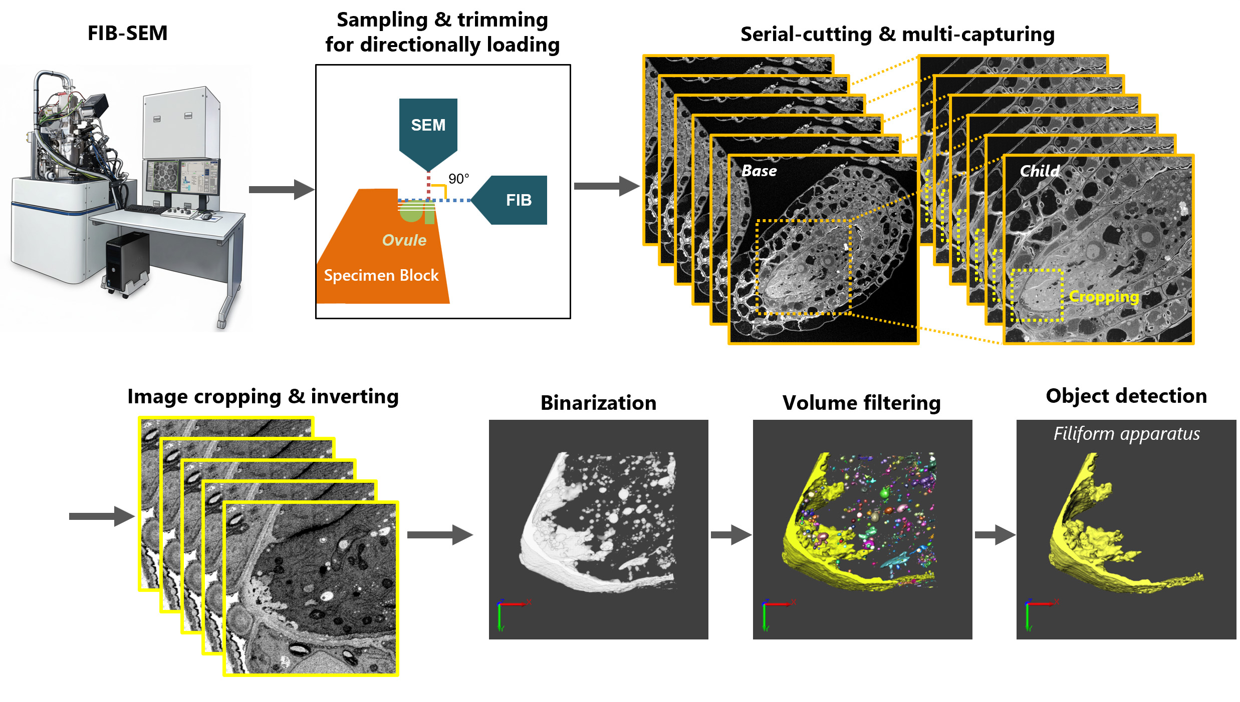
Workflow of 3D reconstruction of filiform apparatus morphology. Focused ion beam (FIB) scanning electron microscopy (SEM) is used for sectioning and observation. Set the ovule embedded in a resin block to align with the beam direction of the FIB and SEM. Serially cut the ovule and capture the SEM images with multiple positions and magnification (Base for a whole ovule and Child for synergid cells). Crop the serial images for the filiform apparatus and invert the grayscale. Detect the bright regions with binarization, filter based on object volumes, and select the largest one for the filiform apparatus without noise.
Background
In the present decade, the three-dimensional (3D) anatomy of fine structures within tissues, cells, and organelles using electron microscopes is becoming widespread, with technological advances in scanning electron microscopy (SEM) [1–3]. The method is based on serially cutting and observing the resin-embedded sample and then reconstructing the original sample structures on the image processing software. Array tomography and serial block face (SBF)-SEM using an ultramicrotome has the advantage of a wide field of view (FOV) but the disadvantage of z-resolution (thickness of cutting >50 nm). Focused ion beam (FIB)-SEM using a finely focused beam of ions (usually, gallium, Ga+) has the advantage of site-specific analysis with ultrafine z-resolution (thickness of cutting >5 nm). Although FIB is unfamiliar to biologists, it is a common instrument in materials science and the semiconductor industry. After the first utilization of FIB-SEM for 3D analysis of biological samples (in brain tissues [4]), it has contributed to clarifying the cellular or subcellular structures of animal and plant tissues or cultured cells [5–8]. FIB-SEM is a powerful tool for observing frequently occurring objects within a sample block (standard mesophyll cells in a leaf [7] or centrifugally concentrated unicellular organisms [8]); however, it is difficult to target the localized object by FIB-SEM due to the limitation of the total volume of milling.
We have studied plant fertilization and previously analyzed the morphology of Arabidopsis ovules using confocal laser scanning microscopy [9,10]. We focused on the filiform apparatus in synergid cells of the ovule [11]. The filiform apparatus [12] is a glandular structure with invaginated cell walls and plasma membranes that has usually been described as a finger-like structure by transmission electron microscopy (TEM) [13,14]. However, previous reports were limited to two-dimensional (2D) cross-sectional information. Therefore, we attempted to reveal the 3D structure of the filiform apparatus at a nanometer scale using FIB-SEM because of its high z-resolution. In a mature Arabidopsis ovule, the target region is confined to the micropylar end of two adjacent synergid cells, each approximately 10 μm wide and 25 μm long. This small and highly localized structure makes precise targeting for FIB-SEM particularly challenging. In this protocol, the ovule was chemically fixed and embedded in resin with a placenta to keep the tissue orientation, and the resin block was trimmed so that the flat surfaces faced the FIB and SEM columns, which guarantees a high probability of showing the target regions after cutting. We also utilized the machine’s Multi Cut & See function in Hitachi MI-4000L [15]; in addition to the Base setting, the Child setting allows the definition of different imaging regions by specifying the FOV and X–Y offsets. This enables sequential SEM imaging of multiple (up to 11) regions at different magnifications during serial sectioning. This protocol sets the Base image for a wider view to check the whole ovule structure and the Child image for a magnified view with high-resolution (4K) capturing to adjust and pick up the space of the whole filiform apparatus structures.
After the successful cutting and observation, we revealed that the 3D filiform apparatus had a porous, sponge-like structure. Because the cross-sectional images of filiform apparatus were complex [11,13,14], manual segmentation (tracing the contours of objects) was difficult, as had been done for organelles (chloroplasts, mitochondria, etc. [6,16]). Machine learning-based image processing was also not well-suited for recognizing the complicated linear shape patterns. Therefore, we also established a method to easily extract the 3D structure by binarization processing using the brightness values observed in the cell walls of the filiform apparatus, which is relatively unstained (bright in TEM images or dark in SEM images) compared to the cytoplasm of synergid cells and integument cells. Those methods can meet various needs for analyzing the 3D structure of localized regions in a sample block with high z-resolution, compensating for the disadvantage of FIB, which has a narrow cutting area.
Materials and reagents
Biological materials
1. Arabidopsis thaliana ecotype Columbia (Col-0), obtained from laboratory seed stocks or a public stock center
Reagents
A. Plant sampling
1. Plant preservative mixture (PPM)TM, 100 mL (Nacalai Tesque, catalog number: 26062-84)
2. Tween 20, 50 mL (Sigma-Aldrich, catalog number: P9416)
3. Murashige and Skoog (MS) plant salt mixture, for 1 L (Wako, catalog number: 392-00591)
4. Sucrose, 500 g (Wako, catalog number: 196-00015)
5. Agar, 500 g (Wako, catalog number: 016-11875)
B. Sample preparation for electron microscopy
1. Sodium cacodylate, EM grade, 250 g (TAAB, catalog number: S007)
2. Paraformaldehyde, EM grade, 100 g (TAAB, catalog number: P001/1)
3. Glutaraldehyde, EM grade, aqueous 25%, 10 mL × 10 (Electron Microscopy Sciences, catalog number: 16220)
4. Osmium tetroxide (OsO4), 1 g (Nissin-EM, catalog number: 300)
5. Thiocarbohydrazide (TCH), 97%, 5 g (Thermo Scientific, Wako catalog number: 584-15321)
6. Toluidine blue O, 25 g (Wako, catalog number: 535-05542)
7. Uranyl acetate (e.g., Electron Microscopy Sciences, catalog number: 541-09-3)
Note: Uranyl acetate is strictly regulated in Japan. Therefore, due to difficulties in purchasing it, the authors used bottles that had been stored in their facilities for several decades.
8. DL-aspartic acid, 25 g (Wako, catalog number: 010-048842)
9. Lead stain solution, 25 mL (Sigma-Aldrich, catalog number: 18-0875-2)
10. Ethanol (99.5%) 500 mL (Wako, catalog number: 057-00456)
11. Propylene oxide 500 mL (Nissin-EM, catalog number: 311)
12. Quetol 651 set (Nissin-EM, catalog number: 370)
13. Molecular sieves 3Å 1/8 (Wako, catalog number: 133-08645)
Working solution compositions
A. Plant sampling
1. Sterilization solution for seed surface: 2% (v/v) PPM, 0.1% (v/v) Tween 20 in distilled water (DW)
2. MS medium containing 1% (w/v) sucrose, in DW
B. Sample preparation for electron microscopy
1. Buffer: 0.05 M cacodylate buffer, pH 7.4, in DW*
2. Primary fixative solution: 4% (w/v) paraformaldehyde + 2% (w/v) glutaraldehyde in the buffer*, Δ
3. OsO4 in buffer: 2% (w/v) OsO4 in the buffer*, Δ
4. OsO4 in DW: 2% (w/v) OsO4 in DW*, Δ
5. TCH solution: 1% (w/v) TCH in DW*, Δ
6. Toluidine blue stain solution: 0.5% (w/v) toluidine blue O in DW
7. Uranium stain solution: 1% (w/v) uranyl acetate in DW*
8. Lead stain solution (use directly)*
9. Graded ethanol series: 50%, 70%, 90%, 100% (v/v) ethanol in DW
10. Ethanol (100%): 99.5% ethanol added molecular sieves for water molecule adsorption
11. Mixture of Quetol 651 (as described in the product manual)
Notes:
1. * indicates hazardous solutions; handle them carefully and dispose of them properly according to the instructions of your organization.
2. Δ indicates that the solution is recommended to be used within a day after preparation.
3. For the electron microscopy sample preparation steps, the authors confirm the reagents used, their working concentrations, buffers, pH values where applicable, temperatures, incubation times, and the order of processing steps. These confirmed working conditions are described in this protocol. However, full preparation recipes for individual solutions are not included because the authors did not directly prepare these solutions.
Equipment
A. Plant sampling
1. Plant germination tray (Showa Seiki Kogyo, catalog number: PG10-HT)
2. Potting mix (SAKATA SEED CORPORATION, catalog number: 72000015)
3. Paper tray (SAKATA SEED CORPORATION, catalog number: 72000016)
4. Soil (SAKATA SEED CORPORATION, catalog number: 72000017)
5. Liquide fertilizer (Hyponex Japan, catalog number: 4977517180036)
6. Light-emitting diode (LED) (TOMY DIGITAL BIOLOGY CO., LTD, model: WPRW01)
7. 27G injection needle, 19 mm length (Terumo, catalog number: NN-2719S)
8. Fine precision tweezers, No. 5-Dumostar (AS ONE, catalog number: 7-562-65)
B. Sample preparation for electron microscopy
1. Double-edged razor blade (FEATHER, catalog number: FA-10)
2. Art knife (OLFA, catalog number: AK-5)
3. Tweezers (KFI, catalog number: K-1 AA)
4. Microtube (Eppendorf, 1.5 mL, catalog number: 3810X)
5. Glass vial (NICHIDEN-RIKA GLASS Co., catalog number: PS-5)
6. Rotary vacuum pump (ULVAC, catalog number: G-25SA)
7. Rotator (PELCO, catalog number: R2 with 1051 Head)
8. Embedding capsules (TAAB, catalog number: 063, polyethylene 8 mm diameter)
9. Embedding film, ACLAR® (Nissin-EM, catalog number: 453)
10. Incubator (DOSAKA EM, catalog number: TD-800)
C. Trimming and setting of the sample block onto the stage
1. Ultramicrotome (Leica, model: EM-UC6)
2. Glass knife maker (Sunkay Laboratories, model: Messer C)
3. Glass plate for glass knife (Nissin-EM, catalog number: 540, 5 × 100 × 100 mm)
4. Diamond knife (DiATOME, model: Code 40-HIS, histo)
5. Loop (SCI Science Center, catalog number: 3512, inoculation loop, Φ 2 mm)
6. Slide glass (Matsunami Glass, catalog number: S1214)
7. Hot plate or slide dryer (e.g., Leica, model: HI 1220, Flattening Table type)
8. Light microscope (Nikon, model: OPTIPHOT-2)
9. Razor (FEATHER, catalog number: FHS-10, Hi-Stainless Single Edge Shaving Blade)
10. Cutting mat (DAISO)
11. Cellophane tape (NICHIBAN, 12 mm) or mending tape (Scotch, 15 mm)
12. Instant adhesive (TOAGOSEI, catalog number: AA489, Aron Alpha 201)
13. Carbon tape (Nissin-EM, catalog number: 7312, w 12 mm × 20 m)
Note: As an alternative to the glass knife maker and glass plate, a diamond knife for trimming (DiATOME, Code TT-90, trim 90) is convenient.
D. Carbon coating
1. Carbon coater (Meiwafosis, model: CADE-E)
2. Carbon fiber (Meiwafosis, NSCF, Ultra-high purity analytical carbon fiber, Φ 3 mm × 6 m)
3. Filter paper (ADVANTEC, No. 1, Φ 70 mm)
4. Carbon tape (Nissin-EM, catalog number: 7312, w 12 mm × 20 m)
5. Masking tape (3M, Scotch 2899, any width, any color except black)
6. Scissors
7. Tweezers
8. Gloves
E. FIB-SEM observation
1. L-shape (FIB column and SEM column are orthogonally arranged) FIB-SEM (Hitachi-High-Tech, model: MI-4000L)
2. Sample holder (Hitachi-High-Tech, model: FIB/SEM type for MI-4000L)
F. 3D image processing
1. Workstation (Dell, model: Precision Tower 3420)
2. Graphics card (NVIDIA, model: Quadro P1000)
Software and datasets
1. MI-40000L (v5.3.5b, Hitachi High-Tech Science)
2. Fiji (v1.50, open source, http://fiji.sc/Fiji) [17]
3. PaintTool SAI (v1, Systemax, Japan) (the license required, https://www.systemax.jp/en/sai/) or any painting software
4. Image-Pro Premier 3D (v9) or Image-Pro 3D (v10) (Media Cybernetics, USA) (the license required, https://mediacy.com/)
Procedure
登录/注册后免费查看全文
文章信息
稿件历史记录
提交日期: Dec 30, 2025
接收日期: Apr 14, 2026
在线发布日期: Apr 30, 2026
出版日期: May 20, 2026
版权信息
© 2026 The Author(s); This is an open access article under the CC BY-NC license (https://creativecommons.org/licenses/by-nc/4.0/).
如何引用
Oi, T., Morikawa, T., Yamazaki, Y., Maruyama, D. and Susaki, D. (2026). 3D Reconstruction of Mature Arabidopsis Ovules Using FIB-SEM to Study Filiform Apparatus Morphology. Bio-protocol 16(10): e5695. DOI: 10.21769/BioProtoc.5695.

 Download Citation in RIS Format
分类
植物科学 > 植物细胞生物学 > 细胞成像
植物科学 > 植物细胞生物学 > 细胞壁
细胞生物学 > 细胞成像 > 电子显微镜
您对这篇实验方案有问题吗?
在此处发布您的问题,我们将邀请本文作者来回答。同时,我们会将您的问题发布到Bio-protocol Exchange,以便寻求社区成员的帮助。
 发布问题
发布问题


