


An In Vitro A-431 Epithelial Cell Infection Model for Studying Fungal Pathogenicity and Immune Responses Associated With Vulvovaginal Candidiasis
用于研究外阴阴道念珠菌病相关真菌致病性与免疫反应的 A-431 上皮细胞体外感染模型

发布: 2026年05月20日第16卷第10期 DOI: 10.21769/BioProtoc.5693 浏览次数: 490
评审: Alba BlesaDamián Lobato-MárquezAnonymous reviewer(s)
 PDF
PDF  Q&A
Q&A
参见作者原研究论文
The authors used this protocol in:

Jul 2025
Advertisement









Abstract
Vulvovaginal candidiasis (VVC), also known as vaginal thrush, is an infection of the vulvovaginal mucosa caused by fungi of the Candida genus. Particularly for patients suffering from recurrent infection, the disease has a significant impact on their quality of life. The still unknown aspects of disease pathogenesis, as well as factors driving the development of infections and recurrence, represent a challenge for both clinical practitioners and patients. Mouse models and patient studies have suggested important roles of the microbiome, deployment of fungal pathogenicity mechanisms in the vagina, and dysregulated immune responses for VVC pathology. Dissecting their individual contributions can reveal specific processes associated with infection and may inspire novel therapeutic strategies. Epithelial in vitro infection models have been playing a key role in dissecting a crucial interaction during VVC, the invasion and infection of the vaginal mucosa. They have been instrumental in characterizing candidalysin as a fungal toxin that damages epithelial cells and elicits initial inflammatory responses to catalyze downstream inflammation. Moreover, they have also revealed potential protective immune pathways. Such a standardized epithelial cell infection model offers high versatility and compatibility with different downstream assays to link epithelial responses with other processes during VVC. This protocol describes a general A-431 vulvovaginal epithelial cell–Candida infection model in detail and provides several adaptations, such as live-cell imaging and mRNA silencing, as well as possible follow-up readouts, like the quantification of cytokine release, cytotoxicity, and neutrophil recruitment to study diverse processes relevant to VVC research.
Key features
• This protocol describes the use of the A-431 epidermoid carcinoma cell line as an in vitro infection model of vulvovaginal candidiasis.
• The protocol can be adapted to answer research questions relevant to vulvovaginal candidiasis (epithelial damage, release of inflammatory cytokines, recruitment and activation of neutrophils).
• Live-cell imaging can be used to study dynamic infection processes.
• mRNA silencing can be applied to interrogate the function of genes of interest in the host–pathogen interaction.
Keywords: Candida albicans (白色念珠菌) Epithelial infection model (上皮感染模型)
Cytotoxicity (细胞毒性)
Cytokine release (细胞因子释放)
Neutrophil recruitment (中性粒细胞募集)
Live-cell imaging (活细胞成像)
A-431 cells (A-431 细胞)
In vitro (体外实验)
Epithelial infection model (上皮感染模型)
Cytotoxicity (细胞毒性)
Cytokine release (细胞因子释放)
Neutrophil recruitment (中性粒细胞募集)
Live-cell imaging (活细胞成像)
A-431 cells (A-431 细胞)
In vitro (体外实验)
Graphical overview
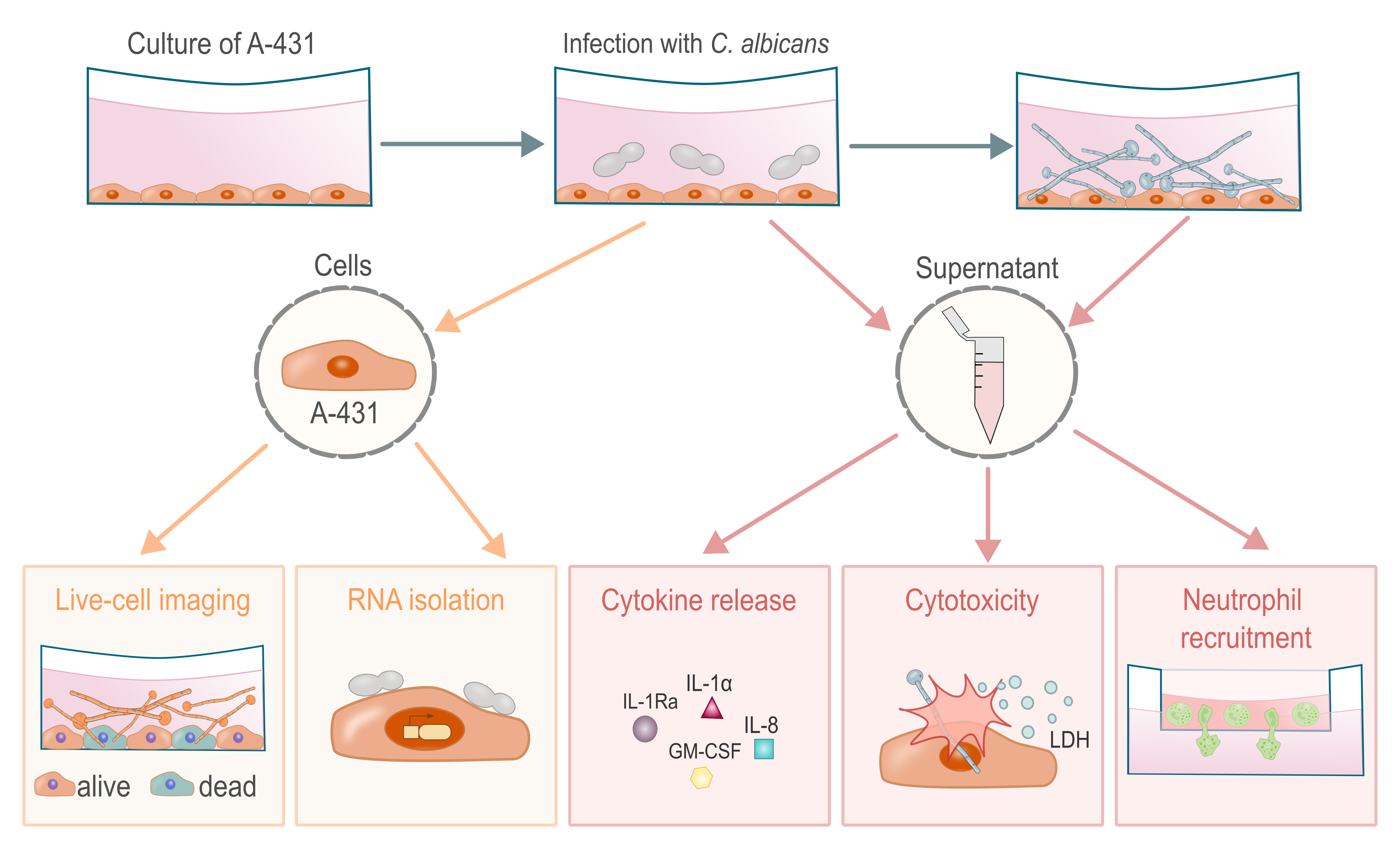
Experimental readouts of the vulvovaginal epithelial infection model with C. albicans. The epithelial cell line A-431 is infected with C. albicans yeast cells, which germinate over the course of infection. During this process, epithelial immune responses can be characterized by directly evaluating the epithelial cells with live-cell imaging and RNA isolation and by analyzing secreted molecules in the supernatant, such as cytokines and cytoplasmic lactate dehydrogenase (LDH) as a marker of cell lysis. Downstream processes on relevant effector cells can be assessed, for example, through neutrophil chemotaxis or activation assays.
Background
Vulvovaginal candidiasis (VVC) is a yeast infection of the vulvar and vaginal mucosa, most commonly caused by Candida albicans [1]. In the majority of women, this infection is linked to predisposing factors like antibiotic use and can be treated by a short course of antifungal therapy [1,2]. Still, 7%–9% of women experience recurrent infection (RVVC), defined by at least four episodes annually [3,4]. Their quality of life, including social and mental health factors, is severely burdened because of the associated discomfort, caused by curdy vaginal discharge, itching, pain, burning, redness, and swelling, as well as persistent medical costs [5–7]. Unlike other fungal infections, the disease severity of VVC is strongly intertwined with exacerbated inflammatory responses during infection, rather than being caused by compromised immunity [8–12]. While RVVC can be treated using fluconazole maintenance therapy to control the fungal burden [13], highly effective diagnostics predicting RVVC flares and sustainable therapeutic strategies are urgently needed [5].
The current state of knowledge on pathogenesis and therapy of (R)VVC has been established based on patient studies [14–22], a seminal study using an experimental human infection model [10], and VVC mouse models [23–35]. Moreover, in vitro infection models have been instrumental in the molecular dissection of cellular processes at the fungal–epithelial interface that cause tissue damage and catalyze inflammation [11,36–43].
The epidermoid carcinoma cell line A-431 is a well-established infection model of human vulvovaginal epithelial cells with Candida species [36–49]. This cell model has contributed to dissecting the molecular pathways underlying candidalysin cytotoxicity and induction of epithelial inflammatory responses driving neutrophil recruitment and activation, which is similarly observed in vivo [34,36,40,41,50,51]. In vitro epithelial infection, similar to the in vivo model, suggests a distinct phenotype of VVC caused by non-albicans species compared to C. albicans [39,41,52]. Similarly, type I interferon signaling has been linked to resistance to C. albicans infection in both in vivo and in vitro epithelial infection models [41,53–55].
A-431 cells are highly versatile due to their easy maintenance, compatibility with RPMI-1640 culture medium (which allows for co-culture with primary human immune cells or transfer of their supernatant), biosafety level, and genetic tractability. Some studies have cross-validated responses of the A-431 vulvovaginal epithelial infection model with primary human epithelial cells [41] or in vivo responses [34,36,56].
Here, we provide a step-by-step protocol based on multiple studies [38,40–42] demonstrating the potential of the A-431 vulvovaginal epithelial cell infection model for studying diverse processes relevant to the VVC research field. In the following protocol, we highlight the capabilities of the A-431 vulvovaginal epithelial infection model in terms of studying tissue damage, fungal burden, inflammatory responses, real-time cell status tracking, neutrophil recruitment, and gene expression. We thereby provide a toolset for further research into improving our understanding of the pathogenesis of VVC and to evaluate novel/candidate anti-infective, anti-virulence, and immunomodulatory therapeutic strategies for VVC.
Materials and reagents
Biological materials
1. A-431 cells (ATCC, catalog number: CRL-1555TM)
2. Candida albicans strain SC5314 (ATCC-MYA-2876) [57], risk group 2, human pathogenic fungus
3. Candida albicans strain CA3153 (kindly provided by Prof. Patrick Van Dijck) [58], risk group 2, human pathogenic fungus
4. Candida albicans strain CA3153::GFP (kindly provided by Prof. Patrick Van Dijck) [59], risk group 2, human pathogenic fungus
5. HUVEC/TERT2 cells [kindly provided by Transfer Group Antiinfectives (Leibniz-HKI); ATCC, catalog number: CRL-4053TM]
Reagents
For cell culture
1. RPMI-1640 (Gibco, catalog number: 11875093)
2. Accutase cell detachment solution (Capricorn, catalog number: ACC-1B)
3. Heat-inactivated fetal calf serum (hiFCS), heat-inactivated for 20 min at 57 °C; South America ultra-low endotoxin triple filtered (0.1 pm) (Bio & Sell, catalog number: FBS.TF.O5OO)
For culturing Candida
4. Yeast extract (Serva, catalog number: 24540.03)
5. Peptone (ThermoFisher, catalog number: 211677)
6. Glucose (Roth, catalog number: X997.2)
7. Agar-agar (Becton Dickinson, catalog number: 257353)
For cytotoxicity assay
8. Hydrochloric acid (HCl) 1 mol/L (Roth, catalog number: K025.1)
For ELISA
9. 1× TMB substrate solution (Invitrogen, catalog number: 00-4201-56)
10. Bovine serum albumin (BSA) fraction V, ≥98%, biotin-free, NZ-Origin (Roth, catalog number: 0163.4)
11. Sodium chloride (NaCl) (Roth, catalog number: 0962.2)
12. Disodium hydrogen phosphate dihydrate (Na2HPO4·2H2O) (Roth, catalog number: T877.1)
13. Potassium dihydrogen phosphate (KH2PO4) (Roth, catalog number: 23Y6.1)
14. Potassium chloride (KCl) (Roth, catalog number: HN02.1)
15. Tween-20 (Roth, catalog number: 9127.1)
For live-cell microscopy
16. SYTOTM Deep Red nucleic acid stain (Invitrogen, catalog number: S34900)
17. SYTOXTM Orange nucleic acid stain (Invitrogen, catalog number: S11368)
For RNA isolation
18. RNaseZap (Sigma-Aldrich, catalog number: R2020)
19. β-mercaptoethanol (ThermoFisher, catalog number: 125472500)
20. Ethanol 70% (Roth, catalog number: T868.3)
21. Oligo-dT (Invitrogen, catalog number: AM5730G)
22. GoTaq® qPCR master mix (Promega, catalog number: A6002)
23. Deoxynucleotide (dNTP) solution mix (New England BioLabs Inc., catalog number: N0447S)
24. RNaseOUTTM recombinant RNase inhibitor (Invitrogen, catalog number: 10777-019)
25. SuperScriptTM III reverse transcriptase (Invitrogen, catalog number: 18080-044), includes 0.1 M DTT and 5× first-strand buffer
For gene silencing
26. 5× siRNA buffer (Dharmacon, catalog number: B-002000-UB-100)
27. DharmaFECTTM transfection reagent 1 (Dharmacon, catalog number: T-2001-02)
28. siRNA target (Dharmacon)
29. siRNA non-targeting (Dharmacon)
For neutrophil chemotaxis
30. EBM® Endothelial Cell Growth Basal Medium (Lonza, catalog number: CC-3121)
31. EGM® Endothelial Cell Growth Medium SingleQuots® kit (Lonza, catalog number: CC-4133)
32. Lymphocyte separation medium, density 1.077 g/mL (Capricorn, catalog number: LSM-A)
33. Collagen A (Sigma, catalog number: C5533)
34. Penicillin-streptomycin, 10,000 U/mL (Gibco, catalog number: 15140122)
35. HBSS (Gibco, catalog number: 14025092)
36. HEPES (Gibco, catalog number: 15630106)
37. Cell Tracking Dye kit, Green, Cytopainter (Abcam, catalog number: ab138891)
38. IL-8 (Immunotools, catalog number: 11349084)
Solutions
1. Phosphate-buffered saline (PBS), pH 7.4 (see Recipes)
2. Cell culture medium RPMI-1640 + hiFCS (see Recipes)
3. Yeast peptone dextrose (YPD) agar plates/liquid medium (see Recipes)
4. ELISA wash buffer 25× (see Recipes)
5. ELISA assay diluent (see Recipes)
Recipes
1. PBS, pH 7.4
| Reagent | Final concentration | Quantity or volume |
|---|---|---|
| Na2HPO4·2H2O | 10 mM | 1.78 g/L |
| KH2PO4 | 1.8 mM | 0.24 g/L |
| NaCl | 140 mM | 8.2 g/L |
| KCl | 2.7 mM | 0.2 g/L |
Fill up with distilled water, autoclave (20 min, 121 °C), and store at room temperature.
2. Cell culture medium RPMI-1640 + hiFCS
| Reagent | Final concentration | Quantity or volume |
|---|---|---|
| RPMI-1640 medium | 90% | 450 mL |
| hiFCS | 10% | 50 mL |
| Total | 100% | 500 mL |
Store at 4 °C for a maximum of 2 weeks.
3. YPD agar plates/medium
| Reagent | Final concentration | Quantity or volume |
|---|---|---|
| Yeast extract | 1% | 10 g/L |
| Peptone | 2% | 20 g/L |
| Glucose | 2% | 20 g/L |
| Agar-agar (only for plates) | 2% | 20 g/L |
Fill up with distilled water and autoclave (20 min, 121 °C).
Liquid medium: Store at room temperature.
Agar plates: Pour 11 mL into a 9 cm culture plate, let it cool down, and store at 4 °C.
4. ELISA wash buffer 25× (1 L)
| Reagent | Final concentration | Quantity or volume |
|---|---|---|
| NaCl | 3.42 M | 200 g/L |
| Na2HPO4·2H2O | 250 mM | 44.5 g/L |
| KH2PO4 | 50 mM | 6.75 g/L |
| KCl | 67 mM | 5 g/L |
| Tween-20 | 12.5 mL/L | 12.5 mL/L |
Fill up with distilled water and store at room temperature. Dilute 1:25 in distilled water before use.
5. ELISA assay diluent (0.5 L)
Dissolve 1% BSA (10 g/L) in PBS (see Recipe 1) and store it at 4 °C. Do not use it for longer than 2 days.
Materials
For cell culture
1. Tissue culture flasks T-75 (Sarstedt, catalog number: 83.3911.002) and T-175 (Sarstedt, catalog number: 83.3912.002)
2. Tissue culture plates 6-well (TPP, catalog number: 92406), 24-well (TPP, catalog number: 92424), 96-well (TPP, catalog number: 92696)
3. Plastic Petri dish 9.2 cm (Sarstedt, catalog number: 82.1473)
4. Counting chamber Neubauer improved 0.1 mm depth (Roth, catalog number: PC72.1)
For cytotoxicity assay
5. Cytotoxicity Detection kit (LDH) (Roche, catalog number: 11644793001)
6. LDH from rabbit muscle (standard for cytotoxicity assay) (Roche, catalog number: 10127884001)
For ELISA
7. Human GM-CSF DuoSet ELISA (R&D Systems, catalog number: DY215)
8. Human IL-1Ra/1F3 DuoSet ELISA (R&D Systems, catalog number: DY280)
9. Human IL-8/CXCL8 DuoSet ELISA (R&D Systems, catalog number: DY208)
10. Human IL-1 alpha/IL-1F1 DuoSet ELISA (R&D Systems, catalog number: DY200)
11. High-binding 96-well ELISA plates, flat bottom, clear (Sarstedt, catalog number: 82.1581.200)
12. Adhesive plate sealing films (Greiner Bio-One, catalog number: 676001)
For RNA isolation
13. RNeasy Mini kit (Qiagen, catalog number: 74106)
14. RNase-Free DNase set (Qiagen, catalog number: 79254)
15. Cell scrapers, 24 cm (TPP, catalog number: 99002)
For neutrophil chemotaxis
16. TC-insert, for 24-well plates (Sarstedt, catalog number: 83.3932.300)
Equipment
1. Live-cell image microscope with filters around 488, 547, and 652 nm [used here: Incucyte SX5 (Sartorius, catalog number: 4816) with SX5 Green/Orange/NIR optical module (Sartorius, catalog number: 4832)]
2. HydroSpeed plate washer (Tecan, catalog number: 30060035)
3. Plate reader with filters for 450 and 570 nm (used here: Absorbance 96 (Byonoy)
4. NanoDrop One (Thermo Fisher, model: ND-ONE-W)
5. CFX Opus 96 Real-Time PCR System (Bio-Rad, catalog number: 12011319)
Software and datasets
1. Plate reader software: Absorbance 96 App (version 2024.08.0)
2. Live-cell imaging microscope (Incucyte 2024B)
3. Microsoft Excel (Microsoft, version 16.0.10417.20095)
4. GraphPad Prism (version 10.5.0)
5. Bio-Rad CFX Manager (version 3.1.1517.0823)
6. ImageJ/FIJI (version 2.16.0/1.54p) 22743772 [60]
Procedure
登录/注册后免费查看全文
文章信息
稿件历史记录
提交日期: Feb 27, 2026
接收日期: Apr 14, 2026
在线发布日期: Apr 30, 2026
出版日期: May 20, 2026
版权信息
© 2026 The Author(s); This is an open access article under the CC BY-NC license (https://creativecommons.org/licenses/by-nc/4.0/).
如何引用
Groß, V. E., Cheng, K. O., Cristóvão, B., Gürel, E., Himmel, M., Fernández-Fernández, C., Schuchardt, J. V., Dietschmann, A., Montaño, D. E. and Gresnigt, M. S. (2026). An In Vitro A-431 Epithelial Cell Infection Model for Studying Fungal Pathogenicity and Immune Responses Associated With Vulvovaginal Candidiasis. Bio-protocol 16(10): e5693. DOI: 10.21769/BioProtoc.5693.

 Download Citation in RIS Format
分类
微生物学 > 微生物-宿主相互作用 > 真菌
细胞生物学 > 基于细胞的分析方法 > 真菌感染
免疫学 > 粘膜免疫学 > 泌尿生殖道
您对这篇实验方案有问题吗?
在此处发布您的问题,我们将邀请本文作者来回答。同时,我们会将您的问题发布到Bio-protocol Exchange,以便寻求社区成员的帮助。
 发布问题
发布问题


